
Bronchopleural fistula: an abnormal communication between the exterior environment and the pleural cavity, often caused entry of bacteria, fluids and other substances into the chest cavity by way of the bronchial tree, for example: bronchial stump breakdown. BPF most commonly occur after large thoracic surgeries such as pneumonectomy but can occur for other reasons such as infection or trauma.
Bronchopleural fistulas (BPF) are a dread complication of thoracic surgery that has (thankfully) become rare in most countries in the last few decades. Treatment of a large bronchopleural fistula can be massive undertaking requiring collaboration and cooperative from multiple specialties including radiology, infectious disease, pulmonology, wound management and plastic surgery.
Patients often endure several months of surgical and wound care treatments prior to undergoing definitive surgical management for this condition. This treatment includes the surgical creation of large open wounds to facilitate drainage of purulent materials, repair of the fistula tract and bronchial stump and debridement / revascularization for proper tissue healing. The case presented today illustrates the devastating emotional, physical and financial costs of bronchopleural fistula as well as the need for interdisciplinary collaboration for definitive surgical repair.
Surgical repair itself carries an elevated risk of morbidity and mortality primarily from respiratory complications, infections/ sepsis and hemorrhage.
More than physical consequences
Bronchopleural fistulas carries more than just the physical consequences of pain and disability for patients and their families. There are also devastating emotional and social effects. Patients can experience a myriad of psychosocial effects from this chronic wound and related treatment. The resultant deformity from many drainage and wound management techniques, in particular, can lead to depression and social ostracism. The development of a bronchopleural fistula can contribute to relationship and intimacy issues. Several of the surgeons interviewed including Dr. Boxiong specifically mentioned both divorce and suicide as being a risk in numerous cases[1].
Case Study
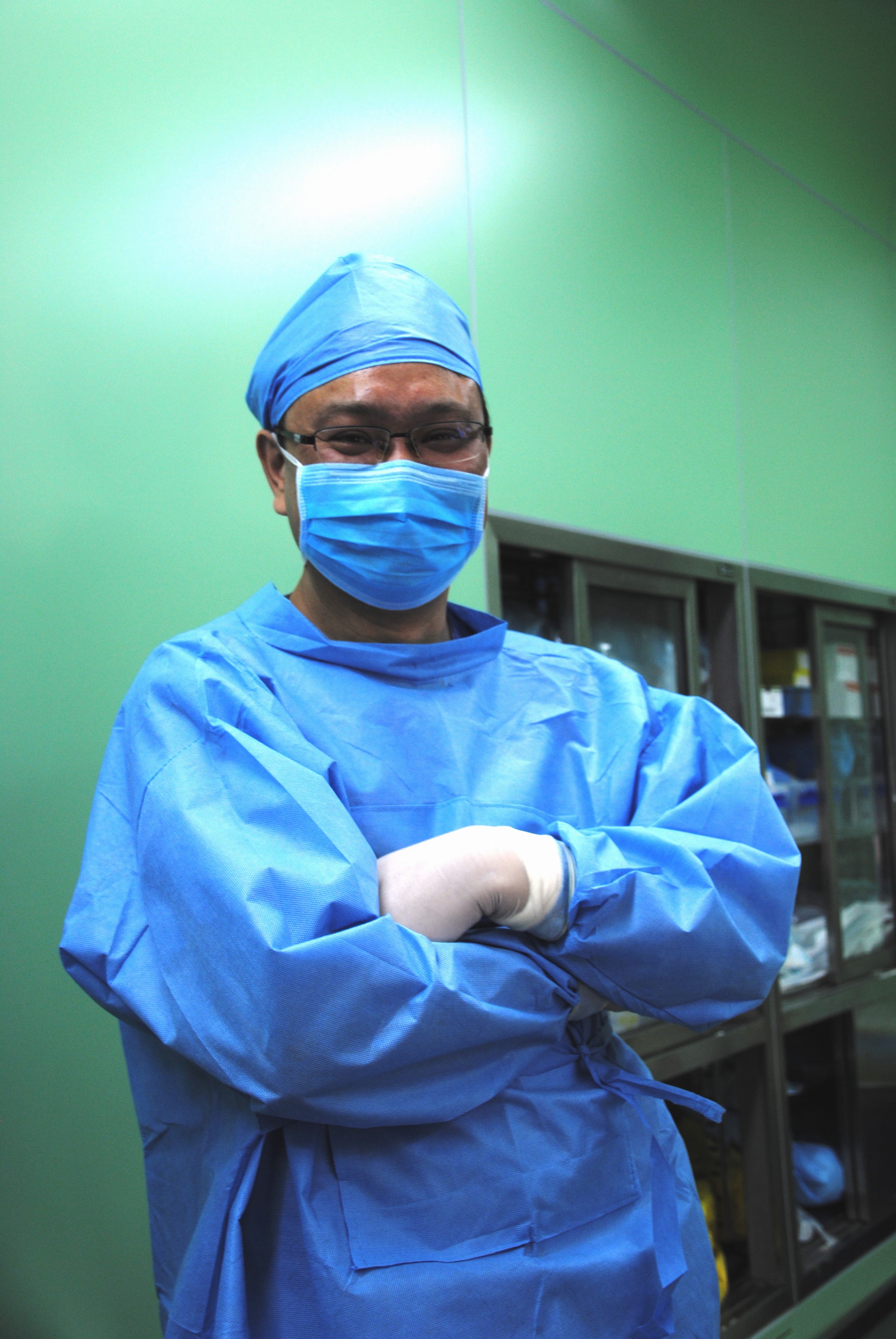
Dr. Boxiong Xie, thoracic surgeon
Dr. Dong Jiasheng & Dr. Zheng, Reconstructive/ Plastic Surgeons
The patient is a young male in his early forties who had undergone a right upper lobectomy for cancer several years prior at a facility in a far away province. He then presented with a large empyema. Initially, conservative treatments were attempted. The patient underwent several drainage procedures, by both open and closed methods. These measures along with attempts to repair the bronchial stump failed due to extensive infection and tissue destruction.
Following the failure of more conservative measures, the patient presented to this facility for specialty care. He had heard about this program, and travelled a long distance to be here at significant difficulty and expense. As his surgeon explained, “it’s his last chance at a normal life.”
Over the continuing course of his treatment, a large opening on the anterior chest was created surgically. Due to the extent of necrotic tissue, this required the removal of anterior sections of ribs #2, 3, 4 and 5, leaving the patient with a very large open cavity, as seen in CT slices (pulmonary and tissue windows).


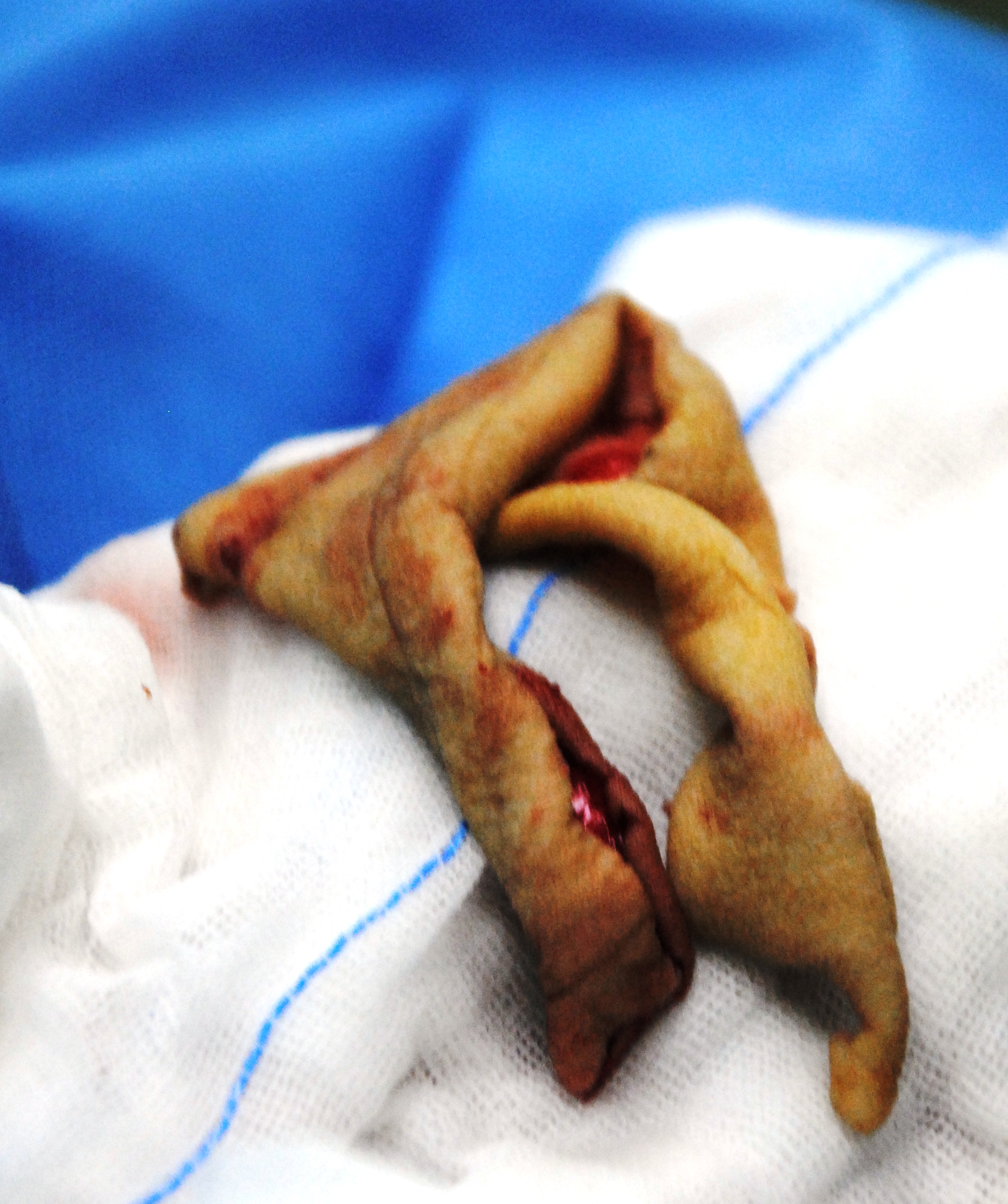
This large cavity was left open for a period of around two years, while infected material was debrided and evacuated, and aggressive wound management was continued. At the time of his presentation to the operating room, the wound bed is dry and pink with a small amount of slough. An opening to the bronchus is visible (with bubbling on respiration at the site of the wound). The wound measures approximately 6 cm X 4 cm. As seen from the CT images above, the wound was also several centimeters in depth.

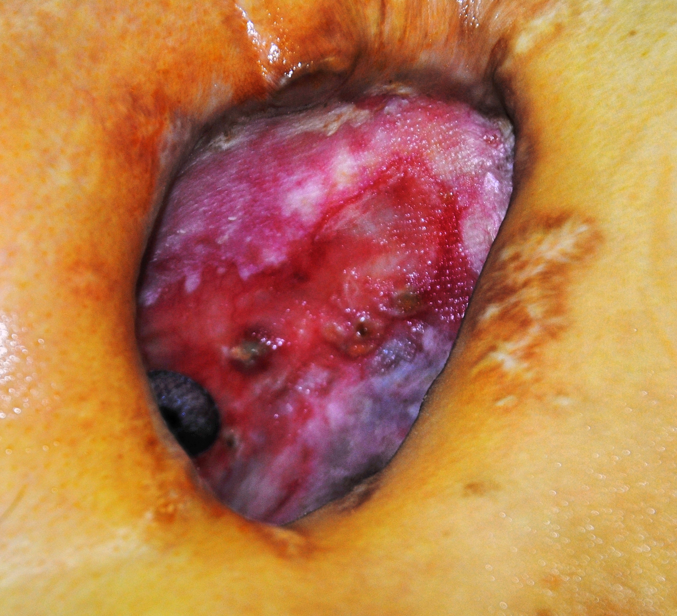
The wound tracks up towards the shoulder, making it deeper and larger than it initially appears on gross visualization. There is a visible pulsation from the border of the cavity, (which may add to the patient and family’s distress).
Surgical procedure:
After the wound is cleaned and prepared with betadine solution, the anesthesiologist introduces a bronchoscope into the airway, for illumination and visualization of the airway. The light from the scope is immediately visible to observation within the chest. At that point, amplatzer patch was inserted into the bronchial stump.

After placement of the patch was confirmed, the patient was re-prepped, and draped. Dr. Boxiong expands the existing wound, and dissects down to healthy bleeding tissue, removing yellow eschar. The wound is lightly packed with moist gauze.
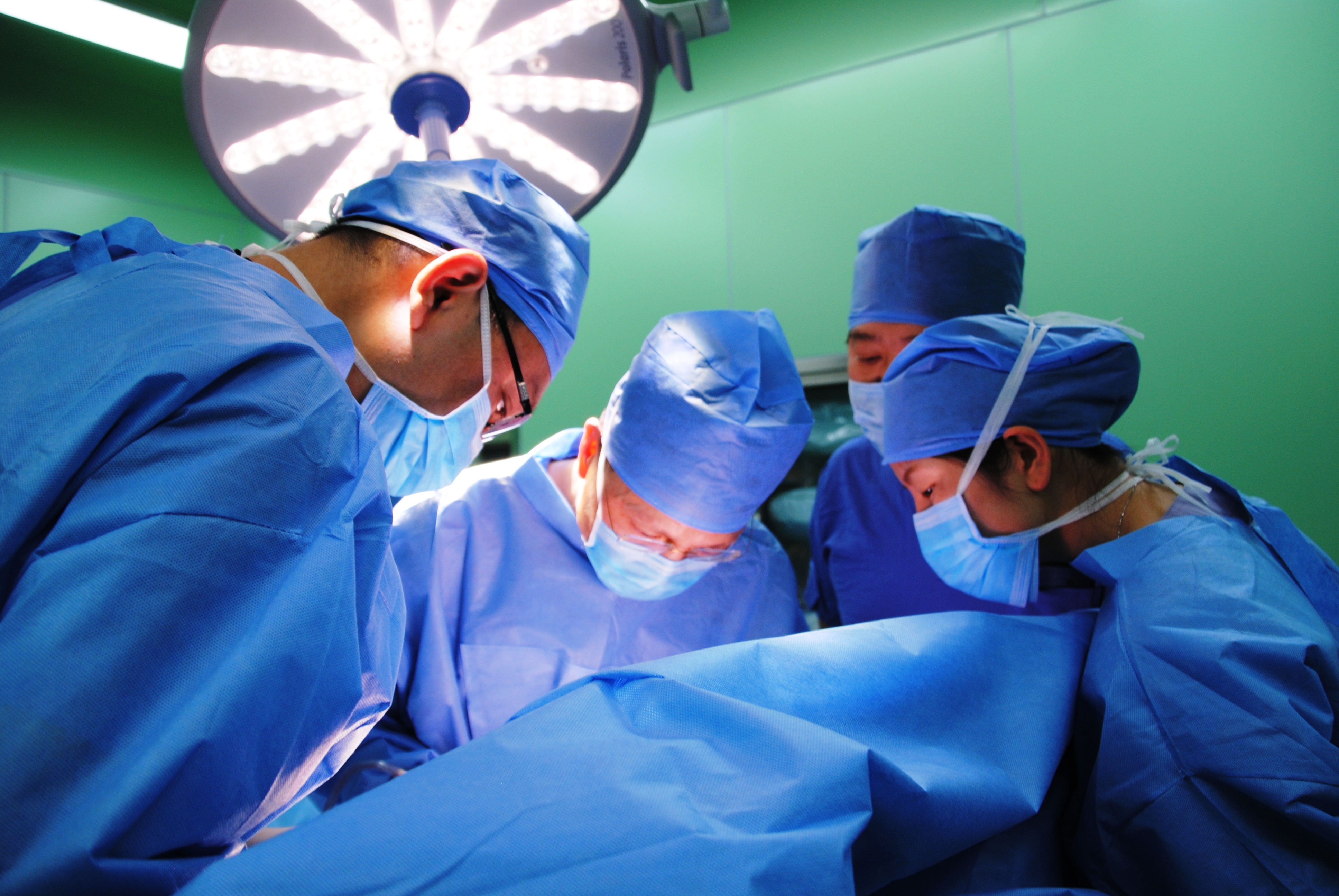
Then Dr. Dong and his assistant surgeon arrive, to start their portion of the operation. Dr. Dong starts another incision approximately 3 cm below the wound area. The incision is extended to the left side of the chest. The surgeon dissects down through skin, adipose and fascia to free the right internal mammary artery to use to ensure that the graft is well vascularized.

Next step: Flap harvesting
Once the IMA was free, it was temporarily secured, and the wound was dressed. The patient was re-positioned, and re-prepped to allow access to the posterior aspect of the left chest. Due to muscle devascularization from the multiple previous surgeries on the right anterior chest, the surgeon harvests the left latissimus dorsi, using a large diamond-shaped incision.

Once the flap was harvested, the patient was left with a large open defect, without enough surrounding skin to cover the area. The surgical site is dressed with a temporary dressing while Dr. Dong moves on to his next surgical site.
Next step: Skin Harvesting
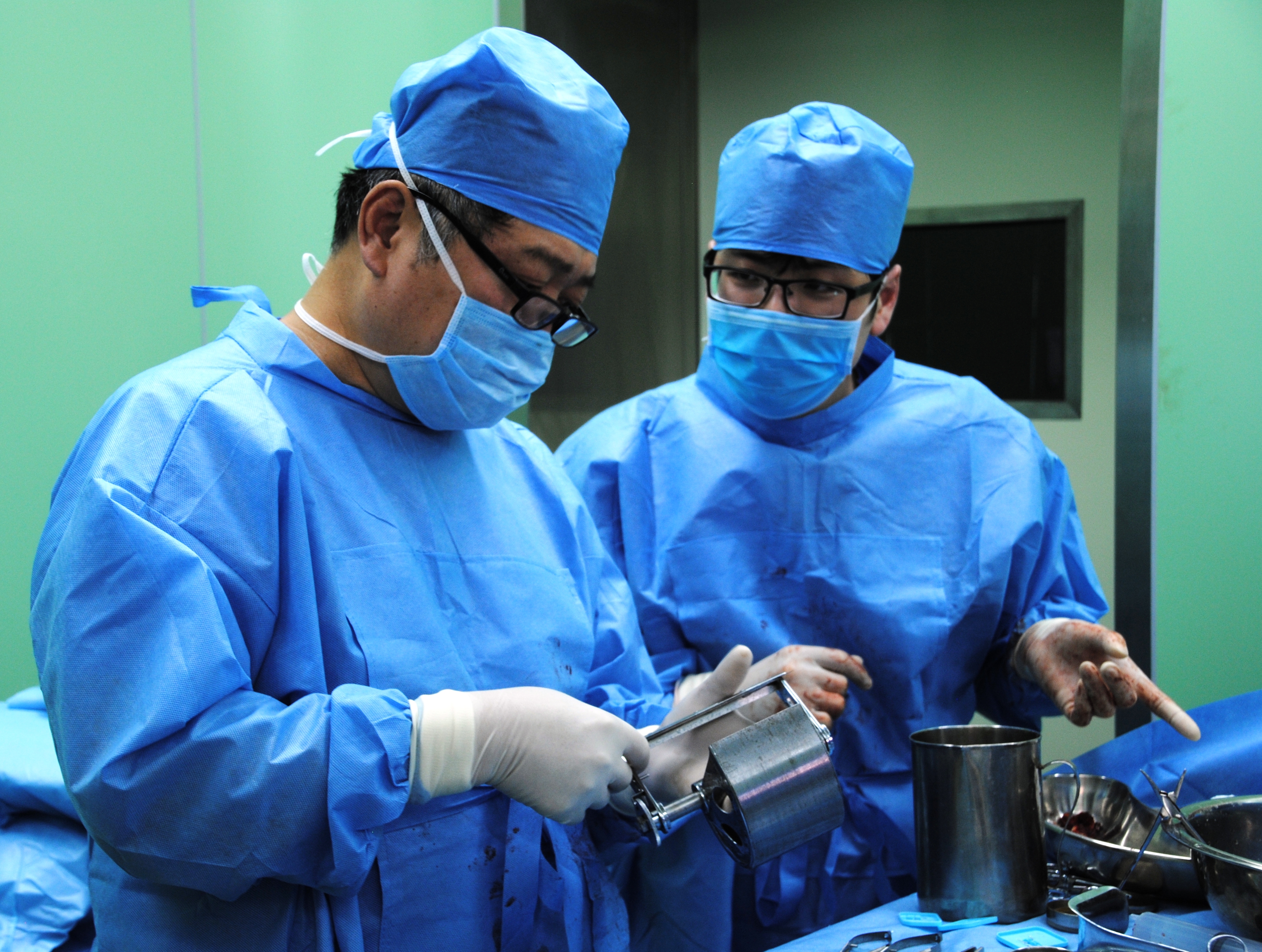
After preparing the patients right thigh, Dr. Dong applied a Padgett dermatome to shave off a thin layer of skin.

After multiple passes, the surgeons have enough skin to cover the defect from the flap site.
Next step: Skin Grafting

The thin strips of skin were applied to the flap site and sutured into place.

Once the sutures were completed, the wound was re-dressed and the patient was re-positioned for the last steps of the operation.
Next step: Anastomosis of mammary artery to flap

Following re-positioning to supine position, the flap was placed within the right chest wound. The flap was loosely sutured into place to maintain a proper position while the painstaking vascular anastomoses were performed. Once the anastomoses were completed, the remaining incisions were carefully closed.
Total surgical time was greater than ten hours.
Discussion
As discussed by Lois and Noppen (2005), BPF management has traditionally been performed in a piece meal or stepwise fashion, with surgical interventions reserved as a last resort. Unfortunately, for some patients, this means that BPF becomes a chronic illness. As a chronic illness, (and all that chronic illness entails such as chronic malnutrition, chronic inflammation, long-term antibiotic therapy), the morbidity and mortality of this condition continues to increase for the duration of the illness. In the case study above, a relatively young, now cancer-free patient had now developed much of the disabilities associated with elderly patients due to the chronic nature of his illness (BPF after a lobectomy ten years prior). This certainly places the patient at significant risk for major complications once a large-scale definitive surgery is performed. Van Schill et al. (2014) notes that better understanding regarding the need for interdisciplinary management including aggressive physical therapy and nutritional support have reduced some of these complications.
While the impact of bronchopleural fistulas are usually discussed in terms of mortality, financial costs (surgical costs) and length of stay,and for this case, we would like to take a closer look at morbidity and quality of life issues raised by the development of this complication.
While BPF is rare, it truly can be a life-altering and destructive diagnosis. In addition to pain, physical debility, there may be gross deformity coupled with chronic wound care. Deformities caused by extensive tissue destruction and removal of several ribs can cause significant emotional and psychological anxiety and stress in both the patient and family members. The visible pulsation (cardiac movement) seen within the wound may exacerbate this anxiety. The stress of this wound combined with additional stressors related to this diagnosis have been observed to lead to a higher rate of marital discord and patient suicide. Patients may also feel a loss of sexuality and personal identity in the presence of this type of disfigurement, similar to some women after radical mastectomy (particularly in female patients).
To add insult to injury, unlike many conditions which can be readily corrected surgically, the creation of myocutaneous flap (and subsequent skin grafting) itself causes additional disfigurement. This patient required a lengthy (ten hour) surgery which resulted in the creation of three new surgical sites in addition to the patient’s original right-sided chest wound. While this is a drastic example, it does serve to highlight the on-going need to consider the psychological and emotional well-being of this patient (and all our patients).
BPF and professional relationships?
This case also reminds of the need for good interdisciplinary relationships. In thoracic surgery, cosmetic outcomes (other that pursuing minimally invasive options when possible) are not usually one of our primary considerations. This leaves us at a disadvantage when managing patients with such a drastic complication. We don’t always have a strong network or relationships with other surgical or medical disciplines outside of oncology or oncology-related fields. We need to take the opportunities available to become more familiar with our local reconstructive surgeons, as well as the latest techniques in reconstructive surgery. It’s not “good enough” to know the name of one of the plastic surgeons we brush elbows with in the surgical waiting lounge. It is not just about referrals and compensation. It is about having an open and free dialogue with surgical colleagues, so that when we do require their assistance, we can work together smoothly and coördinate care.
Consider the need to include social workers, psychologists and other counseling services in both the preoperative and postoperative care of our patients, when necessary for their long-term health and wellness. Unfortunately, due to social stigma, health care/ insurance or financial restrictions as well as provider hesitation**, not enough of our patients receive consultations or referrals to appropriate resources. We can’t change insurance regulations, but by becoming more familiar with our local resources and providers, we can overcome many of the other barriers to supporting our patients emotional health.
[1] I was unable to find literature that specifically cites BPF as a contributing factor to psychosocial complications such as divorce, depression or suicide but the impact of chronic wounds on emotional health, family life and other quality of life indicators are well documented. However, Okonta et. al (2015) and Lois & Noppen (2005) both cite QoL issues in patients with BPF.
** Provider hesitation is a nice term for all the reasons providers sometimes fail to seek mental health referrals for patients; such as fear of embarrassing our patients, believing that counseling is only needed for psychiatric emergencies, failure to understand local resources available, or our own discomfort with mental health “issues”.
References and Additional Readings
Arnold, P. G. & Pairolero, P. C. (1990). Intrathoracic muscle flaps: an account of their use in the management of 100 consecutive patients. Annals of Surgery, 1990; 211(6): 656-660. Study looking at one hundred cases from May 1977 and February 1988. In this potent reminder of the morbidity and mortality that is associated with patients requiring muscle flaps, as well as the advances in medicine over the last two decades, there were 16 operative deaths and 43 additional all-cause deaths in the operative survivors. Interestingly, one of these late-term deaths was due to suicide.
Goyal VD1, Gupta B2, Sharma S3 (2015). Intercostal muscle flap for repair of bronchopleural fistula. Lung India. 2015 Mar-Apr;32(2):152-4. doi: 10.4103/0970-2113.152628. Indian case study of patient presentation of BPF after treatment for spontaneous pneumothorax.
Lois, M. & Noppen, M. (2005). Bronchopleural fistulas: an overview of the problem with special focus on endoscopic management. Chest, 2005, 128: 3955-3965. Joint Belgian- American paper by two pulmonologists discussing management options for bronchopleural fistula. Interesting verbiage but good overview.
Okonta, K. E., Ocheli, E. O. & Gbeneol, T. J. (2015). Surgical management of recalcitrant peripheral bronchopleural fistula with empyema: A preliminary experience. Niger Med J. 2015 Jan- Feb; 56(1): 12-16. Joint Nigerian paper (thoracic surgeons & plastic surgeons) reviewing bronchopleural fistula repair in 5 patients.
Ottevaere, A. et al. (2013). Use of an amplatzer device for endoscopic closure of a large bronchopleural fistula following lobectomy for a stage I squamous cell carcinoma. Case reports in Oncology 2013; 6:550-554. Belgian case report of the successful use of an amplatzer device for endoscopic BPF closure in a patient deemed surgically inoperable.
Van Schil PE1, Hendriks JM1, Lauwers P1 (2014). Focus on treatment complications and optimal management surgery. Transl Lung Cancer Res. 2014 Jun;3(3):181-6. doi: 10.3978/j.issn.2218-6751.2014.06.07. Belgian paper reviewing outcomes of 3,500 surgeries.
Beldon, P. (2007). Technical guide: What you need to know about skin grafts and donor site wounds. Wound Essentials 2007, 149-155. Very nicely written by a British Nurse Consultant working for the NHS as a Tissue Viability Consultant.
Chronic wounds/ quality of life
Green J, Jester R, McKinley R, Pooler A. (2014). The impact of chronic venous leg ulcers: a systematic review. J Wound Care. 2014 Dec;23(12):601-12. doi: 10.12968/jowc.2014.23.12.601. Review
Firth, et. al. Exploring the impact of living with a range of chronic wounds. University of Leeds, Royal College of Nursing presentation.
Levine, L. A. (2013). The clinical and psychosocial impact of Peyronie’s disease. Am J Manag Care. 2013 Mar;19(4 Suppl):S55-61. While unrelated to thoracic surgery, patients with Peyronie’s disease have many of the same emotional and psychological stressors as patients with other chronic wound conditions such as BPF.

In 1985 thoracic surgeons performed an upper right lobectomy to remove a lung cancer tumor. The surgery successfully removed the tumor but before closure the artery to the remaining lobes was accidentally severed. This was repaired and I was sent home. Two weeks later an emergency pneumonectomy was performed because the graft/stent repair clotted. I was sent home. A month and a half later I was admitted for a bpf. A chest tube was inserted, which didn’t work, a Clagett procedure was performed, which didn’t work, an Elloeser flap was created and eventually (after more than a dozen times under anesthesia for cleaning, debridement etc) I was sent home with an open wound. At no time did my surgical “team” make an effort to assist me with the devastating psychological effects of these interventions. I left the hospital on heavy narcotics for excruciating pain that has only (more than a year later) started to be relieved. Three months after coming home I had another surgery to try to repair the BPF again. That failed. The open would is healed and closed but the surgery to repair the BPF was unsuccessful. I am terrified of surgeons and cannot bring myself to let anyone try any further repair.
Because the initial surgery successfully removed the cancer my oncologist considers my surgeries successful. I have been asking for help for depression but my healthcare providers don’t seem to be able to appreciate why I am depressed. The result of undergoing the thoracic surgeries you’ve described isPTSD and severe depression. My short term memory has been destroyed.
Thank you for your article. At least I have some confirmation that I am not crazy, that something truly devastating occurred to me and that my psychological symptoms have some basis in the treatment I received. Patients undergoing who have psychological problems after these surgeries and who are dismissed without help have been done harm….something doctors take an oath to avoid.
I’m sorry, the surgeries were performed October 2015 to January 2016 with follow up surgery June 2016 (not 1985). I was 57 when this started.
I have a BPF also due to an upper right lobectomy. You mention that the surgery to repair your BPF was unsuccessful. I am considering having mine glued via bronchoscopy but I was told I would probably have an external tube for the rest of my life. How are you doing after all this time with your BPF? Thanks.