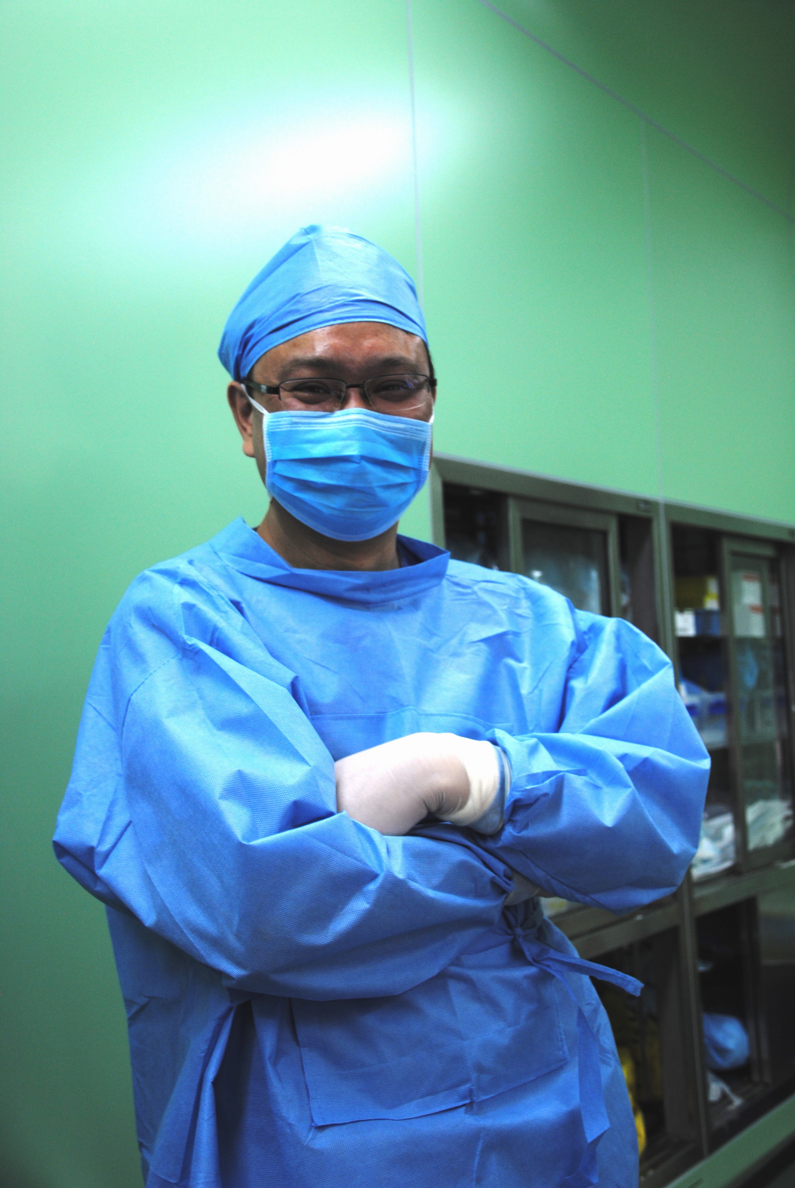
Dr. Giuseppe Aresu of the University Hospital of Udine, Italy presents a case of thymectomy by subxyphoid approach
Article originally published October 31, 2015
We report the case of a thymectomy performed through a subxyphoid vertical single incision port carried out in a 51 years old female myasthenic patient presenting a Masaoka stage I thymoma.
The subxhyphoid approach permits an excellent view of the mediastinal anterior region and of the two pleural spaces giving the surgeon the possibility to perform a very radical and safe dissection of the thymic and peri-thymic fatty tissues.
Considering the position and the 3.5 cm length of the port, it is esthetically excellent. Without a sternal incision, or VATS – associated intercostal nerve injury, the recovery can be faster and less painful than the sternotomy approach or other vats approaches carried out through the intercostal spaces.
Technique
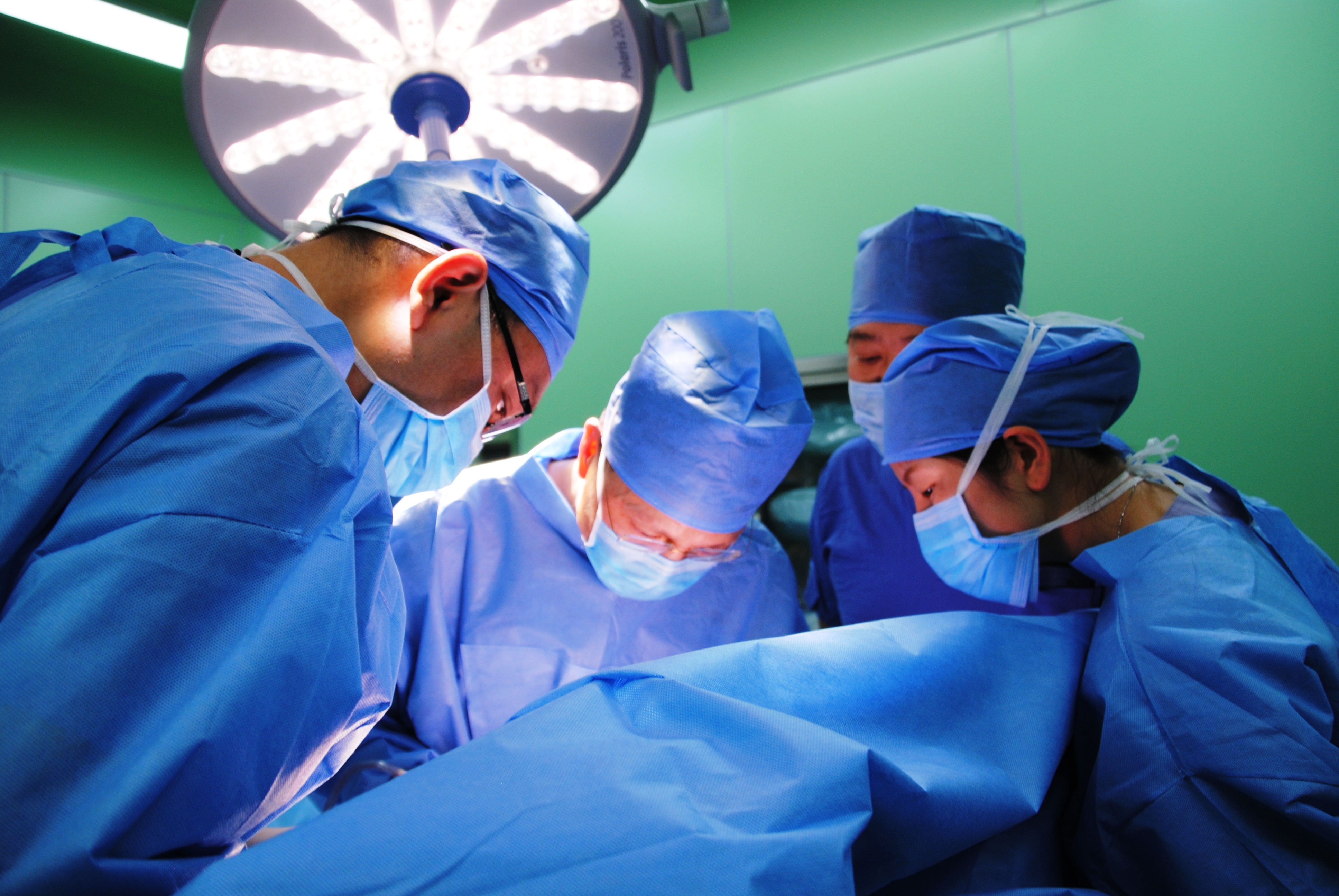
We performed extended thymectomy through a uniportal subxiphoid approach in a 51 years old female presenting a thymoma of 2.5 cm and myasthenia gravis.
The patient was informed about the risks and the benefits of the procedure and the consent to carry on with the operation was obtained.
Under general anesthesia, the patient was intubated with a double-lumen endotracheal tube and artificial ventilation was applied.
The patient was placed in a supine position with a silicon roll positioned below the lower part of the chest in order to lift the subxiphoid region.
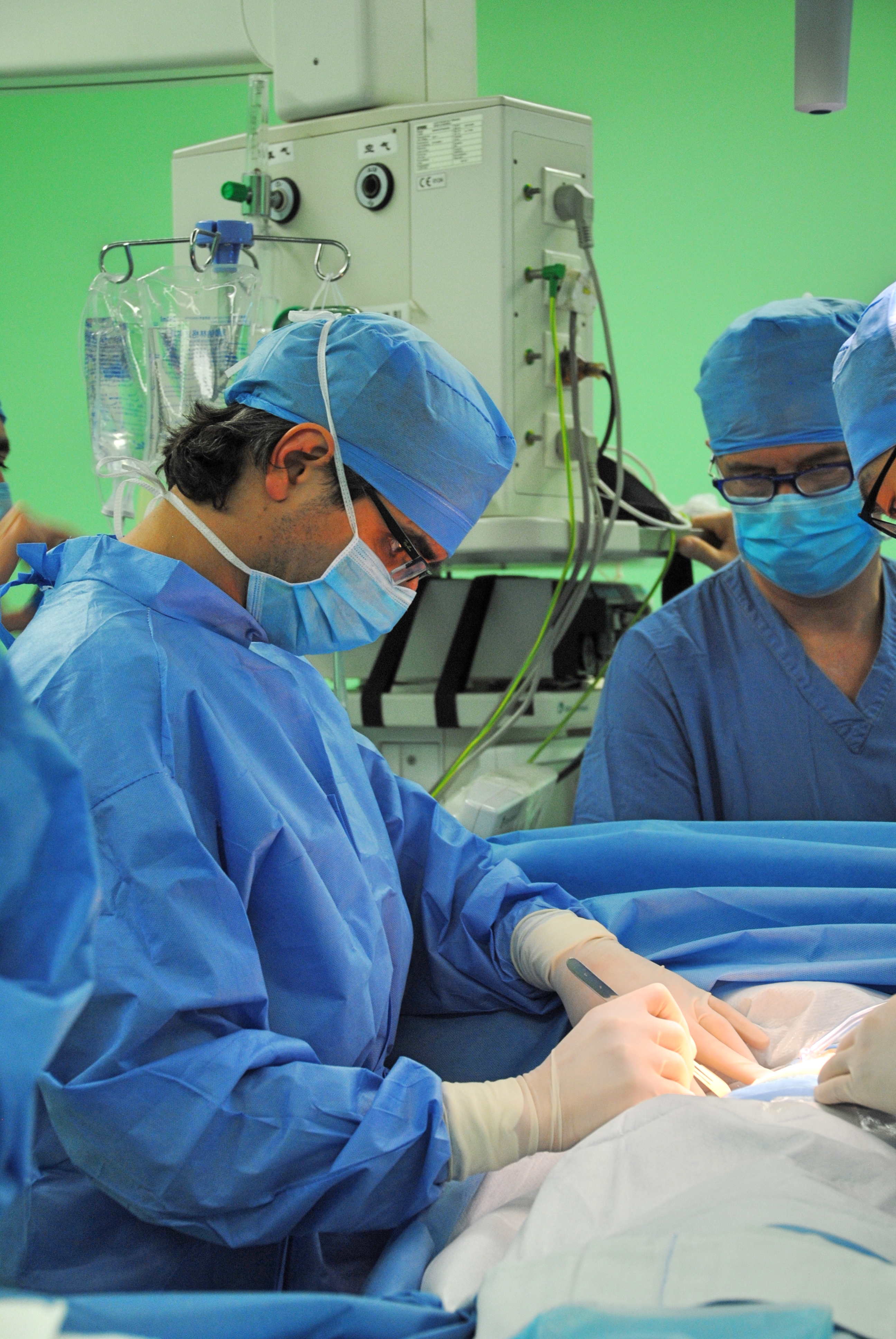
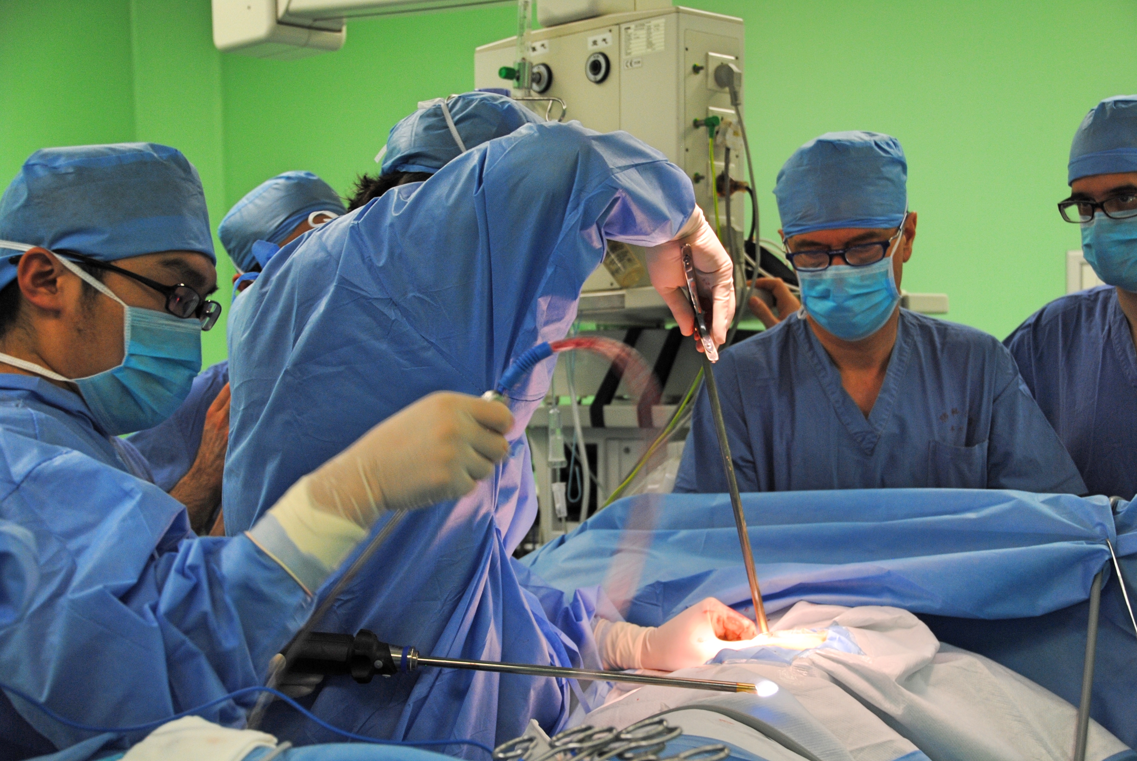
The operating surgeon stood on the right side of the patient, the assistant stood on the patient’s left side and operated the endoscope. The monitor was positioned at the right side of the patient toward the cranial side of the bed.
A 3.5-cm longitudinal muscle sparring incision was made below the xiphoid process between through the linea alba.
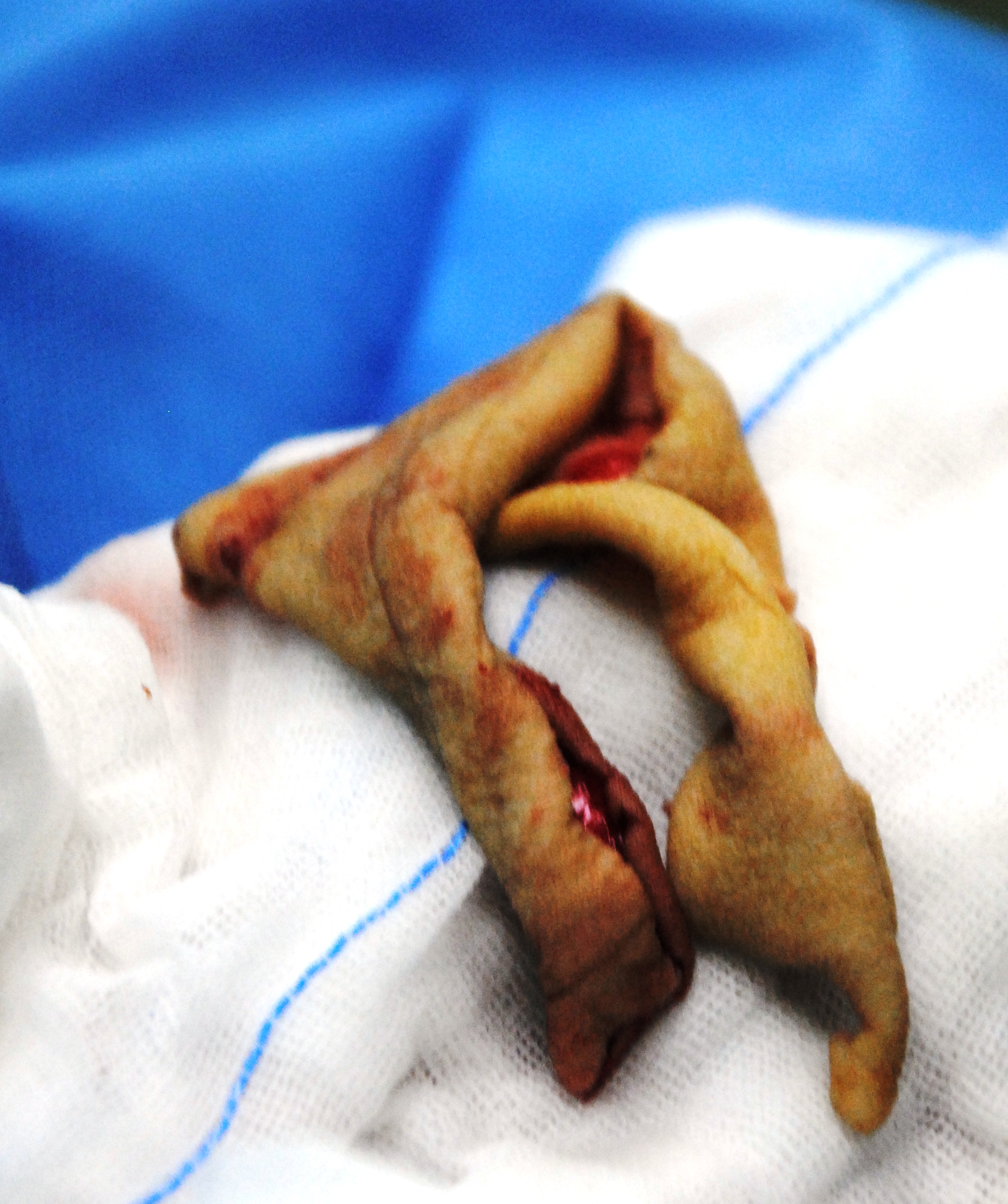
the incision
The xiphoid process was exposed, the inferior part of the sternum was lifted up with a retractor and a blunt dissection was carried out in order to find the pericardial plane.
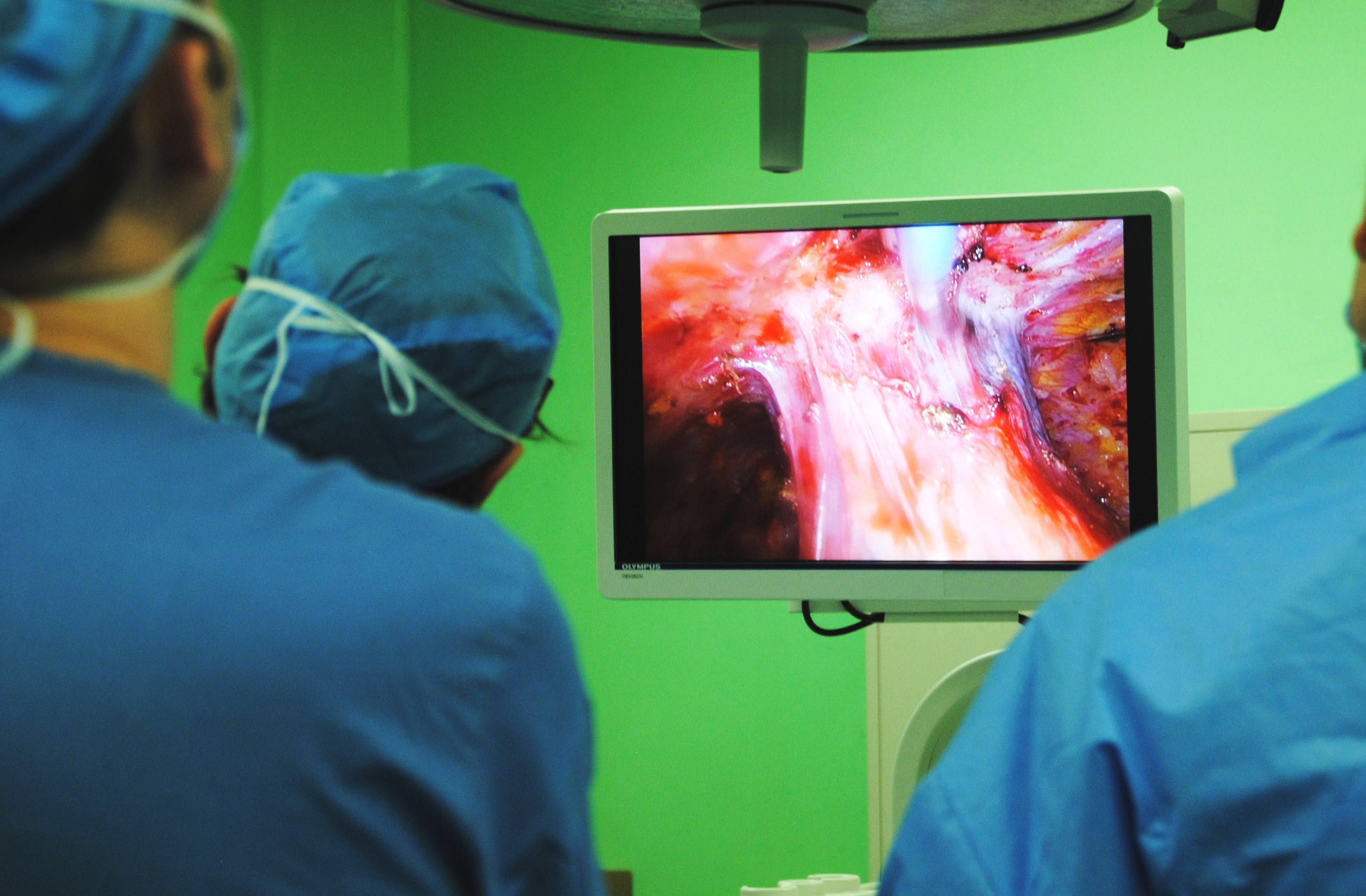
A SILS port (Covidien, Mansfield, MA) was then inserted into the port, and CO2 was insufflated at a maximal pressure of 8 mm Hg. The CO2 insufflation within the mediastinum generates a very useful amount of extra working space within the anterior-superior mediastinum allowing an easier dissection and a better visualization of the mediastinal structures especially toward the cranial part of the mediastinum cephalad to the left innominate vein including the upper poles of the thymus.
Under visual guidance provided through a 10-mm EndoCAMeleon® Telescope, the operator utilized grasping forceps designed for single-incision surgery with his left (SILS Hand Instruments Endo Clinch™ II (Covidien) and performed dissection, coagulation, and division of tissue mainly using the Sonicision™ cordless ultrasonic dissection device (Covidien, Mansfield, MA) and occasionally using a normal straight hook cautery.
The bilateral phrenic nerves and the bilateral mammary arteries and veins were always under optimal control as well as the cranial part of the mediastinum permitting a safe dissection en bloc of the thymus, thymic tumor, and surrounding fatty tissue anterior to the phrenic nerves.
The operation time was about 2 hours and 30 minutes, and blood loss was minimal.
No complications occurred during or after the operation, the drain was taken out after one day and the patient was discharged home 2 days after surgery.
closed incision
Postoperative pain was very low requiring just 1 g X 3 daily of paracetamol during the hospital stay, and no analgesic administration after the discharge.
This case was later published (Dec 14, 2015) at CTSnet. Congratulations Dr. Aresu!
Additional Reading
Suda, T. (2016). Single-port thymectomy using a subxiphoid approach-surgical technique. Ann Cardiothorac Surg. 2016 Jan;5(1):56-8. doi: 10.3978/j.issn.2225-319X.2015.08.02. Review. Free fulll text discussion of a similar case by Japanese surgeon. This article includes a video presentation and a in-depth discussion of technical aspects of the case such as surgeon position and camera access.
aka, “Why we should be nice to plastic surgeons”. This case study highlights the need for close interdisciplinary partnerships among surgeons and also asks the question, “Are we addressing the emotional and psychosocial needs of our patients and their families?”
Bronchopleural fistula: an abnormal communication between the exterior environment and the pleural cavity, often caused entry of bacteria, fluids and other substances into the chest cavity by way of the bronchial tree, for example: bronchial stump breakdown. BPF most commonly occur after large thoracic surgeries such as pneumonectomy but can occur for other reasons such as infection or trauma.
Bronchopleural fistulas (BPF) are a dread complication of thoracic surgery that has (thankfully) become rare in most countries in the last few decades. Treatment of a large bronchopleural fistula can be massive undertaking requiring collaboration and cooperative from multiple specialties including radiology, infectious disease, pulmonology, wound management and plastic surgery.
Patients often endure several months of surgical and wound care treatments prior to undergoing definitive surgical management for this condition. This treatment includes the surgical creation of large open wounds to facilitate drainage of purulent materials, repair of the fistula tract and bronchial stump and debridement / revascularization for proper tissue healing. The case presented today illustrates the devastating emotional, physical and financial costs of bronchopleural fistula as well as the need for interdisciplinary collaboration for definitive surgical repair.
Surgical repair itself carries an elevated risk of morbidity and mortality primarily from respiratory complications, infections/ sepsis and hemorrhage.
More than physical consequences
Bronchopleural fistulas carries more than just the physical consequences of pain and disability for patients and their families. There are also devastating emotional and social effects. Patients can experience a myriad of psychosocial effects from this chronic wound and related treatment. The resultant deformity from many drainage and wound management techniques, in particular, can lead to depression and social ostracism. The development of a bronchopleural fistula can contribute to relationship and intimacy issues. Several of the surgeons interviewed including Dr. Boxiong specifically mentioned both divorce and suicide as being a risk in numerous cases[1].
Case Study
Dr. Boxiong Xie, thoracic surgeon
Dr. Dong Jiasheng & Dr. Zheng, Reconstructive/ Plastic Surgeons
Dr. Boxiong Xie, thoracic surgeon
The patient is a young male in his early forties who had undergone a right upper lobectomy for cancer several years prior at a facility in a far away province. He then presented with a large empyema. Initially, conservative treatments were attempted. The patient underwent several drainage procedures, by both open and closed methods. These measures along with attempts to repair the bronchial stump failed due to extensive infection and tissue destruction.
Following the failure of more conservative measures, the patient presented to this facility for specialty care. He had heard about this program, and travelled a long distance to be here at significant difficulty and expense. As his surgeon explained, “it’s his last chance at a normal life.”
Over the continuing course of his treatment, a large opening on the anterior chest was created surgically. Due to the extent of necrotic tissue, this required the removal of anterior sections of ribs #2, 3, 4 and 5, leaving the patient with a very large open cavity, as seen in CT slices (pulmonary and tissue windows).
packing material can be seen in the right chest cavity.
tissue window showing extent of wound
This large cavity was left open for a period of around two years, while infected material was debrided and evacuated, and aggressive wound management was continued. At the time of his presentation to the operating room, the wound bed is dry and pink with a small amount of slough. An opening to the bronchus is visible (with bubbling on respiration at the site of the wound). The wound measures approximately 6 cm X 4 cm. As seen from the CT images above, the wound was also several centimeters in depth.
overview of wound – which tracks upwards several cm towards shoulder
The wound tracks up towards the shoulder, making it deeper and larger than it initially appears on gross visualization. There is a visible pulsation from the border of the cavity, (which may add to the patient and family’s distress).
Surgical procedure:
After the wound is cleaned and prepared with betadine solution, the anesthesiologist introduces a bronchoscope into the airway, for illumination and visualization of the airway. The light from the scope is immediately visible to observation within the chest. At that point, amplatzer patch was inserted into the bronchial stump.
Amplatzer patch visible in the chest cavity
After placement of the patch was confirmed, the patient was re-prepped, and draped. Dr. Boxiong expands the existing wound, and dissects down to healthy bleeding tissue, removing yellow eschar. The wound is lightly packed with moist gauze.
thoracic surgeons dissect down to healthy tissue (anterior wound site)
Then Dr. Dong and his assistant surgeon arrive, to start their portion of the operation. Dr. Dong starts another incision approximately 3 cm below the wound area. The incision is extended to the left side of the chest. The surgeon dissects down through skin, adipose and fascia to free the right internal mammary artery to use to ensure that the graft is well vascularized.
preparing the internal mammary artery for eventual anastomosis
Next step: Flap harvesting
Once the IMA was free, it was temporarily secured, and the wound was dressed. The patient was re-positioned, and re-prepped to allow access to the posterior aspect of the left chest. Due to muscle devascularization from the multiple previous surgeries on the right anterior chest, the surgeon harvests the left latissimus dorsi, using a large diamond-shaped incision.
Harvesting the myocutaneous flap from posterior chest
Once the flap was harvested, the patient was left with a large open defect, without enough surrounding skin to cover the area. The surgical site is dressed with a temporary dressing while Dr. Dong moves on to his next surgical site.
Next step: Skin Harvesting
Dr. Dong prepares to harvest skin for grafting
After preparing the patients right thigh, Dr. Dong applied a Padgett dermatome to shave off a thin layer of skin.
Harvesting skin from patient’s thigh
After multiple passes, the surgeons have enough skin to cover the defect from the flap site.
Skin harvested to cover flap site
Next step: Skin Grafting
Skin grafting at the myocutaneous flap site
The thin strips of skin were applied to the flap site and sutured into place.
suturing skin graft over flap harvest site (on back)
Once the sutures were completed, the wound was re-dressed and the patient was re-positioned for the last steps of the operation.
Next step: Anastomosis of mammary artery to flap
surgeons using the microscope to complete vascular anastamoses
Following re-positioning to supine position, the flap was placed within the right chest wound. The flap was loosely sutured into place to maintain a proper position while the painstaking vascular anastomoses were performed. Once the anastomoses were completed, the remaining incisions were carefully closed.
Total surgical time was greater than ten hours.
Discussion
As discussed by Lois and Noppen (2005), BPF management has traditionally been performed in a piece meal or stepwise fashion, with surgical interventions reserved as a last resort. Unfortunately, for some patients, this means that BPF becomes a chronic illness. As a chronic illness, (and all that chronic illness entails such as chronic malnutrition, chronic inflammation, long-term antibiotic therapy), the morbidity and mortality of this condition continues to increase for the duration of the illness. In the case study above, a relatively young, now cancer-free patient had now developed much of the disabilities associated with elderly patients due to the chronic nature of his illness (BPF after a lobectomy ten years prior). This certainly places the patient at significant risk for major complications once a large-scale definitive surgery is performed. Van Schill et al. (2014) notes that better understanding regarding the need for interdisciplinary management including aggressive physical therapy and nutritional support have reduced some of these complications.
While the impact of bronchopleural fistulas are usually discussed in terms of mortality, financial costs (surgical costs) and length of stay,and for this case, we would like to take a closer look at morbidity and quality of life issues raised by the development of this complication.
While BPF is rare, it truly can be a life-altering and destructive diagnosis. In addition to pain, physical debility, there may be gross deformity coupled with chronic wound care. Deformities caused by extensive tissue destruction and removal of several ribs can cause significant emotional and psychological anxiety and stress in both the patient and family members. The visible pulsation (cardiac movement) seen within the wound may exacerbate this anxiety. The stress of this wound combined with additional stressors related to this diagnosis have been observed to lead to a higher rate of marital discord and patient suicide. Patients may also feel a loss of sexuality and personal identity in the presence of this type of disfigurement, similar to some women after radical mastectomy (particularly in female patients).
To add insult to injury, unlike many conditions which can be readily corrected surgically, the creation of myocutaneous flap (and subsequent skin grafting) itself causes additional disfigurement. This patient required a lengthy (ten hour) surgery which resulted in the creation of three new surgical sites in addition to the patient’s original right-sided chest wound. While this is a drastic example, it does serve to highlight the on-going need to consider the psychological and emotional well-being of this patient (and all our patients).
BPF and professional relationships?
This case also reminds of the need for good interdisciplinary relationships. In thoracic surgery, cosmetic outcomes (other that pursuing minimally invasive options when possible) are not usually one of our primary considerations. This leaves us at a disadvantage when managing patients with such a drastic complication. We don’t always have a strong network or relationships with other surgical or medical disciplines outside of oncology or oncology-related fields. We need to take the opportunities available to become more familiar with our local reconstructive surgeons, as well as the latest techniques in reconstructive surgery. It’s not “good enough” to know the name of one of the plastic surgeons we brush elbows with in the surgical waiting lounge. It is not just about referrals and compensation. It is about having an open and free dialogue with surgical colleagues, so that when we do require their assistance, we can work together smoothly and coördinate care.
Consider the need to include social workers, psychologists and other counseling services in both the preoperative and postoperative care of our patients, when necessary for their long-term health and wellness. Unfortunately, due to social stigma, health care/ insurance or financial restrictions as well as provider hesitation**, not enough of our patients receive consultations or referrals to appropriate resources. We can’t change insurance regulations, but by becoming more familiar with our local resources and providers, we can overcome many of the other barriers to supporting our patients emotional health.
[1] I was unable to find literature that specifically cites BPF as a contributing factor to psychosocial complications such as divorce, depression or suicide but the impact of chronic wounds on emotional health, family life and other quality of life indicators are well documented. However, Okonta et. al (2015) and Lois & Noppen (2005) both cite QoL issues in patients with BPF.
** Provider hesitation is a nice term for all the reasons providers sometimes fail to seek mental health referrals for patients; such as fear of embarrassing our patients, believing that counseling is only needed for psychiatric emergencies, failure to understand local resources available, or our own discomfort with mental health “issues”.
References and Additional Readings
Arnold, P. G. & Pairolero, P. C. (1990). Intrathoracic muscle flaps: an account of their use in the management of 100 consecutive patients. Annals of Surgery, 1990; 211(6): 656-660. Study looking at one hundred cases from May 1977 and February 1988. In this potent reminder of the morbidity and mortality that is associated with patients requiring muscle flaps, as well as the advances in medicine over the last two decades, there were 16 operative deaths and 43 additional all-cause deaths in the operative survivors. Interestingly, one of these late-term deaths was due to suicide.
Goyal VD1, Gupta B2, Sharma S3 (2015). Intercostal muscle flap for repair of bronchopleural fistula. Lung India. 2015 Mar-Apr;32(2):152-4. doi: 10.4103/0970-2113.152628. Indian case study of patient presentation of BPF after treatment for spontaneous pneumothorax.
Van Schil PE1, Hendriks JM1, Lauwers P1 (2014). Focus on treatment complications and optimal management surgery. Transl Lung Cancer Res. 2014 Jun;3(3):181-6. doi: 10.3978/j.issn.2218-6751.2014.06.07. Belgian paper reviewing outcomes of 3,500 surgeries.
Levine, L. A. (2013). The clinical and psychosocial impact of Peyronie’s disease. Am J Manag Care. 2013 Mar;19(4 Suppl):S55-61. While unrelated to thoracic surgery, patients with Peyronie’s disease have many of the same emotional and psychological stressors as patients with other chronic wound conditions such as BPF.
As Dr. Gonzalez Rivas demonstrates, minimally invasive surgery isn’t just for “easy” cases. Case study with brief discussion and literature review
Uniportal VATS with chest wall resection at Shanghai Pulmonary Hospital
Shanghai, China
Authors: Gonzalez – Rivas, D. & Eckland, K.
Surgeons: Dr. Diego Gonzalez Rivas with Dr. Boxiong Xie assisting.
Case: 66-year-old patient with large left upper lobe mass extending into chest wall, biopsy proven carcinoma.
Pulmonary function tests – within acceptable margins
Imaging:
CT scan – showing a large left-sided lung upper lobe mass with chest wall invasion and rib involvement at the level just beneath the scapula.
Procedure: Uniportal (single incision) VATS with rib resection
Description: at a glance
Determining port placement
Due to tumor location, port placement had to be carefully considered and adjusted.
Vital signs at initiation of operation: HR 78, NSR B/P 95/56 Oxygen saturations: 100% (intubated with double lumen ETT)
First incision: 14:17
Making the initial incision
The tumor was adherent to the chest wall, requiring chest wall resection with rib resection.
performing uniportal VATS for tumor with chest wall invasion
The tumor was palpated thru the 2 cm incision allowing the surgeon the benefit of open surgery despite using a minimally invasive technique.
Palpating the tumor
Ribs were resected using a guillotine designed for minimally invasive use.
Rib resection
Lung resection complete at 17:42. The tumor was removed enblock using a bag system to prevent tumor spillage.
Tumor enblock after removal
Lymph node dissection completed at 17:56
There was a brief run of PVCs lasting about 30 seconds (B/P 83/54) with no desaturations. Patient was otherwise hemodynamically stable for the duration of the case.
Frozen section: clear pleural margins
EBL: 200ml
Discussion:
As noted by Pischik and others, many of the traditional contraindications for VATS procedures are no longer applicable, particularly for surgeons well versed in minimally invasive techniques like uniportal thoracoscopic surgery. In the case above, several of these contraindications were successfully addressed, including multiple adhesions, an incomplete interlobar fissure and a tumor with chest wall involvement.
That being said, this case was technically challenging from start to finish, due to the position of the chest wall tumor that required adjustment of port placement, a lengthy dissection of dense adhesions in addition to a sizeable chest wall mass. Hilar dissection was complicated by anatomical position, and the bronchus was difficult to access. This in addition to an incomplete fissure significantly lengthened the procedure.
VATS resection using a single port approach can be challenging even for experienced surgeons. However, it is a viable alternative for more complicated cases including those requiring a degree of chest wall resection.
This case was just one of numerous cases performed by Dr. Diego Gonzalez Rivas as part of the Uniportal VATS training course at Shanghai Pulmonary Hospital. Dr. Diego Gonzalez Rivas is the inventor of the uniportal technique and Director of Uniportal VATS training program at Shanghai Pulmonary Hospital. He has partnered with the Chinese facility to offer training courses for interested surgeons three times a year, in addition to his ‘wet-lab’ surgical training offerings in his home facility at La Coruna, Spain.
in the operating room with Dr. Diego Gonzalez Rivas for single port thoracoscopic (uniportal) surgery.
Hilar mass resection using single port thoracoscopy with Dr. Diego Gonzalez – Rivas
K. Eckland & Andres M. Neira, MD
Instituto Nacional de Cancerlogia
Bogota, Colombia
Surgeon(s): Dr. Diego Gonzalez Rivas and Dr. Ricardo Buitrago
Dr. Diego Gonzalez Rivas demonstrates single port thoracoscopy
Case History:
59-year-old female with past medical history significant for recurrent mediastinal mass previously resectioned via right VATS. Additional past medical history included prior right-sided nephrectomy.
Pre-operative labs:
CBC: WBC 7230 Neu 73% Hgb:14.1 Hct 37 platelets 365000
Pt 12.1 / INR1.1 PTT: 28.3
Diagnostics:
Pre-operative CT scan: chest
edited to preserve patient privacy
Procedure: Single port thoracoscopy with resection of mediastinal mass and lymph node sampling
After review of relevant patient history including radiographs, patient was positioned for a right-sided procedure. After being prepped, and draped, surgery procedure in sterile fashion. A linear incision was made in the anterior chest – mid clavicular line at approximately the fifth intercostal space. A 10mm port was briefly inserted and the chest cavity inspected. The port was then removed, and the incision was expanded by an additional centimeter to allow for the passage of multiple instruments; including camera, grasper and suction catheter.
Dr. Gonzalez Rivas and Dr. Ricardo Buitrago at National Cancer Institute
The chest cavity, pleura and lung were inspected. The medial mediastinal mass was then identified.
As previously indicated on pre-operative CT scan, the mass was located adjacent and adherent to the vessels of the hilum. This area was carefully dissected free, in a painstaking fashion. After freeing the mediastinal mass from the hilum, the remaining surfaces of the mass were resected. The mass was fixed to the artery pulmonary and infiltrating it) . The mass was removed en-bloc. Care was then taken to identify, and sample the adjacent lymph nodes which were located at stations (4, 7 and 10).
Following removal of the tumor and lymph nodes, the area was re-inspected, and the lung was re-inflated. A 28 french chest tube was inserted in the original incision, with suturing of the fascia, subcutaneous and skin layers.
closing the single port incision
Hemostasis was maintained during the procedure with minimal blood loss.
Patient was hemodynamically stable throughout the case, and maintained appropriate oxygen saturations. Following surgery, the patient was awakened, extubated and transferred to the surgical intensive care unit.
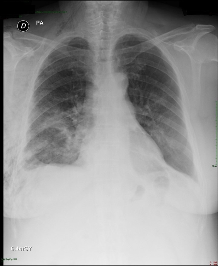
Post-operative: Post-operative chest x-ray confirmed appropriate chest tube placement and no significant bleeding or pneumothorax.
Immediate post-operative film (chest tube visible)
Patient did well post-operatively. Chest tube was discontinued on POD#2 and discharged home.
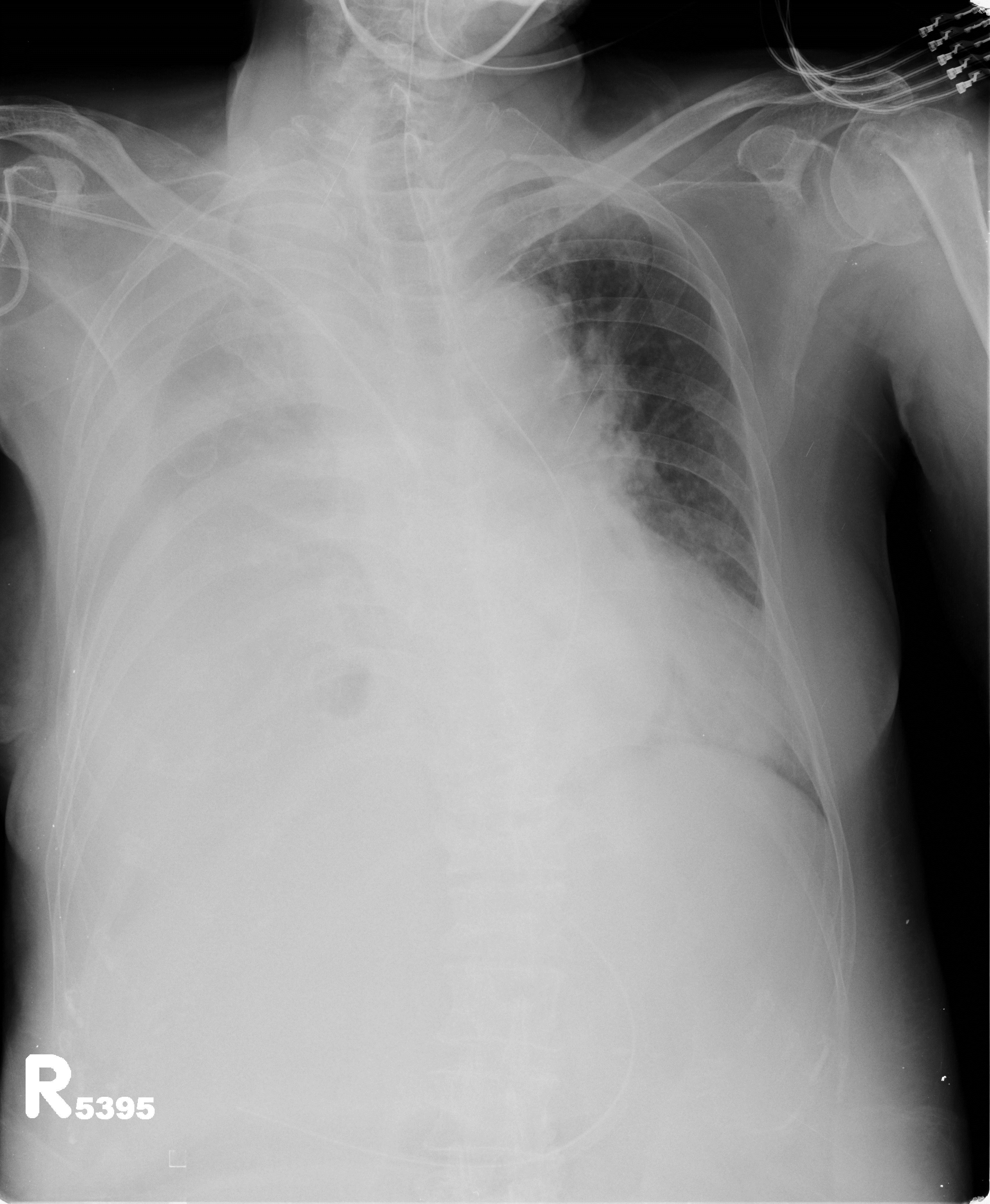
PA & LAT films on post-operative day 2
Discussion: Since the initial published reports of single-port thoracoscopy, this procedure has been applied to an increasing range of cases. Dr. Gonzalez and his team have published reports demonstrating the safety and utility of the single-port technique for multiple procedures including lobectomies, sleeve resections, segmentectomies, pneumonectomies and mediastinal mass resections. Dr. Hanao Chen (Taiwan) has reported several successful esophagectomies using this technical, as well as bilateral pleural drainage using a unilateral single-port approach.
Contrary to popular perception, the use of a single-port versus traditional VATS procedures (three or more) is actually associated with better visibility and accessibility for surgeons. Surgeons using this technical have also reported better ergonomics with less operating fatigue related to awkward body positioning while operating.
The learn curve for this surgical approach is less than anticipated due to the reasons cited above, and has been cited at 5 to 20 cases by Dr. Gonzalez, the creator of this approach.
The main limitations for surgeons using this technique is often related to anticipated (but potentially unrealized) fears regarding the need for urgent conversion to open thoracotomy. In reality, many of the complications that may lead to urgent conversion, such as major bleeding, are manageable thoracoscopically once surgeons are experienced and comfortable with this approach.
Dr. Gonzalez and his colleagues have reported a conversion rate of less than 1% in their practice. Subsequent reports by Dr. Gonzalez and his colleagues have documented these findings.
Other barriers to adoption of this technique are surgeon-based, and may be related to the individual surgeon’s willingness or reluctance to adopt new techniques and technology. Many of these surgeons would be surprised by how this technique mimics open surgery.
The successful adoption of this technique by numerous thoracic surgery fellows shows the feasibility and ease of learning single-port thoracoscopy by surgeons interested in adopting and advancing their surgical proficiency in minimally invasive surgery.
The benefits for utilizing this technique include decreased length of stay, decreased patient discomfort and greater patient satisfaction.
References/ Additional Readings
Bertolaccini, L., Rocco, G., Viti, A. & Terzi, A. (2013). Surgical technique: Geometrical characteristics of uniportal VATS. J. Thorac Dis. 2013, Apr 07. Article from thoracic surgeons at the National Cancer Institute in Naples, Italy explains how the geometric advantages of uniportal VATS improves visibility and spatial perception over traditional VATS, and mimics open surgery.
Calvin, S. H. Ng (2013). Uniportal VATS in Asia. J Thorac Dis 2013 Jun 20. Article discussing the spread of uniportal techniques in Taiwan, China and other parts of Asia.
Chen, Chin-Hao, Lin, Wei-Sha, Chang, Ho, Lee, Shih-Yi, Tzu-Ti, Hung & Tai, Chih-Yin (2013). Treatment of bilateral empyema thoracis using unilateral single-port thoracoscopic approach. Ann Thorac Cardiovasc Surg 2013.
Gonzalez Rivas, D., Fieira, E., Delgado, M., Mendez, L., Fernandez, R. & De la Torre, M. (2013). Surgical technique: Uniportal video-assisted thoracoscopic lobectomy. J. Thorac Dis. 2013 July 4.
Gonzalez Rivas, D., Delgado, M., Fieira, E., Mendez, L. Fernandez, R. & De la Torre, M. (2013). Surgical technique: Uniportal video-assisted thoracoscopic pneumonectomy. J. Thorac Dis. 2013 July 4.
Rocco, G. (2013). VATS and uniportal VATS: a glimpse into the future. J. Thorac Dis. 2013 July 04. After coming across several articles by Dr. Gaetano Rocco, and actively pursuing several other publications by this Italian thoracic surgeon, I have become increasingly convinced that Gaetano Rocco, along with Dr. Gonzalez Rivas is one of the world’s leading innovators in thoracic surgery. Hopefully, cirugia de torax will be able to catch up to Dr. Rocco at some point for an in-depth discussion.
Rocco G. Single port video-assisted thoracic surgery (uniportal) in the routine general thoracic surgical practice. Op Tech (Society of Thoracic and Cardiovascular Surgeons). 2009;14:326–335.
Rocco G, Khalil M, Jutley R. Uniportal video-assisted thoracoscopic surgery wedge lung biopsy in the diagnosis of interstitial lung diseases. J Thorac Cardiovasc Surg. 2005;129:947–948.
5 / Video-assisted thoracic surgery lobectomy: 3-year initial experience with 200 cases. Gonzalez D, De la Torre M, Paradela M, Fernandez R, Delgado M, Garcia J,Fieira E, Mendez L. Eur J Cardiothorac Surg. 2011 40(1):e21-8.
6 / Single-port Video-Assisted Thoracoscopic Anatomical Resection: Initial Experience. Diego Gonzalez , Ricardo Fernandez, Mercedes De La Torre, Maria Delgado, Marina Paradela, Lucia Mendez. Innovations.Vol 6.Number 3. May/jun 2011. Page 165.
Dr. Gaetano Rocco talks about persistent air leaks and the development of a remote-controlled computer assisted suction device.
An air leak lasting longer than 5 to 7 days is considered a ‘prolonged or persistent air leak*’.
A prolonged air leak is one of the most frustrating complications after thoracic surgery for patients and clinicians alike. Far from being life- threatening, a prolonged air leak often occurs in patients that are otherwise stable, healing well and potentially ready for discharge. However, the presence of a persistent air leak can change all that – by limiting patient mobility and prolonging their hospital stay.
Surgeons have attempted to manage this problem in multiple ways in the past; including additional surgery, application of intra-operative glues and other sealants, repeated post-operative pleurodesis and the implantation of long-term devices like the Heimlich valve (to evacuate air while the lung heals).
More radical therapies such as radiation and endobronchial valves (EBV) have also been used with varying degrees of success (Erdoğan Çetinkaya, M. Akif Özgül, Şule Gül, Ertan Çam, Yakup Büyükpolat, 2012).
Ambulatory suction
In this study, Rocco designed a device capable of providing differing levels of suction independent of wall mounted suction**. This in itself, is an important feat since being reliant on wall-mounted suction significantly limits the mobility and activity of otherwise ambulatory patients.
In standard cases, patients are essentially tethered to the suction mount in their rooms by a short length of suction tubing. This prolongs hospitalization and can contribute to the development of additional complications.
The Heimlich valve is often used in these cases to allow patients to be discharged home, despite a persistent air leak. However, while the Heimlich valve relieves patients of this reliance on wall suction, this is also one of it’s limitations. Independent of wall suction, the Heimlich valve prevents the entry of additional air into the pleural space but can not provide active suction to assist in lung healing.
Prior portable suction technologies
In my experience, our hospital had several antiquated portable suction units that allowed for limited ambulation. These units were electric-powered suction units that could be wheeled alongside the patient (similar to wheeled oxygen units.) But these units (dating from the 1950’s – 1960’s and which were found & rehabilitated from an old equipment room) still required the patient to remain in contact with a grounded electrical outlet, though the cord was lengthy. They were used in limited circumstances in the intensive care and step-down units.
Portable suction unit used at Danville Regional Medical Center, Danville, Virginia. Photo by Brian Compton
Dr. Rocco’s device is a significant upgrade from the 1950’s version, and contains computer sensors to detect, and change the level of suction as needed. It also contains a chargeable battery that allows patients to function independent of an electrical outlet for up to 48 hours. This offers considerable freedom, and even permits home use in stable patients.
Continuous patient monitoring
With a laptop computer, both the surgeon and the patient can keep in contact, and monitor progress. The surgeon can also adjust the amount of suction and review the continuously recorded air leak data.
In this case report, Rocco and his colleagues trialed the equipment on a patient with a persistent air leak after a right upper lobectomy with wedge resection of the right lower lobe. The patient was treated and monitored with this device during a stay in the step-down unit, the thoracic floor and finally, in an outpatient setting at a nearby guest house.
While this is a preliminary trial involving a single patient, the potential uses of these technology are considerable – given the frequency of prolonged air leaks post-operatively. This is also important to consider as minimally invasive surgeries make it possible for patients to be medically stable and otherwise eligible for discharge earlier in the post-operative course. Given the inherent risks (and costs) of prolonged hospitalization – this may become a viable option a part of a comprehensive discharge plan for many patients who would otherwise remain tethered to a suction mount in a hospital room.
My apologies to readers – this article was actually published in January of this year, but was somehow overlooked until working on a separate study by Dr. Gaetano Rocco at the National Cancer Institute in Naples, Italy.
Dimos Karangelis, Georgios I Tagarakis, Marios Daskalopoulos, Georgios Skoumis, Nicholaos Desimonas, Vasileios Saleptsis, Theocharis Koufakis, Athanasios Drakos, Dimitrios Papadopoulos, Nikolaos B Tsilimingas (2010). Intrapleural instillation of autologous blood for persistent air leak in spontaneous pneumothorax- is it as effective as it is safe? J Cardiothorac Surg. 2010; 5: 61. Published online 2010 August 17. doi: 10.1186/1749-8090-5-61. The authors investigate the use of blood pleurodesis in fifteen patients and report a 27% success rate.
Erdoğan Çetinkaya, M. Akif Özgül, Şule Gül, Ertan Çam, Yakup Büyükpolat (2012). Treatment of a Prolonged Air Leak with Radiotherapy: A Case Report. Case Rep Pulmonol. 2012; 2012: 158371. Published online 2012 September 27. doi: 10.1155/2012/158371. In this case report, surgeons in Istanbul, Turkey, radiation was applied to a localized area after the probably area of air leak was identified thru ventilation scintigraphy. Patient received two doses of 10 G to a 10 X 10 cm area with resolution of air leak.
Tudor P Toma, Onn Min Kon, William Oldfield, Reina Sanefuji, Mark Griffiths, Frank Wells, Siva Sivasothy, Michael Dusmet, Duncan M Geddes, Michael I Polkey (2007). Reduction of persistent air leak with endoscopic valve implants. Thorax. 2007 September; 62(9): 830–833. doi: 10.1136/thx.2005.044537 Discussion of endobronchial valves (EBV).
The article, entitled, “Treatment of bilateral empyema thoracis using unilateral single port approach,” details one of his recent cases and discusses the use of unilateral single port surgery for the treatment of bilateral conditions. (For the uninitiated – that’s one small incision to treat an infection on both sides of the chest.)
Case report: bilateral empyema
In this case report, a 28 year old male presented with dyspnea, sore throat, malaise, fever and weakness. Patient was admitted with a diagnosis of sepsis and started on antibiotics.
Labs showed an elevated WBC count (19,300), C-reactive protein and D-dimer. Subsequent imaging confirmed the presence of pulmonary emboli, and with serial imaging showing worsening bilateral pleural effusions. Thoracic surgery was consulted for definitive treatment.
Dr. Chen discusses this technique, as well as considerations for using this novel approach.
First look at innovative approach
Other that his recent discussions here at Cirugia de Torax, this is the first time that surgery utilizing this technique has been discussed in a medical forum. This represents a ground-breaking advance in thoracoscopic surgery, single port surgery and thoracic surgery as a whole.
Update: Article published June 18, 2013 in the Annals of Thoracic and Cardiovascular Surgery. A pdf of the full article is available.
a clinical case report from Bogota, Colombia – and the work of Dr. Kalyanam Shivkumar, MD, PhD, Director, UCLA Cardiac Arrhythmia Center on treating ventricular arrhythmias with cardiac sympathetic denervation, including an upcoming clinical trial.
Dr. Ricardo Buitrago, Thoracic Surgeon
Dr. Andres Franco, Thoracic Surgeon
K. Eckland, Nurse Practitioner
Clinica Shaio
Bogota, Colombia
Left cardiac sympathetic denervation for uncontrolled ventricular arrhythmias in a young child
Dr. Andres Franco (right) assists Dr. Ricardo Buitrago (left) during VATS cardiac denervation
Case History: The patient, a 9-year-old Hispanic female child had a history of congenital long QT syndrome*, and had her first AICD (automated internal cardiac defibrillator) placed at two months of age, after receiving the initial diagnosis as a neonate.
After several years and multiple medical regimens were unsuccessful in controlling frequent episodes of ventricular tachycardia, the patient had undergone several additional procedures aimed at reducing the incidence of arrhythmia. The device had been checked thoroughly, evaluated and exchanged twice as part of on-going evaluation to ensure that the device was working properly, and was set at appropriate thresholds.
At the time of the initial referral to thoracic surgery, the child suffered from intense post-traumatic stress disorder symptoms and was being treated for psychiatric disturbances that were believed to be related to the extreme fear and stress related to frequent defibrillations delivered by her device. As a final resort, the department of electrophysiology requested evaluation for Video assisted thoracoscopic (VATS) sympathectomy for cardiac denervation.
After the patient is anesthetized and intubated with a double lumen endotracheal tube, a single anterior thoracic 10 mm port incision is made in the 5th intercostal space of the left chest without rib spreading. A 10mm port is inserted, for camera access to the interior of the chest. The left lung is deflated for easy identification and access to the sympathetic nerves at the T2 – T4 level on the chest wall. After successful identification, the nerves were cauterised. The lung was reinflated, and surgical instruments removed. Chest incision was closed with several layers of suture. The patient was awakened, extubated and transferred to the PACU.
In addition to the standard intra-operative hemodynamic and telemetry monitoring, an electrophysiology cardiologist was present during the case to monitor and treat the patient, if necessary.
This slideshow requires JavaScript.
Following surgery, the patient was transferred to the post-anesthesesia care unit (recovery room). Following an uneventful recovery, she was discharged home. At follow-up surgical visit, her incision was well-healed and family reported no further discharges from her AICD.
At six months, post surgery, the patient has had no further cardiac events or ventricular arrhythmias reported or recorded by her device.
Discussion
The cardiovascular effects of sympathectomy have been well-known and described in the medical literature since the early 20th century. However, limitations in surgical technique prior to the advent of thoracoscopic surgery as well as the potential side effects of sympathectomy procedures have limited the use and research into this technique for the treatment of cardiac conditions.
In recent years, researchers at the David Geffen School of Medicine at the UCLA Cardiac Arrhythmia Center (Ajijola et. al) have published several papers about their experiences treating patients with persistant ventricular arrhythmias. In their work, which is one of the largest studies to date, the authors report their experiences with both selective left-sided cardiac sympathetic denervation (LCSD) alone or bilateral cardiac sympathetic denervation (BCSD) as a last-ditch treatment for persistent ventricular arrhythmias. Many of their patients have previously undergone multiple ablation therapies and/or evaluation for cardiac transplantation.
On-going research and clinical trials
Their results have been so promising, in fact, that they have now made cardiac denervation a routine procedure at UCLA and have designed an international multi-center trial called PREVENT VT to study this procedure in a larger group of patients. Since the publication of their initial work, Aijiola et al. have continued their study, with over 40 cases under their belt. Dr. Kalyanam Shivkumar, MD, PhD, Director, UCLA Cardiac Arrhythmia Center & EP Programs reports that they will be presenting their surgical outcomes at an upcoming conference, the Heart Rhythm Meeting at Denver in May of 2013 (Vaseghi et al Cervicothoracic Sympathectomy In Patients With Refractory Vt: Intermediate And Long Term Follow Up).
Contact information for Dr. Kalyanam Shivkumar:
Kalyanam Shivkumar MD PhD
Professor of Medicine & Radiology
Director, UCLA Cardiac Arrhythmia Center & EP Programs
For both their work, and the purposes of this post, persistent ventricular arrhythmias were defined as repeated episodes of ventricular tachycardia or ventricular fibrillation despite maximal medical therapy with a beta blocker and amiodarone**. (In a related article, Ajijola et al. further define which patients are the best candidates for successful outcomes with this procedure.) In their work, the researchers at UCLA were able to show increased effectiveness with the use of bilateral sympathetomy versus left-sided only.
However, the use of left-sided versus bilateral sympathectomy is also determined by the type of arrhythmia (monomorphic versus polymorphic) as well as previous patient history and medical treatments such as catheter based ablations or extensive scar tissue formation from previous cardiac injury.
Given the high morbidity, mortality and adverse effects on the quality of life for people with uncontrolled ventricular arrhythmias as well as the relative low risk, and ease of VATS procedures to treat this condition, cardiac sympathetic denervation should remain an important clinical tool in the treatment of this life-threatening condition, particularly when other treatments have failed.
*Patients with this condition are at very high risk of sudden cardiac risk.
**In the addition to implanted or external defibrillatory devices.
Wilde AA, Bhuiyan ZA, Crotti L, Facchini M, De Ferrari GM, Paul T, Ferrandi C, Koolbergen DR, Odero A, Schwartz PJ. (2008). Left cardiac sympathetic denervation for catecholaminergic polymorphic ventricular tachycardia. N Engl J Med. 2008 May 8;358(19):2024-9. doi: 10.1056/NEJMoa0708006. Case report of a 17 year old boy. In this article, the authors also talk about the psychological trauma experienced by these patients due to frequent defibrillation from AICDs similar to the patient in Colombia.
Case report with video of SITS (single incision thoracoscopic surgery) repair of diaphragm defect in a case of hepatic hydrothorax resulting from liver cirrhosis with Dr. Chih-Hao Chen, MAckay Memorial Hospital, Taiwan
Case Report: Single incision thoracoscopic repair of diaphragmatic defect in a patient with hepatic hydrothorax
Dr. Chih-Hao Chen, Thoracic Surgeon MAckay Memorial Hospital, Taiwan
Dr. Chen, Thoracic Surgeon
Clinical History:
Patient is an elderly woman who was admitted after a motor-vehicle accident with a traumatic fracture of the humerus and femoral neck. She was brought to our ED immediately and was intubated due to acute respiratory failure.
Subsequent Chest radiograph showed diffuse opacity in right hemithorax and concomitant fracture in left side humerus and femoral neck. Attempt for tapping of the pleural effusion showed clear in nature.
According to previous medical records, she had no relevant history. She was admitted to ICU for further evaluation and management.
Fluid analysis in emergency department showed transudate.
LFTS: Total Bilirubin 2.7 mg/dL AST 116 ALT 68 Albumin 2.3 g/dL Direct Bilirubin H 1.1 mg/dL
Chem panel: BUN 83 mg/dL Creatinine 1.6 mg/dL K 3.2 mEq/LNa 144 mEq/L
Chest radiograph on admission showed a massive right-sided pleural effusion.
Chest radiograph on admission
For symptomatic control, the physician performed intermittent thoracentesis. Because the traumatic site is left aspect of the trunk ( fracture in left side humerus and left side femoral neck ) and right side effusion was very clear.
Hepatic hydrothorax was suspected. Later peritoneal scan confirmed the diagnosis.
Peritoneal scan
The scan showed left side pleural space was sparring from radioisotope. Direct communication between right side pleural cavity and the abdomen. The diagnosis is confirmed with such findings.
CT scans are not diagnostic for this condition, and were not indicated for her other injuries. Therefore, we did not arrange CT scan of the chest / abdomen.
Abdominal ultrasound showed moderate to massive ascites. Along with hepatic encephalopathy, moderate to massive ascites, prolonged PT/PTT, low albumin, higher bilirubin, the extent of cirrhosis is Child’s class C.
Operative Procedure: Single incision thoracoscopic repair of a diaphragmatic defect. Theoretically, with SITS, the wound can be very tiny. However, in our experience (fifteen total cases to date), diaphragm surgery through single port may be a bit difficult because we did not know where the defect is. We have to inspect very carefully and to search for the defect where the fluid came out. In this case, we made one small wound around 2 cm in length at the 6th ICS along the anterior axillary line.
Repair of the diaphragmatic defect was performed using silk suture similar to that used to repair inguinal hernias. Intra-operatively, the defect was 2 -3 mm in diameter.
At the conclusion of the procedure, using the original incision, we placed one Fr.24 chest tube to monitor the drainage and may consider chemical pleurodesis if the drainage persists. The operative procedure was accomplished within 30 minutes.
Post-operative Chest Radiograph
Post-operative condition of the chest film showed near complete resolution of the effusion and lung re-expansion was complete.
Pathology/ Fluid Cytology: fluid analysis and peritoneal scan showed communication between peritoneal space and right side pleural space confirming pre-operative diagnosis. No tissue specimens were taken during this procedure.
Discussion:
Hepatic hydrothorax is the development of a pleural effusion in a patient with liver disease in the absence of cardiopulmonary pathology, making it a diagnosis of exclusion in many cases. It can occur in patients with and without ascites and may be the first presenting symptom in patients with undiagnosed liver disease. Similar to catamenial pneumothorax; hepatic hydrothorax is predominantly a right-sided disease. This is due to an anatomic gutter or diaphragmatic defect that occurs, and allows the passage of material or fluid from the abdominal cavity into the pleural space. This can be seen and identified on peritoneal studies(Peritoneal scan) like the study showed in our case study above. (Similar pathologies can occur in related conditions such as renal failure related hydrothorax due to this defect). Such defect is usually identified in the tendon part of the diaphragm. Peritoneal scan can confirm there is communication between the abdominal cavity and the pleural space. However, the definite location, size and number of defects can not been identified by the scan alone. Thoracoscopic inspection is the only method to search for such defect(s).
Video-assisted thoracoscopic surgery (VATS) has been shown to be a safe and effective method of treating this condition, by allowing surgeons to correct the defect, and thus prevent recurrence (Saito et al. 2012). The cure rate varied greatly in the literature. The key is whether the defect can be repaired. For one to two obvious defects, direct suture repair usually cured the disease. (the cure rate more than 80%) However, for some undetectable defects or defects with fenestration type, the cure rate is very low, ( around 30-50% ). Alternative strategies have to be considered in such condition, such as tissue glue, abrasion pleurodesis, mesh interposition and using sclerosing agents(OK432, bleomycin, Minocin, talc, etc). This is in distinct contrast to the numerous non-surgical drainage procedures such as thoracentesis, which removes accumulated fluid but does not correct the underlying pathology. However, the hallmark of this condition, liver failure predisposes patients to complications such as bleeding, infection and poor wound healing. These risks are one of the primary reasons treatment was often limited to drainage procedures prior to the popularization of lower risk VATS procedures. In the past, patients with Child’s class C liver cirrhosis are basically not proper surgical candidates because of extremely high mortality/morbidity rate. In recent experience of single-port approach, some patients with Child B and C are still safe with minimal postoperative complications. The advance of these minimally invasive technologies such as uni-port thoracoscopy permits fewer and more limited incisions which is believed to further reduce these risks while providing patients with definitive treatment options. More case studies such as this one, along with larger studies are needed to demonstrate the benefits of this technique for hepatic hydrothorax.
References
Doraiswamy V, Riar S, Shrestha P, Pi J, Alsumrain M, Bennet-Venner A, Kam J, Klukowicz A, Miller R. (2011). Hepatic hydrothorax without any evidence of ascites. ScientificWorldJournal. 2011 Mar 7;11:587-91. doi: 10.1100/tsw.2011.68 Case study.
Lee WJ, Kim HJ, Park JH, Park DI, Cho YK, Sohn CI, Jeon WK, Kim BI. (2011). Chemical pleurodesis for the management of refractory hepatic hydrothorax in patients with decompensated liver cirrhosis. Korean J Hepatol. 2011 Dec;17(4):292-8. doi: 0.3350/kjhep.2011.17.4.292. Eleven patient Korean study looking at the effectiveness of pleurodesis in patients with hepatic hydrothorax. While the procedure was successful in 8 patients, the authors noted a high rate of procedural-associated complications. (Notably, the researchers used several different agents for chemical pleurodesis.)
Sawant P, Vashishtha C, Nasa M. (2011). Management of cardiopulmonary complications of cirrhosis. Int J Hepatol. 2011;2011:280569. doi: 10.4061/2011/280569. Epub 2011 Jul 19. Article discussing complications of cirrhosis including hydrothorax.
Chest wall resection with pulmonary segmentectomy for metastatic breast cancer.
a multi-disciplinary approach: plastics, surgical oncology and thoracic surgery
Title:Chest wall resection with pulmonary segmentectomy for metastatic breast cancer
Summary: Breast cancer remains the second leading cause of mortality in females in Mexico, aged 30 to 55, and is usually self-detected in later stages. Due to disparities in health care within the country, even patients with early detection may not receive optimal or timely treatment leading to more recurrent or metastatic disease. Surgery remains the best, but underutilized option for definitive treatment in patients with surgically resectable disease. In this case, a patient with advanced disease was successfully resected using a multi-disciplinary approach.
Authors:K. Eckland, ACNP-BC, Hospital General de Mexicali, Thoracic Surgery
Gabriel Ramos Orozco, MD, Instituto Mexicano Seguro Social (IMSS), Surgical Oncology
Corresponding author: Carlos Ochoa Gaxiola, MD
Email: drcarlosochoa@yahoo.com
Announcement text: a multidisciplinary approach to recurrent metastatic breast cancer with chest wall resection and free flap graft creation.
Subject/ Classification terms: chest wall resection, rib resection, metastatic breast cancer, pulmonary segmentectomy, breast cancer in Mexico
Disclosures: The authors have no disclosures.
History/ Case Summary:
The patient was a 70-year-old Hispanic female with a past medical history significant for local breast cancer in the left breast, initially diagnosed in 1994. This was treated with chemotherapy and radiation. She was then maintained on tamoxifen until 2000. In 2011, she presented with a recurrent mass in the left breast. There was no other history of chronic or active medical conditions such as HTN, CAD or diabetes.
After referral to a surgical oncologist for further evaluation, patient underwent additional evaluation. A PET/ CT scan was positive for a metabolically active lesion in the left breast with an SUV of 9.6 with lytic lesions in anterior ribs with max SUV of 3.0. There was no evidence of distal metastasis to other organs including the brain, lungs, or abdominal cavity on PET or other diagnostic imaging. All pre-operative labs were within normal ranges including alk phos, and serum calcium.
Pre-operative Chest X-ray
After initial surgical evaluation, a multi-disciplinary surgical plan utilizing a general surgical oncologist, thoracic surgery and plastic surgery was devised for surgical resection of breast mass with rib resection and free flap creation.
surgeons planning approach
Operative Course: The left breast including all skin, tissue and lymphatics was excised to the depth of the rib cage. Further dissection and resection of the anterior portion of ribs #2, 3 and #4 was completed.
following rib resection
Following rib resection, upon exploration of the left thoracic cavity, the patient was found to have a large greyish-white lesion, estimated at 3.5 cm in diameter in the left upper lobe. The lesion was hard, and located on the peripheral portion of the upper lobe. No additional lesions were found.
during surgery, a previously undetected pulmonary lesion was discovered
The decision to undertake pulmonary resection was based on the possibility of complete surgical resection of existing disease. At the time, a discussion was undertaken with the patient’s primary surgeon, and the thoracic surgeon on the feasibility of resection by lobectomy versus segmentectomy. The decision was made to proceed with a lung-sparing procedure as the patient’s baseline pulmonary function was not known.
Following successful lung resection and hemostasis, a 32 french chest tube was placed, and surgical mesh was placed for coverage of chest wall / rib defect. After mesh was sutured into place, the patient was re-positioned for harvesting of a free flap from the posterior chest. Abdominal free flap harvest was not undertaken due to patient anatomy. The plastic surgeon involved in the case, Dr. Nastia Gonzalez then proceeded with free flap grafting for breast reconstruction. There was no significant bleeding, hypoxia or hemodynamic instability intraoperatively.
Post-operative Course:
The patient was successfully extubated at the conclusion of the case, and transferred to the post-operative care unit in stable condition. Post-operative course was uncomplicated with the chest tube removed on POD#3, and the patient discharged home on POD#5. The patient’s oxygen saturations were within the normal range (92% or above) and she was discharged home without supplemental oxygen.
Subsequent post-operative visit was uneventful with no evidence of infection, or impaired healing of the graft or harvest site. As of the date of publication, there has been no further evidence of recurrence or metastatic disease.
Conclusions: For patients with metastatic disease limited to adjacent and surgically resectable tissue, surgery remains the best option for longevity and overall survival. However, despite the available and use of advanced imaging studies, surgeons should prepare for and anticipate the possibility of discovery of evidence of additional disease. In this case, a more complete anatomic resection of the newly discovered lung lesion was hindered by the lack of pre-operative evaluation of pulmonary disease.
Chest wall resection and defect closure have been managed with a variety of techniques over the years, including muscle flaps, plastic ribcage creation, mesh closures for stabilization after rib resection (Khalil et al.). In this case, which required a radical mastectomy, surrounding musculature was removed for full resection. Tissue was harvested for free flap grafting but this gives lesser structure than attached muscle, so synthetic mesh was used.
Historically, hardware installation was plagued with a variety of problems including infection and erosion. However, preliminary reports of evolving hardware for oncologic chest resections may change closure techniques in the future (Fabre et al, 2012).
Gharagozloo F, Meyer M, Tempesta BJ, Margolis M, Strother ET, Tummala S. (2012). Robotic en bloc first-rib resection for Paget-Schroetter disease, a form of thoracic outlet syndrome: technique and initial results. Innovations (Phila). 2012 Jan-Feb;7(1):39-44. No free text available. Report on robot-assisted rib resection. (Less relevant but interesting.)
Mohajeri G, Sanei MH, Tabatabaee SA, Hashemi SM, Amjad E, Mohajeri MR, Shemshaki H, Jazi AH, Kolahdouzan M. (2012). Micrometastasis in non-small-cell lung cancer: Detection and staging.Ann Thorac Med. 2012 Jul;7(3):149-52. Using bone marrow biopsy for diagnosis of lytic lesions.
This case study has been published with the gracious consent of the patient. However, in accordance to the patient’s wishes, and privacy – no photos showing the pre-operative site (breast) or the graft after surgery will be published.
This case study was prepared with assistance from Dr. Carlos Ochoa. Since we have been discussing the relevance of case reports and providing tips on case report writing for new academic writers – we have written the following case report in the style advocated by McCarthy & Reilley (2000) using their case report worksheet to demonstrate the ease of doing so in this style.
Since the previous presentation of dual-port thoracoscopy for decortication was missing essential materials, we are presenting a second case report.
Authors: K. Eckland, ACNP-BC, MSN, RN & Carlos Ochoa, MD
Case Report: Dual port thoracoscopy for decortication of a parapneumonic effusion
Abstract: The use of increasingly minimally invasive techniques for the treatment of thoracic disease is becoming more widespread. Dual and even single port thoracoscopy is becoming more frequent in the treatment of parapneumonic effusions and empyema.
Clinical question/problem: the effectiveness and utility of dual port thoracoscopy for parapneumonic effusions.
Analysis of literature review: Despite the increasing frequency of dual and single port thoracoscopic techniques, there remains a dearth of literature or case reports on this topic. Pubmed and related searches reveal only a scattering of reports.
Summary: As the case report suggests, dual port thoracoscopy is a feasible and reasonable option for the treatment of parapneumonic effusion.
Case history: 50-year-old patient with a three-week history of pneumonia, with complaints of right-sided chest pain, cough and increased phlegm production. Additional past medical history is significant for poorly controlled diabetes, hypertension, and obesity with central adiposity. Medications included glyburide and lisinopril.
After being seen and evaluated by an internal medicine physician, the patient was started on oral antibiotics. After three weeks, when his symptoms failed to improve, he was referred by internal medicine to thoracic surgery for out-patient evaluation.
On exam: middle-aged obese diabetic gentleman in no immediate distress, resting comfortable in the exam room. Face appeared moderately flushed, but skin cool and dry to the touch, no evidence of fever.
On auscultation, he had diminished breath sounds over the right lower lobe with egophony over the same area. The remainder of the exam was essentially normal.
Lab studies showed a mildly elevated WBC of 11.6, decreased Hgb of 10.4 / HCT 32.5. Hemoglobin A1c 10.6, Fasting glucose 228, HDL mildly low at 40.
EKG showed slight axis deviation, with slightly prolonged QRS complex (.16) with no evidence of loss of R, St elevation or other abnormalities. He was cleared by internal medicine for surgery.
After risks, benefits and alternatives to VATS decortication were explained to the patient – the patient consented to proceed with surgical decortication. After scheduling surgery, the patient was seen by anesthesia in preparation for the procedure.
Surgical procedure: Dual-port thoracoscopy with decortication of parapneumonic effusion.
Dual port thoracoscopy
After being prepped and drapped in sterile fashion and confirmation of dual lumen endotracheal tube placement, a small 2 cm incision was made for insertion of a 10mm port. Following entry into the chest with the thoracoscope, the right lung was deflated for optimal inspection and decortication of loculations. After completing the majority of the procedure, a second access port was created for better visualization and to ensure that a thorough decortication was completed. The lung and pleural were separated from the chest wall, and diaphragm, and demonstrated good re-expansion with lung re-inflation prior to completion of the procedure.
chest tubes at conclusion of case
At the conclusion of the procedure, two 28 french chest tubes were placed in the existing incisions. These were sutured into place, and connected to a pleurovac drainage system before applying a sterile gauze dressing. The patient remained hemodynamically stable throughout the case, with no episodes of hypoxia or desaturation. Following surgery, the patient was transferred to the PACU in stable condition.
Post-operative course was uncomplicated. Chest tubes were water-sealed on POD#3 and chest tubes were removed POD#4, with the patient being subsequently discharged after chest x-ray.
close up view of dual port thoracoscopy
Literature Review
A literature review was performed on PubMed using “dual port thoracoscopy”, “dual port VATS”, “2 port” as well as minimally invasive thoracoscopic surgery “
Results of search: A limited number of case studies (3) described thoracoscopic surgery with a single port. There was one case found describing cases conducted with two ports, and the majority of reports involved three or more access ports.
Discussion/ Conclusion
While convention medical wisdom dictates a trial and error treatment approach with initial trial of antibiotic therapy followed by chest tube placement (Light, 1995), surgeons have long argued that this delay in definitive treatment places the patient at increased risk of significant morbidity and mortality (Richardson, 1891). Multiple recent reviews of the literature and research comparisons continue to demonstrate optimal outcomes with surgery based approaches versus antibiotics alone, TPA and tube thoracostomy. The ability to perform these procedures in the least invasive fashion (VATS versus thoracotomy approaches) defies the arguments against surgical intervention as advanced by interventionalists (radiologists and pulmonologists.) Successful decortication with the use of dual port thoracoscopy is another example of how technology is advancing to better serve the patient and provide optimal outcomes, and offers a minimally invasive option when single port surgery may not be feasible.
During the case above, visibility and access to the thoracic cavity was excellent. However, in cases requiring additional access, reversion to the standard VATS configuration can be done easily enough with significant delays or additional risks to the patient.
References/ Resources
Foroulis CN, Anastasiadis K, Charokopos N, Antonitsis P, Halvatzoulis HV, Karapanagiotidis GT, Grosomanidis V, Papakonstantinou C. (2012). A modified two-port thoracoscopic technique versus axillary minithoracotomy for the treatment of recurrent spontaneous pneumothorax: a prospective randomized study. Surg Endosc. 2012 Mar;26(3):607-14. [free full text not available.]
Gonzalez – Rivas, D., Fernandez, R., De la Torre, M., & Martin – Ucar, A. E. (2012). Thoracoscopic lobectomy through a single incision.Multimedia manual cardio-thoracic surgery, Volume 2012. This is an excellent article which gives a detailed description, and overview of the techniques used in single incision surgery. Contains illustrations, full color photos and videos of the procedure.
Gonzalez-Rivas D, Paradela M, Fieira E, Velasco C. (2012). Single-incision video-assisted thoracoscopic lobectomy: initial results. J Thorac Cardiovasc Surg. 2012 Mar;143(3):745-7.
Case reports, which are one of the oldest form of medical literature are by and large becoming a lost art. Considered observational, and low-level evidence in a time of ‘pay-for-performance,’ CMS guidelines and ‘evidence-based practice,’ the utility and educational nature of case reports is often overlooked. To many in the medical publishing industry, as the meta-analysis dominates the scientific landscape, the traditional case report is rapidly becoming a thing of the past (Kasim et. al. 2009).
However, to the practitioners, the case report remains an invaluable tool. These anecdotal stories stay with us, and may facilitate recognition of signs and symptoms that would otherwise go unnoticed, particularly by junior medical providers. Also the case report is often the first entry into a career punctuated with scholarly writing.
As such, in honor of the historical, humble case report, we have dedicated this post to providing assistance in authoring high quality case reports. Below are a list of resources to help get you started in your case report writing efforts.
Many readers have also noticed that here at Cirugia de Torax, we have our own section for Case Reports – a practice area, if you will, for budding writers, and others to present cases as we polish our writing skills, and a forum to share information with the thoracic community in a more relaxed and informal way. So collect your films and stories and send them our way.
case report of dual port thoracoscopy for decortication of empyema in a thirty-eight year old woman.
Note: This case report was written with the assistance of Dr. Carlos Cesar Ochoa Gaxiola.
Case Report: Dual port thoracoscopic decortication of empyema
Presentation: A 38-year-old woman presented to the local hospital with fever, pneumonia, chest pain and an elevated leukocyte count of 25,000. Initial chest x-ray showed a large left-sided effusion.
Risk factors: Patient had several traditional risk factors for the development of empyema including heavy ETOH, and malnutrition, poor access to healthcare. Patient HIV, and Hep C negative.
Initial Hospital Course: She was admitted, and started on dual therapy antibiotics (ceftriaxone & levofloxacin). A chest tube was placed with return of frank purulent material. After several days of draining minimal amounts of pus, patient remained febrile.
Subsequent CT scan showed a left-sided empyema with large loculated areas. At that time, thoracic surgery was consulted for additional evaluation and treatment. Surgery was scheduled.
At the time of surgery, patient remained on dual antibiotics with WBC of 19,000. Albumin 1.5 , Hgb 10.2, Hct 33, other labs within normal limits.
Surgical procedure: dual-port VATS with decortication
The initial chest tube was removed, patient was prepped and draped in the traditional sterile fashion. The previous chest tube site was carefully cleaned with a betadine solution, and debrided of purulent material to prevent abscess tract formation, with instrumentation traded out after debridement.
A single additional ten mm thoracoscopy port was created, with visual interior inspection performed. Initial inspection confirmed the presence of a stage IV empyema with large loculations, moderate pleural thickening and the presence of frankly purulent material adhering to the pleural/ chest wall and lung tissue. The pleura was noted to be thickened but malleable, loosely adherent to the pleural and lung surfaces.
A formal decortication was undertaken with separation of the lung from the diaphragm and adhesions to obliterate the empyema cavity. Decortication of visceral pleura was performed until the lung was completely free and able to re-expand. Lavage was performed with evacuation and drainage of copious amounts of purulent materials.
After decortication was complete, two chest tubes were placed*; anteriorly and posteriorly, under thoracoscopic guidance, and the lung was re-inflated.
*Due to the location, and presence of infection/ purulent material in the initial chest tube site, an additional chest tube site (5mm) was created at the time of chest tube insertion to prevent additional infectious complications.
At the conclusion of the case, patient was awakened and extubated prior to being transferred to the PACU as per post-operative protocols.
EBL during the case was minimal.
Post-operative course: Patient’s post-operative course was uncomplicated. On post-operative day #5, anterior chest tube was removed. On post-operative day #7, the posterior chest tube was removed. Patient was discharged post-operative day #8.
Discussion: While convention medical wisdom dictates a trial and error treatment approach with initial trial of antibiotic therapy followed by chest tube placement (Light, 1995), surgeons have long argued that this delay in definitive treatment places the patient at increased risk of significant morbidity and mortality (Richardson, 1891). Indeed, as discussed by Dr. Dov Weissburg (on a previous discussion of empyema and lung abscess) multiple recent reviews of the literature and research comparisons continue to demonstrate optimal outcomes with surgery based approaches versus antibiotics alone, TPA and tube thoracostomy. The ability to perform these procedures in the least invasive fashion (VATS versus thoracotomy approaches) defies the arguments against surgical intervention as advanced by interventionalists (radiologists and pulmonologists.) Successful decortication with the use of dual port thoracoscopy is another example of how technology is advancing to better serve the patient and provide optimal outcomes.
Earlier, not late surgical referral would have been of greater benefit to this patient.
I apologize but I was unable to take films / photographs of this procedure during this case.
References (with historical perspectives)
Andrade – Alegre, R., Garisto, J. D. & Zebede, S. (2008). Open thoracotomy and decortication for chronic empyema.Clinics, 2008; 63 (6), 789 – 93. Color photographs. Panamanian paper discussing the effectiveness of traditional open thoracotomy and decortication for stage III / chronic empyemas in an era of increased reliance on VATS. Observations and recommendations for open thoracotomy approach for chronic empyema based on 33 cases spanning from March 1992 – June 2006, showing safe and effective results with open surgery for more advanced/ chronic empyemas.
Richardson, M. H. (1891). Surgical treatment of acute and chronic empyemas. While surgical techniques have greatly changed in the 100+ years since this paper was initially published (and no one suffers from carbolic acid poisoning anymore), many of the observations of Drs. Richardson and Loomis remain clinically relevant and valid today. (As previously noted by Dr. Weissburg, this was a pre-antibiotic era.)
Balance, H. A. (1904). Seven cases of thoracoplasty performed for the relief of chronic empyema.British medical journal, 10 Dec 1904, 1561 – 1566. Dr. Balance discusses the development of Delnorme’s operation as an alternative to thoracoplasty while presenting several cases from his career. Photographs.
Case report of spontaneous pneumothorax followed by bleb resection and talc pleurodesis.
During my various travels and interviews, I have had the opportunity to meet and talk with thoracic patients from around the world. During a recent trip, I encountered a very nice young woman (in her early 20’s**). This is her story below:
The patient, the aforementioned young woman had no significant past medical history. She initially presented to a small tertiary facility with chest pain. She was evaluated for acute coronary syndrome and discharged from the emergency department. She subsequently miscarried an early pregnancy.
Several days later, her symptoms intensified, and she became short-of-breath so she returned to the emergency department. On chest radiograph, she was found to have a large left-sided pneumothorax. A chest tube was placed but subsequent radiographs showed a persistent pneumothorax. The nearest trauma facility was notified and the patient was transferred for further evaluation and treatment.
On arrival, the patient who was experiencing significant chest and LUQ pain, and breathlessness received a second chest tube. Following chest tube placement in the emergency room, chest radiograph showed the pneumothorax to be unchanged. The patient was admitted to the hospital for further testing.
A CT scan (TAC) of the chest showing chest tubes in good position and several large blebs. Following the CT scan, thoracic surgery was consulted for further treatment and management.
After discussing the risks, benefits and alternatives with the patient and family, the patient elected to proceed with a left-sided VATS (video-assisted thoracoscopic surgery) with blebectomy and talc pleurodesis.
Patient received pre-operative low dose beta blockade for sinus tachycardia. Patient was intubated with a double lumen ETT for uni-lung ventilation. The patient was hemodynamically stable intra-operatively, and the case proceed without incidence.
12 grams of sterile talc was insufflated using an aerosolized technique. A new chest tube was placed at the conclusion of the case. There was minimal to no operative blood loss.
surgeon performing video-assisted thoracoscopy
The patient was awakened, extubated and transferred to the post-operative recovery unit. Chest radiograph in recovery showed the lung to be well expanded on -20cm of suction.
Post-operatively the patient had a small airleak. She was maintained on suction for 48 hours and watersealed. Waterseal trials were successful, and on post-operative day #4, chest tube was removed. Subsequent chest x-ray was negative for pneumothorax. Patient was discharged home with a follow-up appointment and a referral to OB-GYN for additional follow-up.
Discussion: Due to patient’s history of miscarriage in close proximity to first reports of chest pain, special consideration was given to the possibility of catamenial pneumothorax (though this was first instance, and on the left whereas 90% of reported cases occur on the right.) While the literature reports previous episodes of pneumothorax during pregnancy, these reports occurred in later gestation (37 and 40 weeks, respectively.) On further evaluation, patient had no history of abnormal vaginal bleeding, pelvic infections, pelvic inflammatory disease or a previous diagnosis of endometriosis. Thoracoscopic evaluation was negative for the presence of endometrial tissue, and there were no diaphragmatic defects.
Final pathology: no abnormal results, confirming intra-operative findings.
**Note: Since this is a blog, available for public viewing, patient permission was obtained prior to posting. All efforts are made to protect patient privacy, and thus details regarding patient demographics have been changed/ omitted. Also, our gracious thanks to the patient and family for allowing this discussion of the case. If you have an interesting, educational or informative case, contact Cirugia de Torax for publication.