Case Report: Hilar mass resection using single port thoracoscopy with Dr. Diego Gonzalez – Rivas

in the operating room with Dr. Diego Gonzalez Rivas for single port thoracoscopic (uniportal) surgery.

Hilar mass resection using single port thoracoscopy with Dr. Diego Gonzalez – Rivas

K. Eckland & Andres M. Neira, MD

Instituto Nacional de Cancerlogia

Bogota, Colombia

Surgeon(s): Dr. Diego Gonzalez Rivas and Dr. Ricardo Buitrago

Dr. Diego Gonzalez Rivas demonstrates single port thoracoscopy

Case History:

59-year-old female with past medical history significant for recurrent mediastinal mass previously resectioned via right VATS.  Additional past medical history included prior right-sided nephrectomy.

Pre-operative labs:

CBC:  WBC 7230   Neu 73%  Hgb:14.1  Hct 37  platelets 365000

Pt 12.1  / INR1.1  PTT: 28.3

Diagnostics:

Pre-operative CT scan: chest

edited to preserve patient privacy

Procedure:  Single port thoracoscopy with resection of mediastinal mass and lymph node sampling

After review of relevant patient history including radiographs, patient was positioned for a right-sided procedure. After being prepped, and draped, surgery procedure in sterile fashion.  A linear incision was made in the anterior chest – mid clavicular line at approximately the fifth intercostal space.  A 10mm port was briefly inserted and the chest cavity inspected.  The port was then removed, and the incision was expanded by an additional centimeter to allow for the passage of multiple instruments; including camera, grasper and suction catheter.

Dr. Gonzalez Rivas and Dr. Ricardo Buitrago at National Cancer Institute

The chest cavity, pleura and lung were inspected.  The medial mediastinal mass was then identified.

instruments

As previously indicated on pre-operative CT scan, the mass was located adjacent and adherent to the vessels of the hilum.  This area was carefully dissected free, in a painstaking fashion.  After freeing the mediastinal mass from the hilum, the remaining surfaces of the mass were resected.  The mass was fixed to the artery pulmonary and infiltrating it) .  The mass was removed en-bloc.  Care was then taken to identify, and sample the adjacent lymph nodes which were located at stations (4, 7 and 10).

GonzalezRivas 051

Following removal of the tumor and lymph nodes, the area was re-inspected, and the lung was re-inflated.  A 28 french chest tube was inserted in the original incision, with suturing of the fascia, subcutaneous and skin layers.

closing the single port incision

Hemostasis was maintained during the procedure with minimal blood loss.

Patient was hemodynamically stable throughout the case, and maintained appropriate oxygen saturations.  Following surgery, the patient was awakened, extubated and transferred to the surgical intensive care unit.

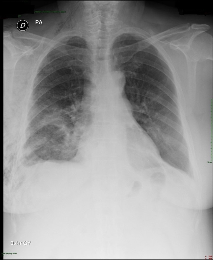
Post-operative:  Post-operative chest x-ray confirmed appropriate chest tube placement and no significant bleeding or pneumothorax.

Immediate post-operative film (chest tube visible)

Patient did well post-operatively.  Chest tube was discontinued on POD#2 and discharged home.

PA & LAT films on post-operative day 2

pod2

Discussion: Since the initial published reports of single-port thoracoscopy, this procedure has been applied to an increasing range of cases.  Dr. Gonzalez and his team have published reports demonstrating the safety and utility of the single-port technique for multiple procedures including lobectomies, sleeve resections, segmentectomies, pneumonectomies and mediastinal mass resections. Dr. Hanao Chen (Taiwan) has reported several successful esophagectomies using this technical, as well as bilateral pleural drainage using a unilateral single-port approach.

Contrary to popular perception, the use of a single-port versus traditional VATS procedures (three or more) is actually associated with better visibility and accessibility for surgeons.  Surgeons using this technical have also reported better ergonomics with less operating fatigue related to awkward body positioning while operating.

The learn curve for this surgical approach is less than anticipated due to the reasons cited above, and has been cited at 5 to 20 cases by Dr. Gonzalez, the creator of this approach.

The main limitations for surgeons using this technique is often related to anticipated (but potentially unrealized) fears regarding the need for urgent conversion to open thoracotomy.  In reality, many of the complications that may lead to urgent conversion, such as major bleeding, are manageable thoracoscopically once surgeons are experienced and comfortable with this approach.

Dr. Gonzalez and his colleagues have reported a conversion rate of less than 1% in their practice.  Subsequent reports by Dr. Gonzalez and his colleagues have documented these findings.

Other barriers to adoption of this technique are surgeon-based, and may be related to the individual surgeon’s willingness or reluctance to adopt new techniques and technology.   Many of these surgeons would be surprised by how this technique mimics open surgery.

The successful adoption of this technique by numerous thoracic surgery fellows shows the feasibility and ease of learning single-port thoracoscopy by surgeons interested in adopting and advancing their surgical proficiency in minimally invasive surgery.

The benefits for utilizing this technique include decreased length of stay, decreased patient discomfort and greater patient satisfaction.

References/ Additional Readings

Bertolaccini, L., Rocco, G., Viti, A. & Terzi, A. (2013).  Surgical technique: Geometrical characteristics of uniportal VATS.  J. Thorac Dis. 2013, Apr 07.  Article from thoracic surgeons at the National Cancer Institute in Naples, Italy explains how the geometric advantages of uniportal VATS improves visibility and spatial perception over traditional VATS, and mimics open surgery.

Calvin, S. H. Ng (2013). Uniportal VATS in Asia.  J Thorac Dis 2013 Jun 20.  Article discussing the spread of uniportal techniques in Taiwan, China and other parts of Asia.

Chen, Chin-Hao, Lin, Wei-Sha, Chang, Ho, Lee, Shih-Yi, Tzu-Ti, Hung & Tai, Chih-Yin (2013).  Treatment of bilateral empyema thoracis using unilateral single-port thoracoscopic approach.  Ann Thorac Cardiovasc Surg 2013.

Gonzalez Rivas, D., Fieira, E., Delgado, M., Mendez, L., Fernandez, R. & De la Torre, M. (2013). Surgical technique: Uniportal video-assisted thoracoscopic lobectomy.  J. Thorac Dis. 2013 July 4.

Gonzalez Rivas, D., Delgado, M., Fieira, E., Mendez, L. Fernandez, R. & De la Torre, M. (2013).  Surgical technique: Uniportal video-assisted thoracoscopic pneumonectomy.  J. Thorac Dis. 2013 July 4.

Rocco, G. (2013). VATS and uniportal VATS: a glimpse into the future.  J. Thorac Dis. 2013 July 04.  After coming across several articles by Dr. Gaetano Rocco, and actively pursuing several other publications by this Italian thoracic surgeon, I have become increasingly convinced that Gaetano Rocco, along with Dr. Gonzalez Rivas is one of the world’s leading innovators in thoracic surgery.  Hopefully, cirugia de torax will be able to catch up to Dr. Rocco at some point for an in-depth discussion.

Rocco, G., Martucci, N., La Manna, C., Jones, D. R., De Luca, G., La Rocca, A., Cuomo, A. & Accardo, R. (2013).  Ten years experience on 644 patients undergoing single-port (uniportal) video-assisted surgery.  Annals of Thoracic Surgery, 2013, Aug, 96(2): 434-438.

Rocco G, Martin-Ucar A, Passera E. Uniportal VATS wedge pulmonary resections. Ann Thorac Surg. 2004;77:726–728.

Rocco G. Single port video-assisted thoracic surgery (uniportal) in the routine general thoracic surgical practiceOp Tech (Society of Thoracic and Cardiovascular Surgeons). 2009;14:326–335.

Rocco G, Khalil M, Jutley R. Uniportal video-assisted thoracoscopic surgery wedge lung biopsy in the diagnosis of interstitial lung diseasesJ Thorac Cardiovasc Surg. 2005;129:947–948.

Rocco G, Brunelli A, Jutley R, et al. Uniportal VATS for mediastinal nodal diagnosis and stagingInteract Cardiovasc Thorac Surg. 2006;5:430–432

Rocco G, La Rocca A, La Manna C, et al. Uniportal video-assisted thoracoscopic surgery pericardial window. J Thorac Cardiovasc Surg. 2006;131:921–922.

Jutley RS, Khalil MW, Rocco G Uniportal vs standard three-port VATS technique for spontaneous pneumothorax: comparison of post-operative pain and residual paraesthesiaEur J Cardiothorac Surg 2005;28:43-46.

Salati M, Brunelli A, Rocco G. Uniportal video-assisted thoracic surgery for diagnosis and treatment of intrathoracic conditions. Thorac Surg Clin. 2008;18:305–310.

Rocco G, Cicalese M, La Manna C, La Rocca A, Martucci N, Salvi R. Ultrasonographic identification of peripheral pulmonary nodules through uniportal video-assisted thoracic surgeryAnn Thorac Surg. 2011;92:1099–1101.

Rocco G, La Rocca A, Martucci N, Accardo R. Awake single-access (uniportal) video-assisted thoracoscopic surgery for spontaneous pneumothorax. J Thorac Cardiovasc Surg. 2011;142:944–945.

Rocco G, Romano V, Accardo R, et al. Awake single-access (uniportal) video-assisted thoracoscopic surgery for peripheral pulmonary nodules in a complete ambulatory setting. Ann Thorac Surg. 2010;89:1625–1627.

Rocco G. (2012). One-port (uniportal) video assisted thoracic surgical resections – a clear advance. J Thorac Cardiovasc Surg.2012;144:S27–S31.

Additional publications on single-port thoracoscopy (Dr. Gonzalez Rivas)

1 / Single-port video-assisted thoracoscopic anatomic segmentectomy and right upper lobectomy.  Gonzalez-Rivas D, Fieira E, Mendez L, Garcia J. Eur J Cardiothorac Surg. 2012 Aug 24

2 / Single-incision video-assisted thoracoscopic lobectomy: Initial results. Gonzalez-Rivas D, Paradela M, Fieira E, Velasco C.J Thorac Cardiovasc Surg. 2012;143(3):745-7

3 / Single-incision video-assisted thoracoscopic right pneumonectomy.  Gonzalez Rivas D, De la Torre M, Fernandez R, Garcia J. Surgical Endoscopy. Jan 11. 2012 (Epub ahead of print)

4 / Single-port video-assisted thoracoscopic left upper lobectomy.  Gonzalez-Rivas D, de la Torre M, Fernandez R, Mosquera VX. Interact Cardiovasc Thorac Surg. 2011 Nov;13(5):539-41

5 / Video-assisted thoracic surgery lobectomy: 3-year initial experience with 200 cases.  Gonzalez D, De la Torre M, Paradela M, Fernandez R, Delgado M, Garcia J,Fieira E, Mendez L. Eur J Cardiothorac Surg. 2011 40(1):e21-8.

6 / Single-port Video-Assisted Thoracoscopic Anatomical Resection: Initial Experience.  Diego Gonzalez , Ricardo Fernandez, Mercedes De La Torre, Maria Delgado, Marina Paradela, Lucia Mendez. Innovations.Vol 6.Number 3. May/jun 2011. Page 165.

Dr. Gonzalez Rivas, Johnson & Johnson and Single-port thoracic surgery

the 2013 S.W.A.T conference, presented by Johnson & Johnson. Featured presenters Dr. Diego Gonzalez Rivas and Dr. Paula Ugalde discuss single port thoracoscopy and topics in minimally invasive surgery

Very pleased that despite the initial difficulties, we are able to provide information regarding the recent conference.

Talking about Single-port surgery in Bogotá, Colombia – 2013 S.W.A.T. Summit

Dr. Diego Gonzalez Rivas and Dr. Paula Ugalde were the headliners at the recent Johnson and Johnson thoracic surgery summit on minimally invasive surgery.  Both surgeons gave multiple presentations on several topics.  They were joined at the lectern by several local Colombian surgeons including Dr. Stella Martinez Jaramillo (Bogotá), Dr. Luis Fernando Rueda (Barranquilla), Dr. Jose Maineri (Venezuela) Dr. Mario Lopez (Bogotá) and Dr. Pardo (Cartagena).

Thoracic surgeons at the 2013 S.W.A.T Summit in Bogota, Colombia. Drs. Gonzalez-Rivas and Dr. Paula Ugalde are center, front-row

Target audience missing from conference

The audience was made up of thirty Latin American surgeons from Colombia, Costa Rica and Venezuela.  This surgeons were hand-picked for this invitation-only event.  Unfortunately, while Johnson and Johnson organized and presented a lovely event; their apparent lack of knowledge about the local (Colombian) thoracic surgery community resulted in the exclusion of several key surgeons including Dr. Mauricio Velasquez, one of Colombia’s earliest adopters of single-port thoracoscopy.  Also excluded were the junior members of the community, including Dr. Castano, Dr. Carlos Carvajal, and current thoracic surgery fellows.  It was an otherwise outstandingand informative event.

However, this oversight represents a lost-opportunity for the parent company of Scanlon surgical instruments, the makers of specialized single port thoracoscopic instrumentation endorsed and designed by Dr. Gonzalez-Rivas himself, including the Gonzalez-Rivas dissector.

The Gonzalez - Rivas dissector, photo courtesy of Scanlon International
The Gonzalez – Rivas dissector, photo courtesy of Scanlon International

As discussed in multiple publications, previous posts as well as during the conference itself, it is these younger members who are more likely to adopt newer surgical techniques versus older, more experienced surgeons.  More seasoned surgeons may be hesitant to change their practices since they are more comfortable and accustomed to open surgical procedures.

Despite their absence, it was an engaging and interesting conference which engendered lively discussion among the surgeons present.

Of course, the highlight of the conference actually occurred the day before, when Dr. Gonzalez- Rivas demonstrated his technique during two separate cases at the National Cancer Institute in Bogotá, Colombia. (Case report).

Dr. Gonzalez-Rivas and Dr. Ricardo Buitrago performing single port thoracoscopy at the National Cancer Institute

Featured presenters:

Dr. Diego Gonzalez – Rivas is a world-renown thoracic surgeon jointly credited (along with Dr. Gaetano Rocco) with the development of single-port thoracoscopic (uni-port) surgery.  He and his colleagues at the Minimally Invasive Surgery Unit in La Coruna, Spain give classes and lectures on this technique internationally.  Recent publications include three papers in July alone detailing the application of this surgical approach, as well as several YouTube videos demonstrating use of this technique for a wide variety of cases.

Dr. Gonzalez Rivas
Dr. Gonzalez Rivas

Dr. Paula Ugalde, a Chilean-borne thoracic surgeon (from Brazil) who gave several presentations on minimally-invasive surgery topics. She is currently affiliated with a facility in Quebec, Canada.

Dr. Paula Ugalde
Dr. Paula Ugalde

Refuting the folklore

Part of the conference focused on refuting the ‘folklore’ of minimally-invasive procedures.   Some of these falsehoods have plagued minimally-invasive surgery since the beginning of VATS (in 1991), such as the belief that VATS should not be applied in oncology cases. The presenters also discussed how uniportal VATS actually provides improved visibility and spatial perception over traditional VATS (Bertolaccini et al. 2013).

However, Gonzalez-Rivas, Ugalde and the other surgeons in attendance presented a wealth of data, and publications to demonstrate:

–          VATS is safe and feasible for surgical resection in patients with cancer. (Like all surgeries, oncological principles like obtaining clear margins, and performing a thorough lymph node dissection need to be maintained).

–          Thorough and complete lymph node dissection is possible using minimally invasive techniques like single-port surgery.  Multiple studies have demonstrated that on average, surgeons using this technique obtain more nodes than surgeons using more traditional methods.

–          Large surgeries like pneumonectomies and sleeve resections are reasonable and feasible to perform with single-port thoracoscopy.  Using these techniques may reduce morbidity, pain and length of stay in these patients.

–          Rates of conversion to open surgery are very low (rare occurrence).  In single-port surgery, “conversion” usually means adding another port – not making a larger incision.

–          Learning curve fallacies:  the learning curve varies with each individual surgeon – but in general, surgeons proficient in traditional VATS and younger surgeons (the “X box generation”) will readily adapt to single-port surgery.

–          Bleeding, even significant bleeding can be managed using single-port thoracoscopy.  Dr. Gonzalez Rivas gave a separate presentation using several operative videos to demonstrate methods of controlling bleeding during single-port surgery – since this is a common concern among surgeons hesitant to apply these advanced surgical techniques.

Additional References / Readings about Single-Port Thoracoscopy

 Scanlon single-port thoracoscopy kits  – informational brochure about specially designed instruments endorsed by Dr. Gonzalez Rivas.

Dr. Diego Gonzalez Rivas – YouTube channel : Dr. Gonzalez Rivas maintains an active YouTube channel with multiple videos demonstrating his surgical technique during a variety of cases.  Below is a full-length video demonstrating the uniportal technique.

Additional posts at Cirugia de Torax about Dr. Diego Gonzalez- Rivas

2012 interview in Santiago, Chile

Dr. Gonzalez-Rivas “TedTalk” –

SITS lobectomy – discussion on previous publication/ case report.

Dr. Gonzalez Rivas and the future of thoracic surgery

Upcoming conference in Florida – information about registering for September conference for hands-on course in single-port thoracoscopic surgery with Dr. Gonzalez-Rivas

Youtube video for web conference on Single-port thoracoscopic surgery

Bertolaccini, L., Rocco, G., Viti, A. & Terzi, A. (2013).  Surgical technique: Geometrical characteristics of uniportal VATSJ. Thorac Dis. 2013, Apr 07.    Article from thoracic surgeons at the National Cancer Institute in Naples, Italy explains how the geometric advantages of uniportal VATS improves visibility and spatial perception over traditional VATS, and mimics open surgery.

Calvin, S. H. Ng (2013). Uniportal VATS in Asia.  J Thorac Dis 2013 Jun 20.  Article discussing the spread of uniportal techniques in Taiwan, China and other parts of Asia.

Gonzalez Rivas, D., Fieira, E., Delgado, M., Mendez, L., Fernandez, R. & De la Torre, M. (2013). Surgical technique: Uniportal video-assisted thoracoscopic lobectomyJ. Thorac Dis. 2013 July 4.

Gonzalez Rivas, D., Delgado, M., Fieira, E., Mendez, L. Fernandez, R. & De la Torre, M. (2013).  Surgical technique: Uniportal video-assisted thoracoscopic pneumonectomy.  J. Thorac Dis. 2013 July 4.

Rocco, G. (2013). VATS and uniportal VATS: a glimpse into the future.  J. Thorac Dis. 2013 July 04.  After coming across several articles by Dr. Gaetano Rocco, and actively pursuing several other publications by this Italian thoracic surgeon, I have become increasingly convinced that Gaetano Rocco, along with Dr. Gonzalez Rivas is one of the world’s leading innovators in thoracic surgery.  Hopefully, cirugia de torax will be able to catch up to Dr. Rocco at some point for an in-depth discussion.

While I advance criticism of this event – it was a fantastic conference.  My only reservations were to the exclusivity of the event.  While this was certainly related to the costs of providing facilities and services for this event – hopefully, the next J & J thoracic event will be open to more interested individuals including young surgeons and nurses.

The cowboys and rodeo stars of thoracic surgery

Discussing Dr. Joseph Coselli and ‘the cowboys of cardiac surgery’ along with some of our own heros of thoracic surgery here at Cirugia de Torax.

There’s a great article in this month’s Annals of Thoracic Surgery, by Dr. Joseph Coselli, from Texas Heart Institute and the Michael DeBakey Department of Surgery at Baylor.   His article, entitled,” My heros have always been cowboys” is more than just a title torn from the song sheets of Willie Nelson.  It’s a look back at both the pioneers of cardiac surgery and his own experiences as a cardiac surgeon.  He also discusses the role of surgeons, and medical practitioners in American society in general and the promises we make to both society at large and our patients.

Here at Cirugia de Torax, I’d like to take a moment to look back at the surgeons that inspired and encouraged me in this and all of my endeavors.  Some of these surgeons knew me, and some of them didn’t – but their encouragement and kindnesses have spurred a career and life that have brought immense personal and professional satisfaction.

Like Dr. Coselli, I too, took inspiration from the likes of Dr. Denton Cooley.  But our stories diverge greatly from there.  I never met Dr. Cooley and I probably never will.  But it was a related story, from my former boss (and cardiothoracic surgeon), Dr. Richard Embrey that led to an email to Dr. Cooley himself.  My boss had too trained under Dr. Cooley, Dr. Debakey and the Texas Heart Institute, the citadel of American heart surgery.   Then, somehow, along the way – Dr. Embrey stopped to work at our little rural Virginia hospital.  We were the remnants of a larger Duke cardiothoracic program but we were a country hospital all the same.

While I learned the ins and outs of surgery from Dr. Embrey (and Dr. Geoffrey Graeber at West Virginia University) on a day-to-day basis, I was also weaned on the folklore of cardiothoracic surgery – stories of the giants of history, like the ones mentioned in Dr. Coselli’s article, as well as local Duke legends who occasionally roamed the halls of our tiny ICU and our two cardiothoracic OR suites; Dr. Duane Davis, Dr. Shu S. Lin and Dr. Peter Smith.  While never working side-by-side, Dr. D’Amico’s name was almost as familiar as my own.  As the sole nurse practitioner in this facility, without residents or fellows, there was no buffer, and little social divide in our daily practice.  Certainly, this changed me – and my perceptions.  I asked the ‘stupid’ questions but received intelligent and insightful answers.  I asked even more questions, and learned even more..

These opportunities fed my mind, and nurtured my ambitions.  Not to be a physician or a doctor, but to learn as much as possible about my specialty; to be the best nurse possible in my field.  It also nurtured a desire to share these experiences, and this knowledge with my peers, my patients and everyone else who ever had an interest.

It was that tiny little email, a gracious three-line reply from Dr. Cooley himself that made me realize that I didn’t have to rely on folklore and second-hand stories to hear more.  That’s critical; because as we’ve seen (here at Cirugia de Torax) there are a quite of few of “Masters of thoracic surgery” or perhaps future giants that haven’t had their stories told.  Dr. Coselli and his fellow writers haven’t written about them yet.. So I will.

Sometimes I interview famous (or semi-famous) surgeons here, but other times, I interview lesser-known but equally talented/ innovative or promising surgeons.  All of them share similar traits; dedication and love for the profession, immense surgical talent and proficiency and sincere belief in the future of technology of surgery.

So, let’s hope that it won’t take forty more years for these surgeons to be recognized for their contributions to thoracic surgery in the way that Cooley, DeBakey and Crawford are heralded in cardiac surgery.

K. Eckland, ACNP-BC

Founder & Editor -in – chief

Awake Epidural Anesthesia for thoracoscopic pleurodesis

Awake epidural anesthesia for thoracoscopic pleurodesis: A prospective cohort study. a new publication from Dr. Mauricio Velasquez and his surgical team reviewing results from their 36 month study

On the heels of a recent announcement on CTSnet.org soliciting surgeon input on their experiences with non-general anesthesia for thoracic surgery procedures, Cirugia de torax is revisiting one of the surgeons we interviewed last year, Dr. Mauricio Velasquez at Fundacion Valle de Lili in Cali, Colombia.

Dr. Velasquez in the operating room with Lina Caicedo Quintero (nurse) Valle de Lili, Cali, Colombia
Dr. Velasquez in the operating room with Lina Caicedo Quintero (nurse) Valle de Lili, Cali, Colombia

The trip to Cali was primarily to discuss Dr. Velasquez’s Thoracic Surgery Registry, and to observe him performing several single port surgery cases.  However, during the trip, Dr. Velasquez also spoke about several other aspects of his current practice including some of his recent cases, and the thoracic surgery program at Fundacion Valle de Lili.

Dr. Mauricio Velasquez after another successful case

We also talked with his wife, (and lead author), the talented Dra. Cujiño, an anesthesiologist who subspecializes in thoracic anesthesia.   Together, they have successfully performed several thoracic cases using thoracic epidural anesthesia on awake patients.

By chance, they published articles in both  Revista Colombianas de anesthesia and Neumologia y cirugía de torax in the last few weeks.

Revista Colombianas de anesthesia

Patients receiving epidural anesthesia received a small dose of midazolam prior to insertion of epidural needle at the T3 – T4 intervertebral space.  During the case, patients received bolus administration via epidural of 0.5% bupivacaine on a prn basis.

Short surgeries, single port approach

All patients, regardless of anesthesia type underwent single port thoracoscopic surgery for the talc pleurodesis procedure.  Surgery times were brief, averaging 30 to 35 minutes  for all cases (range 25 – 45 minutes) with the epidural patient cases being slightly shorter.

Dr. Mauricio Velasquez performing single port thorascopic surgery

Dramatic reduction in length of stay

In their study, patients receiving awake anesthesia had an average length of stay of four days compared with ten days for the general anesthesia group.

Decreased incidence of post-operative complications

There was a marked reduction in the incidence of post-operative respiratory complications (19 in general anesthesia group) versus 3 patients in the awake anesthesia group.  Post-operative mortality was also decreased (six in general anesthesia) versus two deaths in the awake anesthesia group.  However, the mortality statistics may also be impacted by the overall poor prognosis and median survival time of patients presenting with malignant effusions.

Post-operative pain

Study patients also self-reported less post-operative pain in the awake anesthesia group – with only one patient reporting severe pain versus seven patients in the general anesthesia group.

Conclusions

Cujiño, Velasquez and their team found awake thoracic epidural anesthesia (ATEA) was a safe and effective method for intra-operative anesthesia and was associated with a decreased post-operative pain, decreased length of stay (LOS) and decreased incidence of post-operative complications.

Notes

This study was funded by the authors with no relevant disclosures or outside financial support.

References

Indira F. Cujiño,  Mauricio Velásquez,  Fredy Ariza,  Jhon Harry Loaiza (2013).    Awake epidural anesthesia for thoracoscopic pleurodesis: A prospective cohort studyRev Colomb Anestesiol. 2013;41:10-5.  A 36 month study involving 47 cancer patients comparing (standard) general anesthesia outcomes with awake epidural anesthesia.

en Espanol: Anestesia epidural para pleurodesis por toracoscopia: un estudio prospectivo de cohort.

The second article has not been posted online yet.  Look for updates in the coming weeks.

1st Asian Single Port Symposium & Live Surgery

Interested in learning more about single port thoracoscopy, or talking to the inventors of this technique? This March – head to the 1st Asian single port surgery conference in Hong Kong.

It doesn’t look like Cirugia de Torax will be in attendance for this conference, but it’s another opportunity for practicing thoracic surgeons and thoracic surgery fellows to learn more about single port thoracoscopic surgery.

This March (7th – 8th), the Chinese University of Hong Kong, along with the Minimally Invasive Thoracic Surgery Unit (Coruna, Spain), and Duke University are presenting the 1st Asian Single Port Symposium and Live Surgery conference in Hong Kong.

This is your chance to meet the experts – and the inventors of this technique (such as Dr. Diego Gonzalez – Rivas, one of the new masters frequently featured here at Cirugia de Torax.)

conference

“General Thoracic Surgery” is thoracic surgery

what is the future of thoracic surgery education? A new American study asks the if it is time to separate the specialties of cardiac and thoracic surgery.

A new study by Cooke & Wisner performed at a large medical center in California (UC Davis) and published in the Annals of Thoracic Surgery  provides additional weight to the idea that Thoracic Surgery has increasingly developed into it’s own subspecialty away from the traditional cardiothoracic surgery model (seen in the United States and several other countries.)

In an article published in Medical News Today, the authors of the study explained that the increased complexity of (noncardiac) thoracic surgery procedures for general thoracic conditions has led to increased referrals and utilization of general thoracic surgeons (versus cardiac or general surgeons).  This shows a reversal in a previous trend away from specialists – with more patients now receiving “complex” thoracic surgery procedures from specialty trained, board-certified thoracic surgeons.  Previously up to 75% of all thoracic surgery procedures were performed by general surgeons.

As the authors of the study discussed; this has serious implications for the curriculum of thoracic surgery fellowship programs, particularly as the specialty tries to attract more residents to stem an on-going and critical shortage.

With lung cancer rates expected to climb dramatically in North America and Europe, particularly in women – along with esophageal cancer, and   long waits already common, support and on-going discussion about the evolution of resident and fellow education is desperately needed.

Reference

Cooke, D. T. & Wisner, D. H. (2012).  Who performs complex noncardiac thoracic Surgery in United States Academic Medical Centers? Ann Thorac Surg 2012;94:1060-1064. doi:10.1016/j.athoracsur.2012.04.018

In the operating room with Dr. Mauricio Velaquez: Single port thoracoscopy

a day in the operating room with one of Colombia’s New Masters of Thoracic Surgery

Cali, Colombia

Dr. Mauricio Velasquez is probably one of the most famous thoracic surgeons that you’ve never heard of.  His thoracic surgery program at the internationally ranked Fundacion Valle del Lili in Cali, Colombia is one of just a handful of programs in the world to offer single port thoracic surgery.  Dr. Velasquez has also single-handedly created a surgical registry for thoracic surgeons all over Colombia and recently gave a presentation on the registry at a national conference.  This registry allows surgeons to track their surgical data and outcomes, in order to create specifically targeted programs for continued innovation and improvement in surgery (similar to the STS database for American surgeons).

Dr. Mauricio Velasquez after another successful case

Dr. Velasquez is also part of a team at Fundacion Valle del Lili which aims to add lung transplant to the repertoire of services available to the citizens of Cali and surrounding communities.

He is friendly, and enthusiastic about his work but humble and apparently unaware of his growing reputation as one of Colombia’s finest surgeons.

Education and training

After completing medical school at Universidad Pontificia Bolivariana in Medellin in 1997, he completed his general surgery residency at the Universidad del Valle in 2006, followed by his thoracic surgery fellowship at El Bosque in Bogotá.

The Colombia native has also trained with thoracic surgery greats such as Dr. Thomas D’Amico at Duke University in Durham, North Carolina, and single port surgery pioneer, Dr. Diego Gonzalez Rivas in Coruna, Spain.  He is also planning to receive additional training in lung transplantation at the Cleveland Clinic, in Cleveland, Ohio this summer.

Single port surgery

Presently, Dr. Velasquez is just one of a very small handful of surgeons performing single port surgery.  This surgery is an adaptation of a type of minimally invasive surgery called video-assisted thoracoscopy.  This technique allows Dr. Velasquez to perform complex thoracic surgery techniques such as lobectomies and lung resections for lung cancer through a small 2 – 3 cm incision.  Previously, surgeons performed these operations using either three small incisions or one large (10 to 20cm) incision called a thoracotomy.

By using a tiny single incision, much of the trauma, pain and lengthy hospitalization of a major lung surgery are avoided.  Patients are able to recovery and return to their lives much sooner.  The small incision size, and lack of rib spreading means less pain, less dependence on narcotics and a reduced incidence of post-operative pneumonia and other complications caused by prolonged immobilization and poor inspiratory effort.

However, this procedure is not just limited to the treatment of lung cancer, but can also be used to treat lung infections such as empyema, and large mediastinal masses or tumors like thymomas and thyroid cancers.

Dr. Velasquez in the operating room with Lina Caicedo Quintero (nurse)

Team approach

Part of his success in due in no small part to Dr. Velasquez’s surgical skill, another important asset to his surgical practice is his wife, Dr. Indira Cujiño, an anesthesiologist specializing in thoracic anesthesia.  She trained for an additional year in Spain, in order to be able to provide specialized anesthesia for her husband’s patients, including in special circumstances, conscious sedation.  This allows her husband to operate on critically ill patients who cannot tolerate general anesthesia.  While Dr. Cujiño does not perform anesthesia for all of Dr. Velasquez’s cases, she is always available for the more complex cases or more critically ill patients.

In the operating room with Dr. Velasquez

I spent the day in the operating room with Dr. Velasquez for several cases and was immediate struck by the ease and adeptness of the single port approach.  (While I’ve written quite a bit about the literature and surgeons using this technique, prior to this, I’ve had only limited exposure to the technique intra-operatively.)  Visibility and maneuverability of surgical instruments was vastly superior to multi-port approaches.  The technique also had the advantage that it added no time, or complexity to the procedure (unlike robotic surgery).

Dr. Velasquez performing single port thoracoscopy

Cases proceeded rapidly; with no complications.

close up view

Note to readers – some of the content, and information obtained during interviews, conversations etc. with Dr. Velasquez may be used on additional websites aimed at Colombia-based readers.

Recent Publications

Zarama VVelásquez M. (2012). Mainstem Bronchus Transection after Blunt Chest Trauma.  J Emerg Med. 2012 Feb 3.

Single port thoracoscopy for wedge resection – does size matter?

Dr. Chen discusses single port thoracoscopy – and specimen size.

Single port thoracoscopy for wedge resection – does size matter?
Dr. Chih-Hao Chen, Thoracic Surgeon, Mackay Memorial Hospital, Taiwan
Correspondence: musclenet2003@yahoo.com.tw

Case presentation and discussion

A 77 y/o woman was found to have a neoplasm in RML during routine health exam. (Full case presentation here.)
The incision was 2.5 cm. The specimen was removed successfully; patient experienced no complications and was discharged from the hospital without incident. However, when discussing this case with surgeon colleagues a specific question arose.

preparing to remove the specimen through the port

Does size matter? How big is too big to remove through a port incision?
An important issue is the theoretically smallest (and effective) incision size.  The issue is very challenging for most surgeons. In the past, other surgeons have questioned me :
“The specimen to be removed is more than 12-15 cm. How can you remove the specimen from a 3.5 cm incision?”

BUT, the fact is “the smallest size depends mostly on the solid part of the neoplasm “.

The lung is soft in nature. In my experience, a 3.5 cm port is usually enough for any lobe.  The only problem is that if the solid part is huge, pull-out of the specimen may be difficult.
Therefore, the solid part size is probably the smallest incision size we have to make, usually in the last step when dissection completed , if needed.

successful removal of lung specimen thru small port
Port with chest tube in place

Using endo-bags to avoid larger incisions
There is an exception. I have never do this but I think it may be possible.  We usually avoid opening the tumor (or cutting tumor tissue into half ) within the pleural space in order to prevent metastasis from spillage of cancerous tissue.

What if we can protect the tumor from metastasis while we cut it into smaller pieces? For example, into much smaller portions that will easily pass through the small port incisions?
My idea is to use double-layer or even triple layers of endo-bag to contain the specimen. If it is huge, with perfect protection, we cut it into smaller parts within the bag. Then the huge specimen  can be removed through a very tiny incision. This is possible.

We still have to avoid “fragmentations” since this might interfere or confuse the pathologist for accurate cancer staging. Therefore, in theory, we can cut it into smaller parts but not into fragments. However this idea is worthy of consideration, and I welcome debate from my fellow surgeons and medical colleagues.

Thank you to Dr. Chen.

High Altitude Surgery: Carotid Body Tumors

a different kind of case here at Cirugia de Torax

A Carotid paraganglion / carotid body tumor in Bogotá, Colombia

K. Eckland, ACNP-BC, MSN, RN  & Ricardo Buitrago, MD

Case History:  62-year-old Hispanic female who presented with complaints of a right neck mass X 3 years, accompanied by occasional dysphagia, itching and soreness of right neck.  Patient denied history of weight loss, anorexia, aspiration or recent pneumonias.  No history of previous stroke.

Past medical / surgical history:  TAH, 25 years prior, Hypertension, previous DVT of the RLE. Home medications: ASA 100mg po Q day.

On examination, the patient had a palpable, reducible mass over the right carotid, with no bruits on auscultation.

Diagnostics:  carotid duplex showing a right-sided carotid body tumor arising at the bifurcation of the common carotid.  No evidence of hemodynamically significant atherosclerotic plagues, or elevated velocities.  Mass measured at 7cm at widest point.

Labs:  All labs within normal limits including a Hgb 16/ Hct 48

Operative:

After administration of general anesthesia, and endotracheal intubation, the patient was positioned, prepped and draped in sterile fashion.  A 4cm vertical skin incision was made on the right side of the neck.  After blunt dissection and retraction of sternoclastomastoid muscle, the common carotid artery was identified and retracted  with a vessel loop.  Identifying the bifurcation and loosely clamped external and internal carotids.  Care was taken to identify and prevent injury to the hypoglossal nerve and fascial branches.

carotid body tumor, in situ

Extensive ligation of  tributary vessels was undertaken while performing blunt dissection to the avascular plane.  Once the tumor was separated from the carotid bifurcation, it was removed and sent for final pathology.

At the conclusion of the case, a small jp drain was placed.  The patient was awakened in the operating room and extubated.  Patient demonstrated no new neurological deficits.

less than 5% of tumors are malignant

Post-operative:

JP drain was discontinued on the morning of POD#2 and the patient was subsequently discharged.  Patient reported no dysphagia, hoarseness or paresthesia.

Discussion:

The carotid body serves as an important function of the detection and moderation of hypoxia.  This has been shown in several studies of post-operative carotid endartectomy patients who are unable to adjust/ acclimatize to increasing altitude as well as the increased incidence of carotid body tumors (CBT) at altitudes of 2000 meters of greater (Ojeda Parades).

Carotid body tumors occur most frequently at altitudes greater than 2,500 meters but significant differences have been noted in the prevalence, size and other characteristics of carotid body tumors occurring at lower altitudes.  The vast majority (95%) of these tumors are benign but in a minority of these cases, these tumors may represent metastatic disease.

Despite the location of these tumors, (at the bifurcation of the common carotid into the external and internal carotid), the chances for successful excision and resection is high with a mortality of less than 1%.  The most common complication of this procedure is damage to the adjacent nerve, causing hoarseness.

References

Boedecker, C. C. (2011). Paragangliomas and paraganglioma syndromes.  Head and Neck Surgery, 2011 (10).  A nice review article of paragnaglions including paragrangliomas of the head and neck.

Cerecer – Gil, N. Y., Figuera, L. E., Llamas, F. J., et. al. (2010). Mutation of SDHB as a cause of hypoxia related high altitude paraganglion.   Clin Cancer Res 2010; 16: 4148-4154. [free full-text pdf.]

Conde, S. V., Ribeiro, M. J., Obeso, A., Rigual, R., Monteiro, E. C., Gonzalez, C. (2012, recently published research).  Chronic caffeine intake in adult rat inhibits carotid body sensitization produced by chronic hypoxia but maintains intact chemoreflex output. Molecular Pharmacology Fast Forward.  46 page report on animal study.  [full-text available.]

Moore, J. P., Claydon, V. E., Norcliff, L. J., Rivera-Ch, M. C., Leon-Velarde, F., et. al. (2006).  Carotid baroreflex regulation of vascular resistance in high-altitude andean natives with and without chronic mountain sickness.  Experimental Physiology 91(5); 907-913.  [free full-text pdf.]

Ojeda, L. P., Durango, E., Rodriquez, C., & Vivar, N. (1988).  Carotid body tumors at high altitude: Quito, Ecuador, 1987.  World J. Surg. 12: 856- 860.

Park, S. J., Kim, Y. S., Cho, H. R. & Kwon, T. W. (2011).  Huge carotid body ganglion.  J Korean Surg Soc 2011; 81: 291- 294.  Case report. [free full text pdf available.]

Rodriguez – Cuervasm S., Lopez – Garcia, J. & Labastida – Alemandro, S. (1998).  Carotid body tumors in inhabitants of altitudes higher than 2000 meters above sea level.  Head & Neck, Aug 1998: 374-377.  [free full-text pdf available.]

Authors conducted a study of 120 CBT in Mexico City, D.F looking at the incidence and characteristics of all CBTs over a thirty year period (1965 – 1995) in comparison to previously published reports of CBT at lower altitude.  Findings similar to previous and existing literature  with a predominantly female population (89% of cases).  Mean age 49.  Mean size 5.4 cm.   They reported a 20% incidence of cranial nerve injury after resection.

Ruben, R. J. (2007).  The history of the glomus tumors nonchromaffin chemodectoma: a glimpse of biomedical Camelot. Acta Oto-Laryngeologica 2007; 127: 411-416.  If you can get past his fanciful writing style which resembles an ardent love letter, the article gives a nice history of paraganglion tumors (including carotid body tumors).

Case Report: Multidisciplinary approach to metastatic disease, and lessons learned

Chest wall resection with pulmonary segmentectomy for metastatic breast cancer.

a multi-disciplinary approach: plastics, surgical oncology and thoracic surgery

Title:  Chest wall resection with pulmonary segmentectomy for metastatic breast cancer

Summary: Breast cancer remains the second leading cause of mortality in females in Mexico, aged 30 to 55, and is usually self-detected in later stages.  Due to disparities in health care within the country, even patients with early detection may not receive optimal or timely treatment leading to more recurrent or metastatic disease.  Surgery remains the best, but underutilized option for definitive treatment in patients with surgically resectable disease.  In this case, a patient with advanced disease was successfully resected using a multi-disciplinary approach.

Authors: K. Eckland, ACNP-BC, Hospital General de Mexicali, Thoracic Surgery

Carlos Ochoa Gaxiola, MD, Hospital General de Mexicali, Thoracic Surgery

Gabriel Ramos Orozco, MD, Instituto Mexicano Seguro Social (IMSS), Surgical Oncology

Corresponding author: Carlos Ochoa Gaxiola, MD

Email: drcarlosochoa@yahoo.com

Announcement text: a multidisciplinary approach to recurrent metastatic breast cancer with chest wall resection and free flap graft creation.

Subject/ Classification terms: chest wall resection, rib resection, metastatic breast cancer, pulmonary segmentectomy, breast cancer in Mexico

Disclosures:  The authors have no disclosures.

History/ Case Summary:

The patient was a 70-year-old Hispanic female with a past medical history significant for local breast cancer in the left breast, initially diagnosed in 1994.  This was treated with chemotherapy and radiation.  She was then maintained on tamoxifen until 2000.  In 2011, she presented with a recurrent mass in the left breast.  There was no other history of chronic or active medical conditions such as HTN, CAD or diabetes.

After referral to a surgical oncologist for further evaluation, patient underwent additional evaluation.  A PET/ CT scan was positive for a metabolically active lesion in the left breast with an SUV of 9.6 with lytic lesions in anterior ribs with max SUV of 3.0.  There was no evidence of distal metastasis to other organs including the brain, lungs, or abdominal cavity on PET or other diagnostic imaging.  All pre-operative labs were within normal ranges including alk phos, and serum calcium.

Pre-operative Chest X-ray

After initial surgical evaluation, a multi-disciplinary surgical plan utilizing a general surgical oncologist, thoracic surgery and plastic surgery was devised for surgical resection of breast mass with rib resection and free flap creation.

surgeons planning approach

Operative Course:  The left breast including all skin, tissue and lymphatics was excised to the depth of the rib cage.  Further dissection and resection of the anterior portion of ribs #2, 3 and #4 was completed.

following rib resection

Following rib resection, upon exploration of the left thoracic cavity, the patient was found to have a large greyish-white lesion, estimated at 3.5 cm in diameter in the left upper lobe.  The lesion was hard, and located on the peripheral portion of the upper lobe.  No additional lesions were found.

during surgery, a previously undetected pulmonary lesion was discovered

The decision to undertake pulmonary resection was based on the possibility of complete surgical resection of existing disease.  At the time, a discussion was undertaken with the patient’s primary surgeon, and the thoracic surgeon on the feasibility of resection by lobectomy versus segmentectomy.  The decision was made to proceed with a lung-sparing procedure as the patient’s baseline pulmonary function was not known.

Following successful lung resection and hemostasis, a 32 french chest tube was placed, and surgical mesh was placed for coverage of chest wall / rib defect. After mesh was sutured into place, the patient was re-positioned for harvesting of a free flap from the posterior chest. Abdominal free flap harvest was not undertaken due to patient anatomy.  The plastic surgeon involved in the case, Dr. Nastia Gonzalez then proceeded with free flap grafting for breast reconstruction.   There was no significant bleeding, hypoxia or hemodynamic instability intraoperatively.

Post-operative Course:

The patient was successfully extubated at the conclusion of the case, and transferred to the post-operative care unit in stable condition.  Post-operative course was uncomplicated with the chest tube removed on POD#3, and the patient discharged home on POD#5.   The patient’s oxygen saturations were within the normal range (92% or above) and she was discharged home without supplemental oxygen.

Subsequent post-operative visit was uneventful with no evidence of infection, or impaired healing of the graft or harvest site.  As of the date of publication, there has been no further evidence of recurrence or metastatic disease.

Conclusions:  For patients with metastatic disease limited to adjacent and surgically resectable tissue, surgery remains the best option for longevity and overall survival.  However, despite the available and use of advanced imaging studies, surgeons should prepare for and anticipate the possibility of discovery of evidence of additional disease.  In this case, a more complete anatomic resection of the newly discovered lung lesion was hindered by the lack of pre-operative evaluation of pulmonary disease.

Chest wall resection and defect closure have been managed with a variety of techniques over the years, including muscle flaps, plastic ribcage creation, mesh closures for stabilization after rib resection (Khalil et al.).  In this case, which required a radical mastectomy, surrounding musculature was removed for full resection.  Tissue was harvested for free flap grafting but this gives lesser structure than attached muscle, so synthetic mesh was used.

Historically, hardware installation was plagued with a variety of problems including infection and erosion.  However, preliminary reports of evolving hardware for oncologic chest resections may change closure techniques in the future (Fabre et al, 2012).

References/ Additional Information

Akiba T, Takeishi M, Kinoshita S, Morikawa T. (2011).  Vascularized rib support for chest wall reconstruction using Gore-Tex dual mesh after wide sternochondral resection. Interact Cardiovasc Thorac Surg. 2011 Nov;13(5):536-8. Case report of breast ca with sternal reconstruction. Note the one month post-op photo.

Billè A, Okiror L, Karenovics W, Routledge T. (2012). Experience with titanium devices for rib fixation and coverage of chest wall defects.  Interact Cardiovasc Thorac Surg. 2012 Jul 19.  Report of 18 patients with chest wall defects repaired using titanium.  (Majority of cases were trauma related).

Fabre D, El Batti S, Singhal S, Mercier O, Mussot S, Fadel E, Kolb F, Dartevelle PG.  A paradigm shift for sternal reconstruction using a novel titanium rib bridge system following oncological resections. Eur J Cardiothorac Surg. 2012 May 2. No free full text available. Parisian study of titanium bridge system for use after oncological resections.

Gharagozloo F, Meyer M, Tempesta BJ, Margolis M, Strother ET, Tummala S. (2012).  Robotic en bloc first-rib resection for Paget-Schroetter disease, a form of thoracic outlet syndrome: technique and initial results. Innovations (Phila). 2012 Jan-Feb;7(1):39-44.  No free text available.  Report on robot-assisted rib resection. (Less relevant but interesting.)

Khalil el-SA, El-Zohairy MA, Bukhari M.  (2010).  Reconstruction of large full thickness chest wall defects following resection of malignant tumorsJ Egypt Natl Canc Inst. 2010 Mar;22(1):19-27.   Excellent report on series of 18 patients who underwent major chest wall resections for onocologic disease.  Review of available materials and type of resections.

Mohajeri G, Sanei MH, Tabatabaee SA, Hashemi SM, Amjad E, Mohajeri MR, Shemshaki H, Jazi AH, Kolahdouzan M. (2012). Micrometastasis in non-small-cell lung cancer: Detection and staging.  Ann Thorac Med. 2012 Jul;7(3):149-52.  Using bone marrow biopsy for diagnosis of lytic lesions.

Müller AC, Eckert F, Heinrich V, Bamberg M, Brucker S, Hehr T. (2011).  Re-surgery and chest wall re-irradiation for recurrent breast cancer: a second curative approach.  BMC Cancer. 2011 May 25;11:197.

Seki M. (2012). Chest wall reconstruction with a latissimus dorsi musculocutaneous flap via the pleural cavityInteract Cardiovasc Thorac Surg. 2012 Jan;14(1):96-8. Case Report. Transpleural musculocutaneous flap used for defect repair in long term cancer survivor with radiation-related necrosis.

This case study has been published with the gracious consent of the patient.  However, in accordance to the patient’s wishes, and privacy – no photos showing the pre-operative site (breast) or the graft after surgery will be published.

Case Report: Dual port thoracoscopy for decortication, part II

case report on dual port thoracoscopy

This case study was prepared with assistance from Dr. Carlos Ochoa. Since we have been discussing the relevance of case reports and providing tips on case report writing for new academic writers – we have written the following case report in the style advocated by McCarthy & Reilley (2000) using their case report worksheet to demonstrate the ease of doing so in this style.

Since the previous presentation of dual-port thoracoscopy for decortication was missing essential materials, we are presenting a second case report.

Authors: K. Eckland, ACNP-BC, MSN, RN & Carlos Ochoa, MD

Case Report: Dual port thoracoscopy for decortication of a parapneumonic effusion

Abstract:  The use of increasingly minimally invasive techniques for the treatment of thoracic disease is becoming more widespread. Dual and even single port thoracoscopy is becoming more frequent in the treatment of parapneumonic effusions and empyema.

Clinical question/problem: the effectiveness and utility of dual port thoracoscopy for parapneumonic effusions.

Analysis of literature review: Despite the increasing frequency of dual and single port thoracoscopic techniques, there remains a dearth of literature or case reports on this topic.  Pubmed and related searches reveal only a scattering of reports.

Summary: As the case report suggests, dual port thoracoscopy is a feasible and reasonable option for the treatment of parapneumonic effusion.

Case history:  50-year-old patient with a three-week history of pneumonia, with complaints of right-sided chest pain, cough and increased phlegm production.  Additional past medical history is significant for poorly controlled diabetes, hypertension, and obesity with central adiposity.  Medications included glyburide and lisinopril.

After being seen and evaluated by an internal medicine physician, the patient was started on oral antibiotics.  After three weeks, when his symptoms failed to improve, he was referred by internal medicine to thoracic surgery for out-patient evaluation.

On exam: middle-aged obese diabetic gentleman in no immediate distress, resting comfortable in the exam room.  Face appeared moderately flushed, but skin cool and dry to the touch, no evidence of fever.

On auscultation, he had diminished breath sounds over the right lower lobe with egophony over the same area.  The remainder of the exam was essentially normal.

Lab studies showed a mildly elevated WBC of 11.6, decreased Hgb of 10.4 / HCT 32.5.   Hemoglobin A1c 10.6, Fasting glucose 228, HDL mildly low at 40.

EKG showed slight axis deviation, with slightly prolonged QRS complex (.16) with no evidence of loss of R, St elevation or other abnormalities.  He was cleared by internal medicine for surgery.

Radiographic data:

Chest x-ray showing right-sided loculated effusion
CT slices, tissue window

After risks, benefits and alternatives to VATS decortication were explained to the patient – the patient consented to proceed with surgical decortication.  After scheduling surgery, the patient was seen by anesthesia in preparation for the procedure.

Surgical procedure:  Dual-port thoracoscopy with decortication of parapneumonic effusion.

Dual port thoracoscopy

After being prepped and drapped in sterile fashion and confirmation of dual lumen endotracheal tube placement, a small 2 cm incision was made for insertion of a 10mm port.  Following entry into the chest with the thoracoscope, the right lung was deflated for optimal inspection and decortication of loculations.  After completing the majority of the procedure, a second access port was created for better visualization and to ensure that a thorough decortication was completed.  The lung and pleural were separated from the chest wall, and diaphragm, and demonstrated good re-expansion with lung re-inflation prior to completion of the procedure.

chest tubes at conclusion of case

At the conclusion of the procedure, two 28 french chest tubes were placed in the existing incisions.  These were sutured into place, and connected to a pleurovac drainage system before applying a sterile gauze dressing.  The patient remained hemodynamically stable throughout the case, with no episodes of hypoxia or desaturation.  Following surgery, the patient was transferred to the PACU in stable condition.

Post-operative course was uncomplicated.  Chest tubes were water-sealed on POD#3 and chest tubes were removed POD#4, with the patient being subsequently discharged after chest x-ray.

close up view of dual port thoracoscopy

Literature Review

A literature review was performed on PubMed using “dual port thoracoscopy”, “dual port VATS”, “2 port” as well as minimally invasive thoracoscopic surgery “

Results of search:  A limited number of case studies (3) described thoracoscopic surgery with a single port.  There was one case found describing cases conducted with two ports, and the majority of reports involved three or more access ports.

Discussion/ Conclusion

While convention medical wisdom dictates a trial and error treatment approach with initial trial of antibiotic therapy followed by chest tube placement (Light, 1995), surgeons have long argued that this delay in definitive treatment places the patient at increased risk of significant morbidity and mortality (Richardson, 1891). Multiple recent reviews of the literature and research comparisons continue to demonstrate optimal outcomes with surgery based approaches versus antibiotics alone, TPA and tube thoracostomy.  The ability to perform these procedures in the least invasive fashion (VATS versus thoracotomy approaches) defies the arguments against surgical intervention as advanced by interventionalists (radiologists and pulmonologists.)  Successful decortication with the use of dual port thoracoscopy is another example of how technology is advancing to better serve the patient and provide optimal outcomes, and offers a minimally invasive option when single port surgery may not be feasible.

During the case above, visibility and access to the thoracic cavity was excellent.  However, in cases requiring additional access, reversion to the standard VATS configuration can be done easily enough with significant delays or additional risks to the patient.

References/ Resources

Foroulis CN, Anastasiadis K, Charokopos N, Antonitsis P, Halvatzoulis HV, Karapanagiotidis GT, Grosomanidis V, Papakonstantinou C. (2012). A modified two-port thoracoscopic technique versus axillary minithoracotomy for the treatment of recurrent spontaneous pneumothorax: a prospective randomized study.  Surg Endosc. 2012 Mar;26(3):607-14. [free full text not available.]

Gonzalez – Rivas, D., Fernandez, R., De la Torre, M., & Martin – Ucar, A. E. (2012).  Thoracoscopic lobectomy through a single incision.  Multimedia manual cardio-thoracic surgery, Volume 2012This is an excellent article which gives a detailed description, and overview of the techniques used in single incision surgery.  Contains illustrations, full color photos and videos of the procedure.

Gonzalez-Rivas D, Paradela M, Fieira E, Velasco C. (2012).  Single-incision video-assisted thoracoscopic lobectomy: initial results.  J Thorac Cardiovasc Surg. 2012 Mar;143(3):745-7.

Gonzalez-Rivas D, de la Torre M, Fernandez R, Mosquera VX. (2011).  Single-port video-assisted thoracoscopic left upper lobectomyInteract Cardiovasc Thorac Surg. 2011 Nov;13(5):539-41.

Block, Tuffier, Blalock & Gonzalez? Returning to single incision thoracoscopic surgery with Dr. Diego Gonzalez – Rivas

Checking in with Dr. Diego Gonzalez – Rivas and his team in Coruna, Spain – the innovators in single incision thoracic surgery, as Dr. Gonzalez publishes a new report on single incision pneumonectomy.

After speaking with Dr. Raimundo Santolaya last week – I contacted Dr. Diego Gonzalez over at UTCMI in Coruna, Spain  to see what he’s been doing since our last contact, and discuss a possible interview in the future.

The single-port thoracic surgery superstar and his colleagues are certainly keeping busy – and continue to push the edges of modern thoracic surgery firmly into more and more advanced minimally invasive techniques.

Last fall, he published another case report on single incision VATS – lobectomy, and since then he has continued to operate and publish reports on his successes.  Now he has an upcoming case report on a right-sided single-port pneumonectomy, which was largely held as one of the last frontiers in VATS procedures.   (Pneumonectomy by standard VATS, despite being reported in the literature several years ago, remains a relatively uncommon procedure.)

While a common criticism of his work is related to the fact that removing a portion of the lung as large as a lobe, or an entire lung requires a small additional incision at the conclusion of the case – but these criticisms are weak at best – and fail to see the true clinical importance of his continued innovation and investigation in advancing video-assisted thoracoscopic surgery for the benefits of our patients.  Ten years from now – single incision VATS will be a common procedure, and Drs. Gonzalez, Fernandez and De la Torre will be the ones responsible.


References: Single port pneumonectomy

Gonzalez-Rivas D, de la Torre M, Fernandez R, Garcia J. (2012). Single-incision video-assisted thoracoscopic right pneumonectomy.  Surg Endosc. 2012 Jan 11. [Epub ahead of print – abstract re-posted below]

Abstract

BACKGROUND: The most common approach for Video-assisted thoracoscopic (VATS) lobectomy is undertaken with three or four incisions, including a utility incision of about 3-5 cm. However, major pulmonary resections are amenable by using only a single utility incision. This video shows the technical procedure of a right pneumonectomy by single-incision approach with no rib spreading.

METHODSA 52-year-old woman was proposed for single-incision VATS resection of a 5-cm right lower lobe adenocarcinoma. A 4-cm incision was made in the fifth intercostal space. We placed a 30-degree, high-definition, 10-mm thoracoscope in the posterior anterior part of the incision. Digital palpation confirmed that the tumor involved the fissure and the posterior portion of the upper lobe, which indicated the need for right pneumonectomy. We inserted the instruments through the anterior part of the utility incision to start the detachment of the right upper lobe by using a harmonic scalpel. The first step was dissecting the inferior pulmonary vein. The hilar structures were exposed by using harmonic scalpel and a long dissector (Fig. 1A). The upper and middle-lobe pulmonary veins were dissected and transected, allowing visualization of truncus anterior, which was then stapled. The inferior pulmonary vein and the intermediate truncus artery were divided, allowing optimal exposure to the main bronchus, which was stapled. The lung was removed in a protective bag by adding 1 cm to the incision, and a systematic lymph node dissection was performed. A single chest tube was placed in the posterior part of the utility incision.

RESULTS: Total surgery time was 210 min. The chest tube was removed on postoperative day 2, and the patient was discharged home on day 4 with no complications.

CONCLUSIONS:  Single-port VATS pneumonectomy for selected cases is a feasible procedure, especially when performed from a center with previous experience in double-port VATS approach.

DISCUSSION: Recent advances in surgical and video-assisted techniques have allowed minimally invasive pneumonectomy to be undertaken safely. VATS pneumonectomy is not a new procedure and in fact was initially reported 15 years ago and was felt to result in less postoperative pain and a faster return to normal activities [1]. Despite this, there have been only a few case reports or series published of VATS pneumonectomies [2,3].

Additional References/ Resources

Gonzalez – Rivas, D., Fernandez, R., De la Torre, M., & Martin – Ucar, A. E. (2012).  Thoracoscopic lobectomy through a single incision.  Multimedia manual cardio-thoracic surgery, Volume 2012This is an excellent article which gives a detailed description, and overview of the techniques used in single incision surgery.  Contains illustrations, full color photos and videos of the procedure.

Gonzalez-Rivas D, Paradela M, Fieira E, Velasco C. (2012).  Single-incision video-assisted thoracoscopic lobectomy: initial results.  J Thorac Cardiovasc Surg. 2012 Mar;143(3):745-7.

Gonzalez-Rivas D, de la Torre M, Fernandez R, Mosquera VX. (2011).  Single-port video-assisted thoracoscopic left upper lobectomyInteract Cardiovasc Thorac Surg. 2011 Nov;13(5):539-41.

Talking with Dr. Raimundo Santolaya, MD, thoracic and transplant surgeon at the Instituto Nacional del Torax

an interview with thoracic and tranplant surgeon, Dr. Raimundo Santolaya Cohen of Santiago, Chile

Dr. Raimundo Santolaya, Thoracic and Transplant Surgeon

After listening to Dr. Raimundo Santolaya Cohen at the National Conference, where he discussed the diagnosis and management of Pneumothoraces, I immediately contacted him following the lecture to arrange an interview.  I was delighted to be able to spend several hours in the charismatic and elegant Chilean surgeon’s company while we talked about thoracic surgery, and the state of lung transplant in Chile.

Dr. Raimundo Santolaya, is a Valparaiso native who currently practices in Santiago at the Instituto Nacional del Torax and the Clinica Alemana.  He is also a professor and the Chief of the thoracic surgery fellowship program at the Universidad de Chile.

While he reports that while lung transplant programs are fairly small in Chile (in comparison to American institutions) he performs several transplants every year.

In addition to his interest in pulmonary transplant, he is also keenly interested in minimally invasive surgery, including uni-port surgery.  In fact, he is currently arranging for Dr. Diego Gonzalez, the Spanish thoracic surgeon to come to Chile in October to teach uni-port lobectomy.

Like most thoracic surgeons, he performs a wide range of thoracic procedures including lung surgeries, and mediastinal masses.

We also discussed the incidence of Hidatidosis/ hydatidosis, (echinococcus granulosus) which is endemic in Chile.  Hidatidosis is an infection caused by a parasite transmitted by animals (commonly dogs), and is more commonly known as a tapeworm.  Infection with this organism can affect multiple organs, but frequently affects the liver and the lungs, called hepatopulmonary hydatidosis.  Infection is marked by the development of hydatid cysts which are filled with tapeworm larvae.  In the lungs, these cysts can become quite large.

When this occurs, the cysts must be surgically excised in addition to aggressive medical management.

About Dr. Santolaya:

Dr. Raimundo Santolaya completed his thoracic surgery fellowship at the Universidad de Chile before traveling to Madrid, Spain to study lung transplant for an additional year.

Instituto Nacional del Torax

J. M. Infante 717, 4th floor

Santiago, Chile

Telephone (56-2) 340 3462

Clinica Alemana

Manquehue Norte 1410

Centro de Diagnostico, 11th floor

Santiago, Chile

Telephone (56-2) 210 1114

Research update: Recruiting interested surgeons

Cirugia de Torax extends an invitation to all thoracic surgeons interested in participating in clinical research.

If you are interested in participating in our data collection process as a site investigator – please click on the link to fill out one of our secure, encrypted Site Investigator Applications. While we are strongly recruiting surgeons from areas of moderate to high altitude, we are encouraging interested surgeons from any location to consider participating.

Why participate?

As busy surgeons, many of you just don’t have the time to devote to full-time research, academic projects and scholarly writing. That doesn’t mean you aren’t interested in, and can not make a significant contribution to the literature surrounding topics in thoracic surgery. By signing up as a site investigator, you (or your delegates) will use our new electronic applications to upload and submit de-identified data about your patient populations and outcomes for use in our study. (It’s like the STS general thoracic database – but easier!)

In return, you will have made a significant and lasting contribution to the existing body of knowledge in your chosen specialty, and will receive due credit in all subsequent articles and publications based on these contributions.

Fill out my form!

Mexican Surgeons:  See you at the conference in Cancun..

Sandy Ogawa, ACNP and anti-reflux surgery at St. Joseph’s Hospital

Talking with Sandra Ogawa, ACNP about ‘What to do when the purple pill fails?”

Phoenix, Arizona

During my visit with Dr. Bremner at St. Joseph’s Hospital, I had the pleasure of meeting several members of the thoracic surgery team, including Sandy Ogawa.

Ms. Ogawa is an acute care nurse practitioner specializing in Thoracic Surgery.  She initially began working with Dr. Bremner at USC as a nurse coordinator, and has been working with Dr. Bremner since he was a thoracic surgery fellow.  After returning to school for her master’s degree – Ms. Ogawa became a nurse practitioner in thoracic surgery.

Since then she has taken on a wide range of duties and responsibilities caring for thoracic surgery patients, with a keen interest in anti-reflux procedures such as the Nissen fundaplication and the Toupet procedure.

One of the things we talked about was her upcoming presentation on proper patient selection and patient referral, or as Ms. Ogawa states, “What to do when the purple pill fails?” 

Who should consider surgery for reflux?

The best patients for surgical treatment of reflux are patients who have failed first-line medical treatments such as Nexium (or other proton pump inhibitors.)  Patients should explore these options as well as standard medical recommendations such as weight loss, and dietary modification prior to seeking the advice of a surgeon.

Symptoms & Complications of Reflux

Symptoms of GERD are varied and can range from simple heartburn to dysphagia (difficulty swallowing), chest pain, respiratory infections and dental erosion.  Uncontrolled gastric reflux has been shown to negatively impact the patient’s quality of life; through interrupted sleep, impaired eating and other activities of daily living.

Uncontrolled or untreated GERD can lead to serious complications including esophageal ulceration, development of esophageal strictures, pneumonias and scarring of lung tissue (from aspiration of acid contents) and increase the risk of developing esophageal cancer.

At St. Joseph’s, Dr Bremner and his colleagues specialize in both of these procedures  (Nissen fundaplication and Toupet procedure) as well as re-do procedures for patients with re-current symptoms or re-current hernias after surgery.

Pre-surgical Evaluation: Diagnosis & Testing

Having ‘heartburn’ alone isn’t the only factor to consider prior to undergoing an anti-reflux procedure.  The are multiple physiological factors that also help surgeons determine whether surgery is an appropriate treatment, and which surgical procedure is the best surgical option.

As part of their anti-reflux program, all pre-operative evaluation procedures (endoscopy with four quadrant biopsies, barium esophagrams, and manometry are performed in-house.  In fact, the department has their own manometry lab, where they read all of their studies (versus sending patients to multiple departments).  These tests help determine whether reflux is related to different conditions such as the presence of a hiatal hernia, or a malfunctioning esophageal sphincter.  It is also important to rule out other causes of symptoms such as dysphagia such as an esophageal stricture since this condition is treated differently.  If there is acid damaged tissue (tissue changes in the esophagus and stomach due to acid erosion), biopsies of the tissue will be taken to rule out Barrett’s esophagus or esophageal cancer.

Guess we’ll have to wait for the rest of Ms. Ogawa’s presentation to hear more.

Additional Resources: Anti-reflux procedures, GERD and treatment strategies

Overview of GERD, and treatment options from New York Times – health section.

Bansal A, Kahrilas PJ. (2010).  Treatment of GERD complications (Barrett’s, peptic stricture) and extra-oesophageal syndromesBest Pract Res Clin Gastroenterol. 2010 Dec;24(6):961-8. Review.  Does surgery prevent long-term complications from reflux disease?  A review of the literature reveals inconsistant results: bad data, or bad research design?

Davis CS, Baldea A, Johns JR, Joehl RJ, Fisichella PM.  (2010).  The evolution and long-term results of laparoscopic antireflux surgery for the treatment of gastroesophageal reflux diseaseJSLS. 2010 Jul-Sep;14(3):332-41. Review.  Comparison of surgical techniques.

Epstein D, Bojke L, Sculpher MJ; REFLUX trial group. (2009).  Laparoscopic fundoplication compared with medical management for gastro-oesophageal reflux disease: cost effectiveness studyBMJ. 2009 Jul 14;339:b2576.  Surgery emerges as the cheaper option.

Ip S, Chung M, Moorthy D, Yu WW, Lee J, Chan JA, Bonis PA, Lau J.  (2011).  Comparative Effectiveness of Management Strategies for Gastroesophageal Reflux Disease: Update [Internet].  Rockville (MD): Agency for Healthcare Research and Quality (US); 2011 Sep.  Report showing similar efficacy in therapies.

Kripke C. (2010). Medical management vs. surgery for gastroesophageal reflux disease Am Fam Physician. 2010 Aug 1;82(3):244.  Kind of a skimpy statement, which doesn’t really answer the clinical question.

Lippmann QK, Crockett SD, Dellon ES, Shaheen NJ (2009).  Quality of life in GERD and Barrett’s esophagus is related to gender and manifestation of disease.  Am J Gastroenterol. 2009 Nov;104(11):2695-703. Epub 2009 Sep 15

Moraes-Filho JP, Navarro-Rodriguez T, Barbuti R, Eisig J, Chinzon D, Bernardo W; Brazilian Gerd Consensus Group. (2010). Guidelines for the diagnosis and management of gastroesophageal reflux disease: an evidence-based consensusArq Gastroenterol. 2010 Jan-Mar;47(1):99-115

Shan CX, Zhang W, Zheng XM, Jiang DZ, Liu S, Qiu M.  (2010).  Evidence-based appraisal in laparoscopic Nissen and Toupet fundoplications for gastroesophageal reflux diseaseWorld J Gastroenterol. 2010 Jun 28;16(24):3063-71.  Review of literature comparing surgical techniques.

Tessier DJ.  (2009).  Medical, surgical, and endoscopic management of gastroesophageal reflux disease.  Perm J. 2009 Winter;13(1):30-6.  Review article aimed at Primary care physicians.  Excellent overview article of GERD and treatment options.

The Clamshell Incision

Talking about the clamshell incision in thoracic, cardiac and vascular surgery. Also, soliciting surgical photographs.

The Clamshell Incision

The clamshell incision, also known as a transverse sternotomy, is one of my favorite surgical incisions utilized in thoracic surgery.  As surgical procedures become more and more minimally invasive with the use of robotics, and thoracoscopy, we sometimes forget the beauty of the clamshell incision for large-scale thoracic surgeries.

As a frequent observer in the operating rooms of a variety of surgeons employing an array of surgical techniques and operative philosophies, I have a greater opportunity than most to consider the distinct advantages and disadvantages of this surgical approach.

The clamshell incision is shaped like a curved ‘W”, and is typically performed in the anatomic skin / rib groove below the breasts.  The surgeon then dissects through the tissue, and intercostal muscles to enter the pleural space bilaterally.   It allows greater access than the traditional sternotomy, and has become more popular for bilateral lung transplantation (Durrleman & Massard, 2006).  This technique has also been used to salvage lungs in donors that would have otherwise gone unused due to previous sternotomies (Zuin, Marulli, Loy & Rea, 2008).

This incision gives the surgeon easy access to the heart and both lungs, and the great vessels and is sometimes used by cardiovascular surgeons for repair of the thoracic aorta and aortic arch. (Doss et al. 2003).

example of surgical exposure with hemi-clamshell incision

Somewhat ironically, one of the things I like best about this very large surgical incision is the cosmetic results after a successful surgery. Due to the location of anatomical skin folds beneath the breasts, particularly in females – the incision is minimized, and more cosmetically pleasing that standard sternotomy incisions.  (There is a nice photo of a healed clamshell incision at this neighboring blog.)

This disadvantages of this incision are obvious; as a large chest incision; longer hospital stays, infection/ poor wound healing, and increased pain but when compared to a similar incision such as a sternotomy, the clamshell may prove superior.

* If you have any medical photographs showing clamshell incisions that you would like featured at cirugia de torax.org, please contact me.

References

Doss M, Woehleke T, Wood JP, Martens S, Greinecker GW, Moritz A.  (2003).  The clamshell approach for the treatment of extensive thoracic aortic disease.  J Thorac Cardiovasc Surg. 2003 Sep;126(3):814-7.

Doss, M., Woehlecke, T., Wood, J. P., Martens, S., Greinecker, G. W. & Moritz, A. (2003).  The clamshell approach for the treatment of extensive thoracic aortic disease.  Journal of Thoracic & Cardiovascular Surgery, 2003 Sept., 123(3); 814 -817.

Durrleman, N. & Massard, G. (2006).  Clamshell and hemi-clamshell incisionsMulti-media manual of cardio-thoracic surgery, 2006, Issue 0810.  Full-text article with discussion of incisions, and intra-operative photos.

Wise, D., Davies, G., Coats, T., Lockey, D., Hyde, J. & Good, A.  (2005).  Emergency thoracotomy: How to do itEmergency Medical Journal, 2005;22:22-24. Full-text article with photos showing planned incision and exposure created by the clamshell incision.

Dr. Ross Bremner, and the state of thoracic surgery in Arizona

Talking with Dr. Ross Bremner, Chief of Thoracic Surgery and Chair of Thoracic Disease & Transplant at St. Joseph’s Hospital in Phoenix, Arizona.

Dr. Ross Bremner

St. Joseph’s Hospital

After talking to Dr. Bremner of the phone, I felt compelled to come down to Phoenix and meet him in person.  I am glad I did.  While St. Joseph’s is a large 607 bed hospital – it’s just one of many large healthcare facilities in the Phoenix area.  The same can not be said of their robust thoracic surgery program.  They have a surprising range of thoracic surgery subspecialties, and sub-specialty programs including transplant, anti-reflux surgery, minimally invasive surgery, esophageal surgery program and robotic surgery.  As you can imagine, I felt a bit like a kid in a candy store – so overwhelmed by the array of services, that my mind was just bursting with questions.  (I rounded with the group and got to see the full spectrum of patients – including four recent post-transplant patients.)  They also have a pediatric thoracic surgery program and plan to start a pediatric transplant program soon.

Dr. Ross Bremner & Dr. Mike Smith, Heart & Lung Institute

The head of the program, Dr. Ross Bremner is one of five thoracic surgeons at the Heart & Lung Institute of St. Joseph’s Hospital in Phoenix, Arizona which is currently the state’s largest thoracic surgery program.  A native of Johannesburg, Dr. Bremner maintains international ties to his home country by staying active in the South African Cardiothoracic Surgery Society.  He began his thoracic surgery career at University of Southern California (USC) where he met and recruited both Dr. Michael Smith, MD and Sandra Ogawa, ACNP.

As Arizona has grown, so has thoracic surgery.  Despite the relatively small population of Arizona overall, both the esophageal surgery program and the lung transplant program maintain volumes that are competitive with the big-name east coast institutions.

With over 45 lung transplants last year – and the University of Arizona currently out of the running, Dr. Bremner* and his team are set to boost those numbers this year.  They have already done ten transplants here in the first quarter of 2012, and anticipate doing fifty to sixty this year.  (If you remember from our previous posts about lung transplantation – even very large institutions are not doing huge numbers of transplants.  In fact, you can check the numbers at the Organ Procurement and Transplant Network if you’re interested*.)

St. Joseph’s also has an esophageal surgery program which maintains the high volumes of esophagectomies needed for optimal outcomes.  Dr. Bremner reports that they perform on average 50 – 60 esophagectomies for esophageal cancer ever year in addition to their benign esophageal surgery program.  (As we discussed with Dr. Molena, ‘benign’ is a bit of a misnomer for esophageal conditions since achalasia, esophageal strictures and other non-cancerous conditions of the esophagus may have a huge negative impact on the individual’s quality of life.)

The Heart & Lung Institute also offers training courses for surgeons and residents in minimally invasive surgery – in fact, they are teaching a course the weekend of my visit.

As a practicing surgeon in Phoenix, Arizona, Dr. Bremner also sees numerous cases of Coccidoidomycosis** (or Valley Fever) which is endemic to this area of the country.  In fact, Maricopa county, which encompasses the city of Phoenix sees more cases annually than the California valley the disease was originally named for.

* At the site, you can create data reports by organ, region, outcome, waiting period, etc..  For example – using this data table – we can see that there were a total 1,516 isolated lung transplants in the United States in 2011 which is actually a decrease from 2010 and 2009.

** Readers can anticipate a future article on this topic

More about Dr. Ross Bremner, MD, PhD

Dr. Bremner is a genial gentleman and a ready conversationalist.  Our interview was relaxed, but informative.  He welcomed my questions on a variety of topics and was generous with his time.  In fact, I had ready access to multiple members of his team, and spent the entire afternoon with the department of thoracic surgery.  It was an engaging afternoon, and highlighted one of the reasons I pursue interviews and opportunities to speak to my colleagues within thoracics; it was an opportunity to learn more about the specialty, and the care of thoracic surgery patients.

Dr. Bremner is a board-certified thoracic surgeon.  After obtaining his baccalaureate degree and medical school training at Witwatersrand University in South Africa, he continued his education in the United States.

He completed his general surgery residency, PhD research and thoracic surgery residency at the University of Southern California. He was the Director of the Hastings Thoracic Oncology Research Laboratory on the USC campus.  At this lab, surgeons along with researchers from multiple disciplines conduct research on the diagnosis and treatment of lung cancer including research in gene therapies prior to coming to Arizona.

He has several YouTube videos talking about his current research projects at St. Joseph’s.

He also has an informational series for patients about Lung Transplant over at EmpowHer.com

Dr. Ross M. Bremner, MD, PhD

Chief of Thoracic Surgery

Chair of the Center for Thoracic Disease & Transplantation

Heart & Lung Institute – St. Joseph’s Hospital and Medical Center

500 W. Thomas Road, Suite 500

Phoenix, Arizona 85013

Tele: (602) 406 4000

Fax: (602) 406 3090

Selected publications (not a full listing)

Jacobs JV, Hodges TN, Bremner RM, Walia R, Huang J, Smith MA. (2011). Hardware preservation after sternal wound infection in a lung transplant recipient. Ann Thorac Surg. 2011 Aug;92(2):718-20. [no free text available].

Felton VM, Inge LJ, Willis BC, Bremner RM, Smith MA. (2011). Immunosuppression-induced bronchial epithelial-mesenchymal transition: a potential contributor to obliterative bronchiolitis.  J Thorac Cardiovasc Surg. 2011 Feb;141(2):523-30.  [no free text available].

Gotway MB, Conomos PJ, Bremner RM. (2011)  Pleural metastatic disease from glioblastoma multiforme.  J Thorac Imaging. 2011 May;26(2):W54-8. [no free text available].

Coon KD, Inge LJ, Swetel K, Felton V, Stafford P, Bremner RM.  (2010).  Genomic characterization of the inflammatory response initiated by surgical intervention and the effect of perioperative cyclooxygenase 2 blockade.  J Thorac Cardiovasc Surg. 2010 May;139(5):1253-60, 1260.e1-2.  [no free text available].

Wu C, Hao H, Li L, Zhou X, Guo Z, Zhang L, Zhang X, Zhong W, Guo H, Bremner RM, Lin P. (2009).  Preliminary investigation of the clinical significance of detecting circulating tumor cells enriched from lung cancer patients.  J Thorac Oncol. 2009 Jan;4(1):30-6. [no free full-text available].

Backhus LM, Bremner RM. (2006).  Images in clinical medicine. Intrathoracic splenosis after remote trauma.  N Engl J Med. 2006 Oct 26;355(17):1811.

Backhus LM, Sievers E, Lin GY, Castanos R, Bart RD, Starnes VA, Bremner RM.  (2006).  Perioperative cyclooxygenase 2 inhibition to reduce tumor cell adhesion and metastatic potential of circulating tumor cells in non-small cell lung cancer.  J Thorac Cardiovasc Surg. 2006 Aug;132(2):297-303. [no free full-text available].

Backhus LM, Sievers EM, Schenkel FA, Barr ML, Cohen RG, Smith MA, Starnes VA, Bremner RM.  (2005).  Pleural space problems after living lobar transplantation.  J Heart Lung Transplant. 2005 Dec;24(12):2086-90.  [no free text available].

Backhus LM, Petasis NA, Uddin J, Schönthal AH, Bart RD, Lin Y, Starnes VA, Bremner RM. (2005).  Dimethyl celecoxib as a novel non-cyclooxygenase 2 therapy in the treatment of non-small cell lung cancer.  J Thorac Cardiovasc Surg. 2005 Nov;130(5):1406-12.  [no free full-text available].

Sievers EM, Bart RD, Backhus LM, Lin Y, Starnes M, Castanos R, Starnes VA, Bremner RM.  (2005).  Evaluation of cyclooxygenase-2 inhibition in an orthotopic murine model of lung cancer for dose-dependent effect.  J Thorac Cardiovasc Surg. 2005 Jun;129(6):1242-9.  [no free full-text available].

Bowdish ME, Barr ML, Schenkel FA, Woo MS, Bremner RM, Horn MV, Baker CJ, Barbers RG, Wells WJ, Starnes VA.  (2004).  A decade of living lobar lung transplantation: perioperative complications after 253 donor lobectomies.  Am J Transplant. 2004 Aug;4(8):1283-8.  [no free full-text available].

Starnes VA, Bowdish ME, Woo MS, Barbers RG, Schenkel FA, Horn MV, Pessotto R, Sievers EM, Baker CJ, Cohen RG, Bremner RM, Wells WJ, Barr ML.  (2004).  A decade of living lobar lung transplantation: recipient outcomes.  J Thorac Cardiovasc Surg. 2004 Jan;127(1):114-22.   [no free full-text available].

Haddy SM, Bremner RM, Moore-Jefferies EW, Thangathurai D, Schenkel FA, Barr ML, Starnes VA.  (2002).  Hyperinflation resulting in hemodynamic collapse following living donor lobar transplantation.  Anesthesiology. 2002 Nov;97(5):1315-7.

DiPerna CA, Bowdish ME, Weaver FA, Bremner RM, Jabbour N, Skinner D, Menendez LR, Hood DB, Rowe VL, Katz S, Kohl R.  (2002).  Concomitant vascular procedures for malignancies with vascular invasion.  Arch Surg. 2002 Aug;137(8):901-6; discussion 906-7.

Bremner RM, Hoeft SF, Costantini M, Crookes PF, Bremner CG, DeMeester TR. (1993).  Pharyngeal swallowing. The major factor in clearance of esophageal reflux episodesAnn Surg. 1993 Sep;218(3):364-9; discussion 369-70.

Mediastinitis: a potentially lethal infection

Exploring the causes of mediastinitis in non-cardiac surgery patients with review of recent cases in the literature.

Mediastinitis is a serious, and potentially life-threatening infection of the mid-chest area (or mediastinum.) While it most commonly occurs after cardiac surgery* (and is a dreaded complication of), it can also occur after large thoracic procedures or blunt trauma. 

Sternotomy incisions, along with clamshell/ hemi-clamshell incisions may be utilized for large anterior mediastinal tumor resections, which places patients at the same risk of mediastinitis as traditional cardiac surgery procedures.

Blunt trauma can include injuries such as an esophageal tear that allows bacterial/ food/ fluids to seep from the torn esophagus into the chest.   In rare cases, it can occur due to the spread of an infection affecting the head /neck.  Recently, there have been several case reports of mediastinitis resulting from necrotizing fascitis which is particularly devastating, with cases originating as dental infections.

If untreated, mediastinitis can rapidly progress to sepsis (a systemic bloodstream infection causing numerous serious sequelae) and death.  Mortality related to the development of mediastinitis ranges from 21 – 60% (depending on sources).

Additional Risk Factors for the development of mediastinitis

Any condition that delays or impedes healing can promote the development of mediastinitis – particularly in post-surgical patients.  This includes diabetes, patients taking immunosuppressive therapies (such as Rheumatoid arthritis treatments, COPD and other patients on prednisone (and other steroids), transplant recipients and cancer patients receiving chemotherapy.)  This is why care of sternotomy or large chest incisions should be taken very seriously. 

Prevention of mediastinitis in patients with sternotomy incisions:  (s/p thymectomies, mediastinal mass resections etc.)

Patients should be sure to follow all lifting or movement restrictions (sternotomy precautions) and report any drainage from their incisions.  Patients should also contact their surgeons if they develop any wound dehiscence (wound edges come apart) or development fluctuance or swelling around the surgical site.  Fever following discharge from the hospital should be reported to the surgical service, particularly if it accompanies any signs of wound breakdown.

Patients with diabetes or elevated serum blood glucose need to be aggressive in the treatment of hyperglycemia.  Even patients who were previously well controlled on oral agents may require basal or correction insulins post-operatively to prevent elevated glucose, and increased risk of infection. 

Large breasted female patients, or obese males should wear a support bra to keep gravity from pulling breast tissue apart, and placing stress on the healing incision.  This is one of the most common reasons for poor wound healing of sternotomies.  (This will also significantly reduce post-operative pain.)

Post-thoracic surgery patients receiving radiation early in the course of their surgical recovery are also at risk, even from smaller procedures such as mediastinoscopies and Chamberlain procedures.  Aggressive surveillence and regular inspection of all wounds may help detect early signs of superficial infection/ wound breakdown to prevent the development of more serious complications.

Do not apply creams, lotions or ointments to incisions without speaking to your surgeon.  Avoid overly harsh anti-bacterial soaps and scrubs.  These products may actually damage the delicate tissues and promote infection.

Diagnosis may require CT scans of the chest to detect the development of a fluid collection within the chest.

Treatment of Acute Mediastinitis

Early treatment and surgical debridement of infected material (dead tissue, pus, etc) are essential for optimal results.  Intravenous antibiotics are a crucial part of this treatment to help prevent / and treat possible sepsis.  In patients presenting with more advanced infection – fluid resuscitation and treatment of underlying sepsis and sepsis related complications (organ failure) may be required along with other supportive measures.  Urgent evacuation of mediastinal space should remain a priority, even in the critically ill and unstable patient to prevent further spread of infection, particularly if necrotizing organisms are suspected.

* Sternotomy or the surgical division of the sternum was actually first adopted by a thoracic surgeon to access the anterior chest and mediastinum almost fifty years prior to its use in cardiac surgery.  Sternotomy remains one the primary ways (along with clamshell and hemi-clamshell incisions) that surgeons can access the anterior mediastinum for large tumor resections.

Additional References and Information about Mediastinitis

New York Times article on mediastinitis

Banazadeh M.  (2011).  Successful management of acute necrotizing mediastinitis with trans-cervical drainage.  Ann Thorac Cardiovasc Surg. 2011 Oct 25;17(5):498-500. Epub  2011 Jul 13. 

Dajer-Fadel, et al (2012).  Thoracic necrotizing fasciitis due to snake ointment that progressed to a mediastinitis. Interact Cardiovasc Thorac Surg. 2012 Jan;14(1):94-5. Epub  2011 Nov 18.  Story of fatal case of mediastinitis in Mexico City, Mexico.  Please note: photos are fairly graphic.

Kim, et. al. (2011).  Application of radiographic images in diagnosis and treatment of deep neck infections with necrotizing fasciitis: a case report.  Imaging Sci Dent. 2011 Dec;41(4):189-93. Epub  2011 Dec 19.  Discussion of case of serious, necrotizing infection originating from a dental infection- with CT images showing close proximity of infection to thoracic cavity. [Patient in case report did not develop mediastinitis.]

Mahmodlou (2011).  Aggressive surgical treatment in late-diagnosed esophageal perforation: a report of 11 cases.  ISRN Surg. 2011;2011:868356. Epub  2011 Jun 22.  Iran case reports of mediastinitis after esophageal injury.

Saha et. al (2011).  Perils of prolonged impaction of oesophageal foreign bodies.  ISRN Surg. 2011;2011:621682. Epub  2011 Jun 13.  2 Cases (with color photos and CT scans) of mediastinitis after foreign body ingestion.

Blebs, Bullae and Spontaneous Pneumothorax

Overview of spontaneus pneumothorax and treatment modalities.

There are multiple classifications of pneumothoraces – primary, secondary, iatrogenic, traumatic, tension etc.  This article is a limited overview of the most common type(s) of pneumothorax, and methods of treatment.

What are blebs? 

The lung is made up of lung tissue itself (consisting of alveoli, bronchi and bronchioles) and a thin, membranous covering called the pleura.  This covering serves to prevent inhaled air from travelling from the lung to the area inside the thoracic cavity.  ‘Blebs’ are blister-like air pockets that form on the surface of the lung.  Bulla (or Bullae for pleural) is the term used for air-filled cavities within the lung tissue.

Who gets/ who has blebs and/or bullae?

Blebs and bullae may be related to an underlying disease process such as emphysema / chronic obstructive pulmonary disease, but they (blebs in particular) may also be found in young, healthy people with no other medical issues.  Indeed, the ‘classic’ scenario for a primary spontaneous pneumothorax is a young adult male (18 – 20’s), tall and thin in appearance and no other known medical history who presents with complaints of shortness of breath or dyspnea.

Smoking, and smoking cannabis have been implicated in the development of spontaneous pneumothorax in young (otherwise healthy) patients.

Bullae, or air pockets within the lung tissue are more commonly associated with chronic disease processes such as chronic obstructive pulmonary disease (emphysema).  It can be also part of the clinical picture in cystic fibrosis and other lung diseases.

How do blebs cause a pneumothorax?

When these blebs rupture or ‘pop’ inhaled air is able to travel from the airways to the thoracic cavity, creating a pneumothorax or lung collapse.

The symptoms of a pneumothorax depend on the amount of lung collapse and the baseline respiratory status of the patient.   In young, otherwise healthy patients, the symptoms may be more subtle even with a large pneumothorax.  In patients with limited reserve (chronic smokers, COPD, pulmonary fibrosis, sarcoidosis) patients may experience shortness of breath, dyspnea/ difficulty breathing, chest and chest wall pain.  With large pneumothoraces or complete collapse of a lung, patients may become cyanotic, or develop respiratory distress.

In cases of pneumothorax caused by external puncture of the lung, or other traumatic circumstances, a patient may develop a life-threatening condition from a tension pneumothorax.  This can happen with a simple, primary lung collapse from bleb rupture, but it is uncommon. 

How is this treated?

Simple (or first-time) pneumothorax

Oxygen therapy – traditional treatment for small pneumothorax in asymptomatic or minimally symptomatic patients was oxygen via a face mask or non-rebreather.  Much of the more recent literature has discredited this as an effective treatment.

Tube thoracostomy  (aka chest tube placement) – a chest tube is placed to evacuate air from the thoracic cavity, to allow the lung to re-expand.  The chest tube is initially placed to suction until the lung surface heals, and the lung is fully expanded.  After a waterseal trial, the chest tube is removed.

Recurrent pneumothorax / other circumstances;

Blebectomy via:

  1. VATS (video-assisted thoracoscopy)
  2. Open thoracotomy or mini-thoracotomy

As we have discussed previously, the VATS procedure / open thoracotomy and mini-thoracotomy are not really stand alone procedures but are the surgical approaches or techniques used to gain entry into the chest.  Using a VATS technique involves the creation of one or more ‘ports’ or opening for the use of thoracoscopic surgical tools, and a thoracoscope (or camera.)  There are rigid and flexible scopes available; but most thoracic surgeons prefer the rigid scopes for better visibility and control of tissue during the operation[1].

blebs seen during VATS procedure

Open thoracotomy or mini-thoracotomy incisions may be used to gain access to the lung, particularly for resection of bullae (lung volume reduction) surgeries for the treatment of chronic disease.

During this procedure, fibrin sealants may be used.  Investigational use of both radio-frequency and other ablative therapies have also been used (Linchevskyy, Makarov & Getman, 2010, Funai, Suzuki, Shimizu & Shiiya 2011**).

Treatment Guidelines

British Thoracic Surgeons 2010 treatment guidelines

American College of Chest Physicians – a bit dated (2001)

Linchevskyy, Makarov & Getman, 2010.  Lung sealing using the tissue-welding technology in spontaneous pneumothorax.  Eur J Cardiothorac Surg (2010) 37(5): 1126-1128.

Funai, Suzuki, Shimizu & Shiiya (2011).  Ablation of weak emphysematous visceral pleura by an ultrasonically activated device for spontaneous pneumothorax. Interact CardioVasc Thorac Surg (2011) 12(6): 908-911. 

Pleurodesis may also be used – in combination with either tube thoracostomy or surgical resection.  Pleurodesis can be performed either mechanically, chemically or both.  Mechanical pleurodesis is accomplished by irritated the pleura by physical means (such as scratching or rubbing the pleura with the bovie scratch pad or surgical brushes.  A chest tube also produces a small amount of mechanical pleurodesis as the tube rubs on the chest wall during patient movement.

Chemical pleurodesis is the instillation of either sterile talc or erythromycin to produce irritation or inflammation of the pleura.  With bedside pleurodesis or tube thoracostomy pleurodesis, sterile talc is mixed with lidocaine and sterile water to create a talc slurry.  (If you like your patient, carry it in your pocket for 10 – 20 minutes to allow the solution to warm to at least room temperature.  This will help reduce the discomfort during instillation.)  The mixture should be in a 60cc syringe or similar delivery device – shake briskly before use.  The mixture is then instilled via the existing thoracostomy tube.  The chest tube is clamped for 30 – 60 minutes (dwell time) and the patient is re-positioned every 10 to 20 minutes. Despite the lidocaine, the talc will produce a burning sensation, so pre-medication is desirable.  This procedure has largely fallen out of fashion in many facilities.  Post-pleurodesis, pleural inflammation may cause a brief temperature elevation.  This is best treated with incentive spirometry, and pulmonary toileting.

Chemical pleurodesis can also be performed in the operating room.  Loose sterile talc can be insufflated, or instilled using multiple delivery devices including aerosolized talc.  As discussed in previous articles, pleurodesis can also be used for the treatment of pleural effusions.

Sepehripour, Nasir and Shah (2011).  Does mechanical pleurodesis result in better outcomes than chemical pleurodesis for recurrent primary spontaneous pneumothorax?  Interact CardioVasc Thorac Surg ivr094 first published online December 18, 2011 doi:10.1093/icvts/ivr094

Alayouty, Hasan,  Alhadad Omar Barabba (2011).  Mechanical versus chemical pleurodesis for management of primary spontaneous pneumothorax evaluated with thoracic echography.                     Interact CardioVasc Thorac Surg (2011) 13(5): 475-479 

Special conditions and circumstances related to Pneumothorax:

Catamenial pneumothorax – this a pneumothorax that occurs in menstruating women.  It usually occurs on the right-side and is associated with endometriosis, and defects in the diaphragm. A related case study can be viewed here.  Several recent studies suggest catamenial pneumothorax may be more common that previously believed and should be suspected in all women presenting with right-sided pneumothorax, particularly if pneumothorax occurs within 48 – 72 hours of menstrual cycle.  This may be the first indication of underlying endometrial disease.

Additional References

For more reference citations and articles about the less common causes  – see More Blebs, Bullae and Spontaneous Pneumothorax

Pneumothorax: an update – gives a nice overview of the different types of pneumothorax, and causes of each.

Medscape overview of pneumothorax – this is a good article with radiographs with basic information about pneumothoraces.

More on the difference between blebs and bullae – from learning radiology.com

Lung resection for bullous emphysema

Japanese study suggesting Fibulin-5 protein deficiency in young people with pneumothoraces.

VATS versus tube thoracostomy for spontaneous pneumothorax

What’s worse than a spontaneous pneumothorax?  Bilateral pneumothoraces – a case report.

Early article suggesting VATS for treatment of spontaneous pneumothorax (1997)

Blebs, Pneumothorax and chest drains


[1] Flexible scopes are usually preferred for GI procedures such as colonoscopy, where the camera is inserted into a soft tissue orifice.  By comparison, the thoracic cavity with the bony rib cage is more easily navigated with the use of a firm instrument.

** I have contacted the primary authors on both of these papers for more information.

Like all materials presented on this site, this paper is presented for information only.  It should not be considered medical advice or treatment.  Also, all information provided is generalized information and (outside of clinical case presentations) is not intended to treat of diagnose any disease or condition.  If you have questions about the content, please contact us.  If you have medical questions, please consult your thoracic surgeon or pulmonologist.

Sociedad Mexicana de Neumologia y Cirugia de Torax

Cirugia de Torax.org heads south of the border for the upcoming Sociedad Mexicana de Neumologia y Cirugia de Torax congreso (conference) this April. It’s also a chance for surgeons to find out more about the high altitude project.

The title of this post is apt in more ways that one.  The Mexican Society of Pulmonologists and Thoracic Surgeons is meeting for their 2012 annual meeting this April, and yes, Cirugia de Torax.org is going to be there.  We’re hoping to interview and talk to some of Mexico’s greatest innovators and researchers in thoracic surgery during our visit this year.

We will be also talking about the high altitude lung surgery project with interested and potential participants – including prospective timelines, data collection tools (and validity of measurements), and expected responsibilities/ duties of site participants.

This year’s conference is being held in Cancun, from April 9th thru April 13th, 2012.  Check back in April for more news and conference coverage.

Early detection of esophageal cancer

A review of recently published articles on the risk factors and early detection of esophageal cancer.

Last year, while researching a book in Latin America, I had the good fortune to meet Dr. Fabian Emura.  Unlike most physicians profiled here at Cirugia de Torax.org, Dr. Emura is not a thoracic surgeon.  Dr. Emura is a gastroenterologist specializing in the early detection of digestive cancers, including esophageal cancer.  Dr. Emura, and other doctors like him, use a diagnostic technique called chromoendoscopy to identify early gastric, esophageal and colonic lesions in high risk patients.  This is particularly important in gastric cancers such as esophageal and stomach cancers.  These cancers are usually not detected until late stage disease when the patients develop symptoms such as dysphagia (inability to eat), anorexia, weight loss, nausea, early satiety or a feeling of abdominal fullness.

However, the development of chromoendoscopy, which is a fairly inexpensive technique that involves using a dye (Lugol’s) to detect abnormal cells in esophageal (and gastric mucosa.)  The areas of abnormality will fail to change color when dye is applied.  This technique, combined with narrow band imaging and other diagnostic modalities can aid in the early diagnosis of esophageal cancers.

Who should get tested?

As we discussed in a previous post, the incidence of adenocarcinoma based esophageal cancers is rising dramatically.  Unlike esophageal cancer from squamous cell carcinoma, the risk factors for squamous cell type vary from the traditional risk factors of smoking, alcohol ingestion, history of Barrett’s esophagus and geographic factors.

Hippisley – Cox and Copeland attempt to address and identify these risk factors with an algorithm created to assist primary care providers in identifying at- risk patients. An study by Jessri et al. looked at the risks of esophageal cancer related to dietary malnutrition. Jessri found that a plant rich or vegetarian diet may lower the risk of esophageal cancer.

In comparison, a study by Yu et. al. found that contrary to common belief, coffee did not contribute to the development of esophageal (and other cancers) and this meta-analysis of over 500 published studies showed that coffee may actually be beneficial.

Dawsey et al. in their investigation of 109 cases of esophageal cancer in patients under the age of 30, found that family history of esophageal cancer was one of the biggest risk factors.

Anyone with the above mentioned risk factors of smoking, heavy alcohol ingestion, frequent or uncontrolled gerd (acid reflux), or a family history of esophageal cancer should consider additional testing.  Anyone with unexplained weight loss, loss of appetite, dysphagia (difficulty eating or swallowing), or abdominal pain should seek prompt medical attention.

Neither this article or any information of this site should be used in lieu of medical attention/ evaluation or advice from a licensed medical provider.

References:

Antonio Barros Lopes and  Renato Borges Fagundes.  Esophageal squamous cell carcinoma – precursor lesions and early diagnosis.  World J Gastrointest Endosc. 2012 January 16; 4(1): 9–16.

Ide E, Maluf-Filho F, Chaves DM, Matuguma SE, Sakai P.  Narrow-band imaging without magnification for detecting early esophageal squamous cell carcinoma.  World J Gastroenterol. 2011 Oct 21;17(39):4408-13.  Comparison between diagnostic techniques.

Talking with Dr. Daniela Molena

at John Hopkins, talking with Dr. Daniela Molena about minimally invasive thoracic surgery.

Baltimore, Maryland

John Hopkin’s newest recruit to the Department of Thoracic Surgery, Dr. Daniela Molena is a bright point in the future of thoracic surgery.  While she has only been at Hopkins for a few short months (since September) she is already innovating and bringing positive changes to the institution.  In fact, there is too much to say about this fascinating and charming surgeon in just one article.

A practicing general surgeon in her native Italy, Dr. Molena rapidly became interested and proficient in the surgical treatment of benign esophageal diseases.  As a specialist in diseases of the esophagus, she has extensive training in both gastrointestinal and thoracic surgery.    Pursuit of this education brought her to the United States.

Once here, Dr. Molena took advantage of the opportunities to train with some of the most renown surgeons in the country; with Dr Marco Patti in San Francisco, with Dr Peters Jeffrey in Rochester, Memorial Sloan Kettering Cancer Center in NY with Dr Rusch Valerie and  Dr. James Luketich at the University of Pittsburgh Medical Center, and now here at John Hopkins with Dr. Stephen Yang.  She believes this gave her a better appreciation for all the different techniques and schools of though in thoracic surgery.  “I don’t just use a specific surgeon’s approach, I can use the best I have learnt from each mentor  and apply it to best fit the individual patient and their needs.”

Once she arrived here, she hit the ground running; gathering research on esophageal surgery; starting a lung cancer screening program for some of Baltimore’s more poverty-stricken communities, arranging for patient outreach sessions for cancer patients, working with Dr. Avo Meneshian’s robot-assisted thoracic surgery (RATS) program at the John Hopkins Bayview facility, and quickly advancing, promoting and heading a new program for minimally invasive esophageal surgery (including minimally invasive esophagectomy (MIE) for esophageal cancer).  She favors the Ivor-Lewis style procedure but performs it via thoracoscopy and laparoscopic approaches.  She thinks it is important to stress that minimally invasive surgery is just the approach or the tool to gain access to the chest to complete a surgical procedure.  Thus, a minimally invasive procedure does not mean a lesser or inferior resection.  “It’s how we get in – once we get in [to the chest], we can do whatever surgery is needed, respecting oncologic principles.”

While the MIE program is young, Dr. Daniela Molena hopes to grow this program with time, as part of a multi-disciplinary program for esophageal cancer patients. This holistic approach which combines diagnosticians, oncologists, dietitians, nutritionists, nurses, and surgeons is also an immensely practical one.  This multi-specialty clinic ensures that the patient/ and their family is able to meet with, consult with and work collaboratively with all of these specialties to determine their course of treatment on the same day during a single trip.  This alleviates much of the financial and transportation hardships experienced by many of the families travelling from around the country (and around the world[1]) for treatment at John Hopkins.

Dr. Molena takes this holistic approach to thoracic disease very seriously.  As she explains, “Even benign (non-cancerous) esophageal diseases are terrible for patients and their families.  They have to learn to adapt and accept that even with treatment, life may never be the same”.  She feels that it is essential that we speak to patients openly, and honestly and set realistic expectations, stating “People, especially our patients, are remarkably resilient if we communicate clearly with them during this process.” She also feels that as a surgeon she is here to do more for her patients that operate, that it’s not just about cutting.  She is here to help patients (and their loved ones) find all the resources they need to regain optimal health and promote wellness.  “It is more than just surviving the surgery.  It’s about health & wellness,” she finishes.

It’s a strong, profound statement and a reminder for surgery but one that reflects the thoughts and feelings of many of the wonderful thoracic surgeons I have met, interviewed or worked with.  But in Medicine, with many of it’s rigid definitions and delineations; too often the surgeons themselves, their motivations, and their desire to heal gets lost among the surgeries, and the procedures.

More about Dr. Daniela Molena

John Hopkins – Department of Thoracic Surgery, Surgeon profile

Medical School: Faculty of Medicine University of Padova (Padova Italy)
(1996)

General Surgery residencies:

University of Rochester Medical Center (Rochester NY) – General Surgery (2009)

Faculty of Medicine University of Padova (Padova Italy) – General Surgery (2001)

Faculty of Medicine University of Padova (Padova Italy) – General Surgery (1999)

Fellowships:

Memorial Sloan-Kettering Cancer Center (New York NY) – Cardiothoracic Surgery (2011)

University of Pittsburgh Medical Center (Pittsburgh PA) – Cardiothoracic Surgery (2010)

New York Presybterian Hospital (New York NY) – Cardiothoracic Surgery (2011)

Memorial Sloan-Kettering Cancer (New York NY) – Cardiothoracic Surgery (2009)

Universita Degli Studi di Padova (Padova Italy) – Minimally Invasive Surgery (2002)

[1] John Hopkins has a separate department called the International Patient Center which is devoted to detangling and simplifying the health care process for overseas patients.

Contact Information:

The Johns Hopkins Hospital

600 N. Wolfe Street

Baltimore, MD 21287

Phone: 410-614-3891

Appointment Phone: 410-933-1233

Selected publications

Dubecz A, Molena D, Peters JH.  Modern surgery for esophageal cancer.  Gastroenterol Clin North Am. 2008 Dec;37(4):965-87, xi. Review.

Zaninotto G, Annese V, Costantini M, Del Genio A, Costantino M, Epifani M, Gatto G, D’onofrio V, Benini L, Contini S, Molena D, Battaglia G, Tardio B, Andriulli A, Ancona E.  Randomized controlled trial of botulinum toxin versus laparoscopic heller myotomy for esophageal achalasia. Ann Surg. 2004 Mar;239(3):364-70.

Zaninotto G, Costantini M, Portale G, Battaglia G, Molena D, Carta A, Costantino M, Nicoletti L, Ancona E. Etiology, diagnosis, and treatment of failures after laparoscopic Heller myotomy for achalasia.  Ann Surg. 2002 Feb;235(2):186-92.

Patti MG, Molena D, Fisichella PM, Whang K, Yamada H, Perretta S, Way LW.  Laparoscopic Heller myotomy and Dor fundoplication for achalasia: analysis of successes and failures.  Arch Surg. 2001 Aug;136(8):870-7.

Williams VA, Watson TJ, Gellersen O, Feuerlein S, Molena D, Sillin LF, Jones C, Peters JH.  Gastrectomy as a remedial operation for failed fundoplication.  J Gastrointest Surg. 2007 Jan;11(1):29-35. [no free full-text available].

In the operating room with Dr. Carlos Ochoa

Dr. Ochoa (left) & Dr. Vasquez (right)

After my first encounter with the young, energetic thoracic surgeon in Mexicali in November of 2011, I’ve been waiting for an opportunity to return to Mexico to learn more about Dr. Ochoa and his practice.  After spending an exhausting 48 hours with Dr. Ochoa, I must say that my first impressions regarding this surgeon were correct.  He is tireless in his dedication to his patients, and his efforts to treat the people of Mexicali with the most modern surgical treatments available are impressive.

He carries a small black backpack with him everywhere he goes.  After following him to the operating room for the first case; I know why.  He brings much of his own, privately purchased sterile equipment with him – especially when he is operating at the public hospital.  Out of the bag came sterile packages of double lumen endotracheal tubes*.  Sterile packages of surgical instruments.  His own freshly laundered surgical scrubs.  (The hospital does provide its own sterile surgical gowns, gloves and patient drapes.)

Dr. Ochoa’s black bag filled with sterile supplies

He knows he could ‘do better’ and make more money in a larger city at a more affluent hospital in Mexico, but as he explains – there are only three thoracic surgeons for all of Northern Mexico[1].  He says this without pretension, or expectations. The people of Mexicali need him – even if they don’t know it.  Prior to his arrival, affluent patients went to Tijuana or the United States for treatment.  Poorer patients often went without.

Dr. Carlos Ochoa, at Hospital General de Mexicali

After two cases that morning, and another that evening, we round at several hospitals seeing patients – finishing well past midnight.  He remains unflagging, unhesitating – even swinging past the emergency department at the General hospital to make sure there were no chest trauma cases arriving before finally signing out for the night[2].

We meet again, the next morning and it starts all over again – rounds, clinic visits, surgery, and more rounds.  It is well past ten pm when we finish.  In my brief 48 hours in Mexico during this trip – I’ve spent most of it in the company of Dr. Ochoa while he strives to build his practice and his reputation as a new surgeon.  Watching him, I am frankly, fatigued but he’s ready to continue for as long as he’s needed.

It’s an impressive start to what I anticipate to be a long and rewarding career in thoracic surgery.

* This isn’t as strange as it sounds, especially since he is the only thoracic surgeon in the area (thus the only surgeon using double lumen ET tubes in the city.)


[1] Despite high rates of thoracic diseases such as empyema and XDR tuberculosis.

[2] ‘Signing out’ simply means he is no longer on/ or in the nearby vicinity.  He remains on-call 24/7.