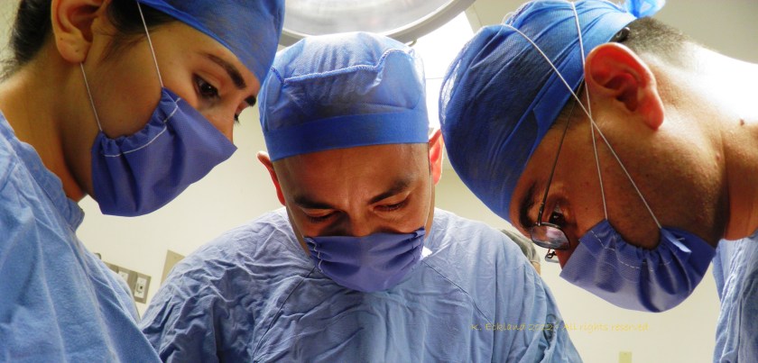
Case Report: Hilar mass resection using single port thoracoscopy with Dr. Diego Gonzalez – Rivas

in the operating room with Dr. Diego Gonzalez Rivas for single port thoracoscopic (uniportal) surgery.

Hilar mass resection using single port thoracoscopy with Dr. Diego Gonzalez – Rivas

K. Eckland & Andres M. Neira, MD

Instituto Nacional de Cancerlogia

Bogota, Colombia

Surgeon(s): Dr. Diego Gonzalez Rivas and Dr. Ricardo Buitrago

Dr. Diego Gonzalez Rivas demonstrates single port thoracoscopy

Case History:

59-year-old female with past medical history significant for recurrent mediastinal mass previously resectioned via right VATS.  Additional past medical history included prior right-sided nephrectomy.

Pre-operative labs:

CBC:  WBC 7230   Neu 73%  Hgb:14.1  Hct 37  platelets 365000

Pt 12.1  / INR1.1  PTT: 28.3

Diagnostics:

Pre-operative CT scan: chest

edited to preserve patient privacy

Procedure:  Single port thoracoscopy with resection of mediastinal mass and lymph node sampling

After review of relevant patient history including radiographs, patient was positioned for a right-sided procedure. After being prepped, and draped, surgery procedure in sterile fashion.  A linear incision was made in the anterior chest – mid clavicular line at approximately the fifth intercostal space.  A 10mm port was briefly inserted and the chest cavity inspected.  The port was then removed, and the incision was expanded by an additional centimeter to allow for the passage of multiple instruments; including camera, grasper and suction catheter.

Dr. Gonzalez Rivas and Dr. Ricardo Buitrago at National Cancer Institute

The chest cavity, pleura and lung were inspected.  The medial mediastinal mass was then identified.

instruments

As previously indicated on pre-operative CT scan, the mass was located adjacent and adherent to the vessels of the hilum.  This area was carefully dissected free, in a painstaking fashion.  After freeing the mediastinal mass from the hilum, the remaining surfaces of the mass were resected.  The mass was fixed to the artery pulmonary and infiltrating it) .  The mass was removed en-bloc.  Care was then taken to identify, and sample the adjacent lymph nodes which were located at stations (4, 7 and 10).

GonzalezRivas 051

Following removal of the tumor and lymph nodes, the area was re-inspected, and the lung was re-inflated.  A 28 french chest tube was inserted in the original incision, with suturing of the fascia, subcutaneous and skin layers.

closing the single port incision

Hemostasis was maintained during the procedure with minimal blood loss.

Patient was hemodynamically stable throughout the case, and maintained appropriate oxygen saturations.  Following surgery, the patient was awakened, extubated and transferred to the surgical intensive care unit.

Post-operative:  Post-operative chest x-ray confirmed appropriate chest tube placement and no significant bleeding or pneumothorax.

Immediate post-operative film (chest tube visible)

Patient did well post-operatively.  Chest tube was discontinued on POD#2 and discharged home.

PA & LAT films on post-operative day 2

pod2

Discussion: Since the initial published reports of single-port thoracoscopy, this procedure has been applied to an increasing range of cases.  Dr. Gonzalez and his team have published reports demonstrating the safety and utility of the single-port technique for multiple procedures including lobectomies, sleeve resections, segmentectomies, pneumonectomies and mediastinal mass resections. Dr. Hanao Chen (Taiwan) has reported several successful esophagectomies using this technical, as well as bilateral pleural drainage using a unilateral single-port approach.

Contrary to popular perception, the use of a single-port versus traditional VATS procedures (three or more) is actually associated with better visibility and accessibility for surgeons.  Surgeons using this technical have also reported better ergonomics with less operating fatigue related to awkward body positioning while operating.

The learn curve for this surgical approach is less than anticipated due to the reasons cited above, and has been cited at 5 to 20 cases by Dr. Gonzalez, the creator of this approach.

The main limitations for surgeons using this technique is often related to anticipated (but potentially unrealized) fears regarding the need for urgent conversion to open thoracotomy.  In reality, many of the complications that may lead to urgent conversion, such as major bleeding, are manageable thoracoscopically once surgeons are experienced and comfortable with this approach.

Dr. Gonzalez and his colleagues have reported a conversion rate of less than 1% in their practice.  Subsequent reports by Dr. Gonzalez and his colleagues have documented these findings.

Other barriers to adoption of this technique are surgeon-based, and may be related to the individual surgeon’s willingness or reluctance to adopt new techniques and technology.   Many of these surgeons would be surprised by how this technique mimics open surgery.

The successful adoption of this technique by numerous thoracic surgery fellows shows the feasibility and ease of learning single-port thoracoscopy by surgeons interested in adopting and advancing their surgical proficiency in minimally invasive surgery.

The benefits for utilizing this technique include decreased length of stay, decreased patient discomfort and greater patient satisfaction.

References/ Additional Readings

Bertolaccini, L., Rocco, G., Viti, A. & Terzi, A. (2013).  Surgical technique: Geometrical characteristics of uniportal VATS.  J. Thorac Dis. 2013, Apr 07.  Article from thoracic surgeons at the National Cancer Institute in Naples, Italy explains how the geometric advantages of uniportal VATS improves visibility and spatial perception over traditional VATS, and mimics open surgery.

Calvin, S. H. Ng (2013). Uniportal VATS in Asia.  J Thorac Dis 2013 Jun 20.  Article discussing the spread of uniportal techniques in Taiwan, China and other parts of Asia.

Chen, Chin-Hao, Lin, Wei-Sha, Chang, Ho, Lee, Shih-Yi, Tzu-Ti, Hung & Tai, Chih-Yin (2013).  Treatment of bilateral empyema thoracis using unilateral single-port thoracoscopic approach.  Ann Thorac Cardiovasc Surg 2013.

Gonzalez Rivas, D., Fieira, E., Delgado, M., Mendez, L., Fernandez, R. & De la Torre, M. (2013). Surgical technique: Uniportal video-assisted thoracoscopic lobectomy.  J. Thorac Dis. 2013 July 4.

Gonzalez Rivas, D., Delgado, M., Fieira, E., Mendez, L. Fernandez, R. & De la Torre, M. (2013).  Surgical technique: Uniportal video-assisted thoracoscopic pneumonectomy.  J. Thorac Dis. 2013 July 4.

Rocco, G. (2013). VATS and uniportal VATS: a glimpse into the future.  J. Thorac Dis. 2013 July 04.  After coming across several articles by Dr. Gaetano Rocco, and actively pursuing several other publications by this Italian thoracic surgeon, I have become increasingly convinced that Gaetano Rocco, along with Dr. Gonzalez Rivas is one of the world’s leading innovators in thoracic surgery.  Hopefully, cirugia de torax will be able to catch up to Dr. Rocco at some point for an in-depth discussion.

Rocco, G., Martucci, N., La Manna, C., Jones, D. R., De Luca, G., La Rocca, A., Cuomo, A. & Accardo, R. (2013).  Ten years experience on 644 patients undergoing single-port (uniportal) video-assisted surgery.  Annals of Thoracic Surgery, 2013, Aug, 96(2): 434-438.

Rocco G, Martin-Ucar A, Passera E. Uniportal VATS wedge pulmonary resections. Ann Thorac Surg. 2004;77:726–728.

Rocco G. Single port video-assisted thoracic surgery (uniportal) in the routine general thoracic surgical practiceOp Tech (Society of Thoracic and Cardiovascular Surgeons). 2009;14:326–335.

Rocco G, Khalil M, Jutley R. Uniportal video-assisted thoracoscopic surgery wedge lung biopsy in the diagnosis of interstitial lung diseasesJ Thorac Cardiovasc Surg. 2005;129:947–948.

Rocco G, Brunelli A, Jutley R, et al. Uniportal VATS for mediastinal nodal diagnosis and stagingInteract Cardiovasc Thorac Surg. 2006;5:430–432

Rocco G, La Rocca A, La Manna C, et al. Uniportal video-assisted thoracoscopic surgery pericardial window. J Thorac Cardiovasc Surg. 2006;131:921–922.

Jutley RS, Khalil MW, Rocco G Uniportal vs standard three-port VATS technique for spontaneous pneumothorax: comparison of post-operative pain and residual paraesthesiaEur J Cardiothorac Surg 2005;28:43-46.

Salati M, Brunelli A, Rocco G. Uniportal video-assisted thoracic surgery for diagnosis and treatment of intrathoracic conditions. Thorac Surg Clin. 2008;18:305–310.

Rocco G, Cicalese M, La Manna C, La Rocca A, Martucci N, Salvi R. Ultrasonographic identification of peripheral pulmonary nodules through uniportal video-assisted thoracic surgeryAnn Thorac Surg. 2011;92:1099–1101.

Rocco G, La Rocca A, Martucci N, Accardo R. Awake single-access (uniportal) video-assisted thoracoscopic surgery for spontaneous pneumothorax. J Thorac Cardiovasc Surg. 2011;142:944–945.

Rocco G, Romano V, Accardo R, et al. Awake single-access (uniportal) video-assisted thoracoscopic surgery for peripheral pulmonary nodules in a complete ambulatory setting. Ann Thorac Surg. 2010;89:1625–1627.

Rocco G. (2012). One-port (uniportal) video assisted thoracic surgical resections – a clear advance. J Thorac Cardiovasc Surg.2012;144:S27–S31.

Additional publications on single-port thoracoscopy (Dr. Gonzalez Rivas)

1 / Single-port video-assisted thoracoscopic anatomic segmentectomy and right upper lobectomy.  Gonzalez-Rivas D, Fieira E, Mendez L, Garcia J. Eur J Cardiothorac Surg. 2012 Aug 24

2 / Single-incision video-assisted thoracoscopic lobectomy: Initial results. Gonzalez-Rivas D, Paradela M, Fieira E, Velasco C.J Thorac Cardiovasc Surg. 2012;143(3):745-7

3 / Single-incision video-assisted thoracoscopic right pneumonectomy.  Gonzalez Rivas D, De la Torre M, Fernandez R, Garcia J. Surgical Endoscopy. Jan 11. 2012 (Epub ahead of print)

4 / Single-port video-assisted thoracoscopic left upper lobectomy.  Gonzalez-Rivas D, de la Torre M, Fernandez R, Mosquera VX. Interact Cardiovasc Thorac Surg. 2011 Nov;13(5):539-41

5 / Video-assisted thoracic surgery lobectomy: 3-year initial experience with 200 cases.  Gonzalez D, De la Torre M, Paradela M, Fernandez R, Delgado M, Garcia J,Fieira E, Mendez L. Eur J Cardiothorac Surg. 2011 40(1):e21-8.

6 / Single-port Video-Assisted Thoracoscopic Anatomical Resection: Initial Experience.  Diego Gonzalez , Ricardo Fernandez, Mercedes De La Torre, Maria Delgado, Marina Paradela, Lucia Mendez. Innovations.Vol 6.Number 3. May/jun 2011. Page 165.

Living legends and Cirugia de Torax

writing about Dr. Diego Gonzalez Rivas and the other living legends in thoracic surgery and connecting people to the world of thoracic surgery

Readers at Cirugia de Torax have certainly noticed that there are numerous articles regarding the work of Dr. Diego Gonzalez Rivas.  This week in particular, after a recent thoracic surgery conference and an afternoon in the operating room – there is a lot to say about the Spanish surgeon.

It’s also hard to escape that fact that I regard him in considerable awe and esteem for his numerous contributions to thoracic surgery and prolific publications.  I imagine that this is similar to how many people felt about Drs. Cooley, Pearson or Debakey during their prime.

Making thoracic surgery accessible

But the difference is Dr. Diego Gonzalez Rivas himself.  Despite the international fame and critical surgical acclaim, he remains friendly and approachable. He has also been extremely supportive of my work, at a time when not many people in thoracic surgery see the necessity or utility of a nurse-run website.

After all, the internet is filled with other options for readers; CTSnet.org, multiple societies like the Society of Thoracic Surgeons (STS), and massive compilations like journal-based sites (Annals of Thoracic Surgery, Journal of Thoracic Disease, Interactive Journal of Cardiothoracic Surgery).

But the difference between Cirugia de Torax and those sites is like the difference between Dr. Gonzalez Rivas and many of the original masters of surgery: Approach-ability and accessibility.

This site is specifically designed for a wider range of appeal, for both professionals in thoracic surgery, and for our consumers – the patients and their families.  Research, innovation, news and development matters to all of us, not just the professionals in the hallowed halls of academia.  But sometimes it doesn’t feel that way.

Serving practicing surgeons

For practice-based clinicians, and international surgeons publication in an academia-based journal requires a significant effort.  These surgeons usually don’t have research assistants, residents and government grants to support their efforts, collect their data and clean up their grammar.   Often English is a second or third language.  But that doesn’t mean that they don’t make valuable contributions to their patients and the practice of thoracic surgery.   This is their platform, to bring their efforts to their peers and the world.

Heady aspirations

That may sound like a lofty goal, but we have readers from over a 110 countries, with hundreds of subscribers along with over 6,000 people with Cirugia de Torax directly on their smart phone.  Each month, we attract more hits and more readers.

Patient-focused information

That’s important for the other half of our mission – connecting our patients with the world of thoracic surgery. Discussing research findings, describing procedures and presenting information to the people who are actually undergoing the procedures we are writing about.  Letting them know what’s new, what’s changed – and what to expect.  

Every day, at least 200 people read “Blebs, Bullae and Spontanous Pneumothorax”.  Why?  Because it’s a concise article that explains what blebs are, how a pneumothorax occurs and how it’s treated.  Another hundred people usually go on to read the accompanying case report about blebectomy, for similar reasons.  There are links for more information, CT scans and intra-operative photos included, so that people can find exactly what they need with a minimum of effort.

Avoiding ‘Google overload’

With the massive volume of information available on the internet, high-quality, easily understood, applicable information has actually become even more difficult for patients to find than ever before.  Patients spend hours upon hours browsing through academic jargon, commercial websites and biased materials while attempting to sift through the reams of information for pertinent and easily understandable information.  There is also a lot of great material out there – so we provide links to reputable sites, recommend well-written articles and discuss related research.

Connecting patients to surgeons

We also provide patients with more information about the people they are entrusting their bodies, their hopes and their lives to.  It’s important that they know about the Dr. Benny Wekslers, the Dr. Hanao Chens, and the Dr. Diego Gonzalez Rivas out there.

Update:  June 2019

After multiple reader requests from this site, we have launched a service to assist readers in pursuiting minimally invasive thoracic surgery, uniportal surgery, HITHOC and other state-of-the-art thoracic surgery procedures with the modern masters of thoracic surgery.  We won’t talk a lot about this on the site, but we do want readers to know that we are here to help you.  If you are wondering what surgery costs like with one of the world’s experts – it’s often surprisingly affordable.

If you are interested in knowing more, please head to our sister site, www.americanphysiciansnetwork.org or send an email to kristin@americanphysiciansnetwork.org.

Keeping it ‘real’

Looking over the shoulder of Dr. Gonzalez Rivas in the operating room

As much as I may admire the work and the accomplishments of Dr. Gonzalez-Rivas – it’s important not to place him on a pedestal.  He and his colleagues are real, practicing surgeons who operating on regular people, not just heads of state and celebrities.  So when we interview these surgeons and head to the OR, it’s time to forget about the accolades, the published papers and the fancy titles. It’s time to focus on the operations, the techniques, the patients and the outcomes because ‘master of thoracic surgery’ or rural surgeon – the operation and patient are all that really matters.

K. Eckland

The cowboys and rodeo stars of thoracic surgery

Discussing Dr. Joseph Coselli and ‘the cowboys of cardiac surgery’ along with some of our own heros of thoracic surgery here at Cirugia de Torax.

There’s a great article in this month’s Annals of Thoracic Surgery, by Dr. Joseph Coselli, from Texas Heart Institute and the Michael DeBakey Department of Surgery at Baylor.   His article, entitled,” My heros have always been cowboys” is more than just a title torn from the song sheets of Willie Nelson.  It’s a look back at both the pioneers of cardiac surgery and his own experiences as a cardiac surgeon.  He also discusses the role of surgeons, and medical practitioners in American society in general and the promises we make to both society at large and our patients.

Here at Cirugia de Torax, I’d like to take a moment to look back at the surgeons that inspired and encouraged me in this and all of my endeavors.  Some of these surgeons knew me, and some of them didn’t – but their encouragement and kindnesses have spurred a career and life that have brought immense personal and professional satisfaction.

Like Dr. Coselli, I too, took inspiration from the likes of Dr. Denton Cooley.  But our stories diverge greatly from there.  I never met Dr. Cooley and I probably never will.  But it was a related story, from my former boss (and cardiothoracic surgeon), Dr. Richard Embrey that led to an email to Dr. Cooley himself.  My boss had too trained under Dr. Cooley, Dr. Debakey and the Texas Heart Institute, the citadel of American heart surgery.   Then, somehow, along the way – Dr. Embrey stopped to work at our little rural Virginia hospital.  We were the remnants of a larger Duke cardiothoracic program but we were a country hospital all the same.

While I learned the ins and outs of surgery from Dr. Embrey (and Dr. Geoffrey Graeber at West Virginia University) on a day-to-day basis, I was also weaned on the folklore of cardiothoracic surgery – stories of the giants of history, like the ones mentioned in Dr. Coselli’s article, as well as local Duke legends who occasionally roamed the halls of our tiny ICU and our two cardiothoracic OR suites; Dr. Duane Davis, Dr. Shu S. Lin and Dr. Peter Smith.  While never working side-by-side, Dr. D’Amico’s name was almost as familiar as my own.  As the sole nurse practitioner in this facility, without residents or fellows, there was no buffer, and little social divide in our daily practice.  Certainly, this changed me – and my perceptions.  I asked the ‘stupid’ questions but received intelligent and insightful answers.  I asked even more questions, and learned even more..

These opportunities fed my mind, and nurtured my ambitions.  Not to be a physician or a doctor, but to learn as much as possible about my specialty; to be the best nurse possible in my field.  It also nurtured a desire to share these experiences, and this knowledge with my peers, my patients and everyone else who ever had an interest.

It was that tiny little email, a gracious three-line reply from Dr. Cooley himself that made me realize that I didn’t have to rely on folklore and second-hand stories to hear more.  That’s critical; because as we’ve seen (here at Cirugia de Torax) there are a quite of few of “Masters of thoracic surgery” or perhaps future giants that haven’t had their stories told.  Dr. Coselli and his fellow writers haven’t written about them yet.. So I will.

Sometimes I interview famous (or semi-famous) surgeons here, but other times, I interview lesser-known but equally talented/ innovative or promising surgeons.  All of them share similar traits; dedication and love for the profession, immense surgical talent and proficiency and sincere belief in the future of technology of surgery.

So, let’s hope that it won’t take forty more years for these surgeons to be recognized for their contributions to thoracic surgery in the way that Cooley, DeBakey and Crawford are heralded in cardiac surgery.

K. Eckland, ACNP-BC

Founder & Editor -in – chief

Single port thoracoscopy for wedge resection – does size matter?

Dr. Chen discusses single port thoracoscopy – and specimen size.

Single port thoracoscopy for wedge resection – does size matter?
Dr. Chih-Hao Chen, Thoracic Surgeon, Mackay Memorial Hospital, Taiwan
Correspondence: musclenet2003@yahoo.com.tw

Case presentation and discussion

A 77 y/o woman was found to have a neoplasm in RML during routine health exam. (Full case presentation here.)
The incision was 2.5 cm. The specimen was removed successfully; patient experienced no complications and was discharged from the hospital without incident. However, when discussing this case with surgeon colleagues a specific question arose.

preparing to remove the specimen through the port

Does size matter? How big is too big to remove through a port incision?
An important issue is the theoretically smallest (and effective) incision size.  The issue is very challenging for most surgeons. In the past, other surgeons have questioned me :
“The specimen to be removed is more than 12-15 cm. How can you remove the specimen from a 3.5 cm incision?”

BUT, the fact is “the smallest size depends mostly on the solid part of the neoplasm “.

The lung is soft in nature. In my experience, a 3.5 cm port is usually enough for any lobe.  The only problem is that if the solid part is huge, pull-out of the specimen may be difficult.
Therefore, the solid part size is probably the smallest incision size we have to make, usually in the last step when dissection completed , if needed.

successful removal of lung specimen thru small port
Port with chest tube in place

Using endo-bags to avoid larger incisions
There is an exception. I have never do this but I think it may be possible.  We usually avoid opening the tumor (or cutting tumor tissue into half ) within the pleural space in order to prevent metastasis from spillage of cancerous tissue.

What if we can protect the tumor from metastasis while we cut it into smaller pieces? For example, into much smaller portions that will easily pass through the small port incisions?
My idea is to use double-layer or even triple layers of endo-bag to contain the specimen. If it is huge, with perfect protection, we cut it into smaller parts within the bag. Then the huge specimen  can be removed through a very tiny incision. This is possible.

We still have to avoid “fragmentations” since this might interfere or confuse the pathologist for accurate cancer staging. Therefore, in theory, we can cut it into smaller parts but not into fragments. However this idea is worthy of consideration, and I welcome debate from my fellow surgeons and medical colleagues.

Thank you to Dr. Chen.

High Altitude Surgery: Carotid Body Tumors

a different kind of case here at Cirugia de Torax

A Carotid paraganglion / carotid body tumor in Bogotá, Colombia

K. Eckland, ACNP-BC, MSN, RN  & Ricardo Buitrago, MD

Case History:  62-year-old Hispanic female who presented with complaints of a right neck mass X 3 years, accompanied by occasional dysphagia, itching and soreness of right neck.  Patient denied history of weight loss, anorexia, aspiration or recent pneumonias.  No history of previous stroke.

Past medical / surgical history:  TAH, 25 years prior, Hypertension, previous DVT of the RLE. Home medications: ASA 100mg po Q day.

On examination, the patient had a palpable, reducible mass over the right carotid, with no bruits on auscultation.

Diagnostics:  carotid duplex showing a right-sided carotid body tumor arising at the bifurcation of the common carotid.  No evidence of hemodynamically significant atherosclerotic plagues, or elevated velocities.  Mass measured at 7cm at widest point.

Labs:  All labs within normal limits including a Hgb 16/ Hct 48

Operative:

After administration of general anesthesia, and endotracheal intubation, the patient was positioned, prepped and draped in sterile fashion.  A 4cm vertical skin incision was made on the right side of the neck.  After blunt dissection and retraction of sternoclastomastoid muscle, the common carotid artery was identified and retracted  with a vessel loop.  Identifying the bifurcation and loosely clamped external and internal carotids.  Care was taken to identify and prevent injury to the hypoglossal nerve and fascial branches.

carotid body tumor, in situ

Extensive ligation of  tributary vessels was undertaken while performing blunt dissection to the avascular plane.  Once the tumor was separated from the carotid bifurcation, it was removed and sent for final pathology.

At the conclusion of the case, a small jp drain was placed.  The patient was awakened in the operating room and extubated.  Patient demonstrated no new neurological deficits.

less than 5% of tumors are malignant

Post-operative:

JP drain was discontinued on the morning of POD#2 and the patient was subsequently discharged.  Patient reported no dysphagia, hoarseness or paresthesia.

Discussion:

The carotid body serves as an important function of the detection and moderation of hypoxia.  This has been shown in several studies of post-operative carotid endartectomy patients who are unable to adjust/ acclimatize to increasing altitude as well as the increased incidence of carotid body tumors (CBT) at altitudes of 2000 meters of greater (Ojeda Parades).

Carotid body tumors occur most frequently at altitudes greater than 2,500 meters but significant differences have been noted in the prevalence, size and other characteristics of carotid body tumors occurring at lower altitudes.  The vast majority (95%) of these tumors are benign but in a minority of these cases, these tumors may represent metastatic disease.

Despite the location of these tumors, (at the bifurcation of the common carotid into the external and internal carotid), the chances for successful excision and resection is high with a mortality of less than 1%.  The most common complication of this procedure is damage to the adjacent nerve, causing hoarseness.

References

Boedecker, C. C. (2011). Paragangliomas and paraganglioma syndromes.  Head and Neck Surgery, 2011 (10).  A nice review article of paragnaglions including paragrangliomas of the head and neck.

Cerecer – Gil, N. Y., Figuera, L. E., Llamas, F. J., et. al. (2010). Mutation of SDHB as a cause of hypoxia related high altitude paraganglion.   Clin Cancer Res 2010; 16: 4148-4154. [free full-text pdf.]

Conde, S. V., Ribeiro, M. J., Obeso, A., Rigual, R., Monteiro, E. C., Gonzalez, C. (2012, recently published research).  Chronic caffeine intake in adult rat inhibits carotid body sensitization produced by chronic hypoxia but maintains intact chemoreflex output. Molecular Pharmacology Fast Forward.  46 page report on animal study.  [full-text available.]

Moore, J. P., Claydon, V. E., Norcliff, L. J., Rivera-Ch, M. C., Leon-Velarde, F., et. al. (2006).  Carotid baroreflex regulation of vascular resistance in high-altitude andean natives with and without chronic mountain sickness.  Experimental Physiology 91(5); 907-913.  [free full-text pdf.]

Ojeda, L. P., Durango, E., Rodriquez, C., & Vivar, N. (1988).  Carotid body tumors at high altitude: Quito, Ecuador, 1987.  World J. Surg. 12: 856- 860.

Park, S. J., Kim, Y. S., Cho, H. R. & Kwon, T. W. (2011).  Huge carotid body ganglion.  J Korean Surg Soc 2011; 81: 291- 294.  Case report. [free full text pdf available.]

Rodriguez – Cuervasm S., Lopez – Garcia, J. & Labastida – Alemandro, S. (1998).  Carotid body tumors in inhabitants of altitudes higher than 2000 meters above sea level.  Head & Neck, Aug 1998: 374-377.  [free full-text pdf available.]

Authors conducted a study of 120 CBT in Mexico City, D.F looking at the incidence and characteristics of all CBTs over a thirty year period (1965 – 1995) in comparison to previously published reports of CBT at lower altitude.  Findings similar to previous and existing literature  with a predominantly female population (89% of cases).  Mean age 49.  Mean size 5.4 cm.   They reported a 20% incidence of cranial nerve injury after resection.

Ruben, R. J. (2007).  The history of the glomus tumors nonchromaffin chemodectoma: a glimpse of biomedical Camelot. Acta Oto-Laryngeologica 2007; 127: 411-416.  If you can get past his fanciful writing style which resembles an ardent love letter, the article gives a nice history of paraganglion tumors (including carotid body tumors).

Case Report: Multidisciplinary approach to metastatic disease, and lessons learned

Chest wall resection with pulmonary segmentectomy for metastatic breast cancer.

a multi-disciplinary approach: plastics, surgical oncology and thoracic surgery

Title:  Chest wall resection with pulmonary segmentectomy for metastatic breast cancer

Summary: Breast cancer remains the second leading cause of mortality in females in Mexico, aged 30 to 55, and is usually self-detected in later stages.  Due to disparities in health care within the country, even patients with early detection may not receive optimal or timely treatment leading to more recurrent or metastatic disease.  Surgery remains the best, but underutilized option for definitive treatment in patients with surgically resectable disease.  In this case, a patient with advanced disease was successfully resected using a multi-disciplinary approach.

Authors: K. Eckland, ACNP-BC, Hospital General de Mexicali, Thoracic Surgery

Carlos Ochoa Gaxiola, MD, Hospital General de Mexicali, Thoracic Surgery

Gabriel Ramos Orozco, MD, Instituto Mexicano Seguro Social (IMSS), Surgical Oncology

Corresponding author: Carlos Ochoa Gaxiola, MD

Email: drcarlosochoa@yahoo.com

Announcement text: a multidisciplinary approach to recurrent metastatic breast cancer with chest wall resection and free flap graft creation.

Subject/ Classification terms: chest wall resection, rib resection, metastatic breast cancer, pulmonary segmentectomy, breast cancer in Mexico

Disclosures:  The authors have no disclosures.

History/ Case Summary:

The patient was a 70-year-old Hispanic female with a past medical history significant for local breast cancer in the left breast, initially diagnosed in 1994.  This was treated with chemotherapy and radiation.  She was then maintained on tamoxifen until 2000.  In 2011, she presented with a recurrent mass in the left breast.  There was no other history of chronic or active medical conditions such as HTN, CAD or diabetes.

After referral to a surgical oncologist for further evaluation, patient underwent additional evaluation.  A PET/ CT scan was positive for a metabolically active lesion in the left breast with an SUV of 9.6 with lytic lesions in anterior ribs with max SUV of 3.0.  There was no evidence of distal metastasis to other organs including the brain, lungs, or abdominal cavity on PET or other diagnostic imaging.  All pre-operative labs were within normal ranges including alk phos, and serum calcium.

Pre-operative Chest X-ray

After initial surgical evaluation, a multi-disciplinary surgical plan utilizing a general surgical oncologist, thoracic surgery and plastic surgery was devised for surgical resection of breast mass with rib resection and free flap creation.

surgeons planning approach

Operative Course:  The left breast including all skin, tissue and lymphatics was excised to the depth of the rib cage.  Further dissection and resection of the anterior portion of ribs #2, 3 and #4 was completed.

following rib resection

Following rib resection, upon exploration of the left thoracic cavity, the patient was found to have a large greyish-white lesion, estimated at 3.5 cm in diameter in the left upper lobe.  The lesion was hard, and located on the peripheral portion of the upper lobe.  No additional lesions were found.

during surgery, a previously undetected pulmonary lesion was discovered

The decision to undertake pulmonary resection was based on the possibility of complete surgical resection of existing disease.  At the time, a discussion was undertaken with the patient’s primary surgeon, and the thoracic surgeon on the feasibility of resection by lobectomy versus segmentectomy.  The decision was made to proceed with a lung-sparing procedure as the patient’s baseline pulmonary function was not known.

Following successful lung resection and hemostasis, a 32 french chest tube was placed, and surgical mesh was placed for coverage of chest wall / rib defect. After mesh was sutured into place, the patient was re-positioned for harvesting of a free flap from the posterior chest. Abdominal free flap harvest was not undertaken due to patient anatomy.  The plastic surgeon involved in the case, Dr. Nastia Gonzalez then proceeded with free flap grafting for breast reconstruction.   There was no significant bleeding, hypoxia or hemodynamic instability intraoperatively.

Post-operative Course:

The patient was successfully extubated at the conclusion of the case, and transferred to the post-operative care unit in stable condition.  Post-operative course was uncomplicated with the chest tube removed on POD#3, and the patient discharged home on POD#5.   The patient’s oxygen saturations were within the normal range (92% or above) and she was discharged home without supplemental oxygen.

Subsequent post-operative visit was uneventful with no evidence of infection, or impaired healing of the graft or harvest site.  As of the date of publication, there has been no further evidence of recurrence or metastatic disease.

Conclusions:  For patients with metastatic disease limited to adjacent and surgically resectable tissue, surgery remains the best option for longevity and overall survival.  However, despite the available and use of advanced imaging studies, surgeons should prepare for and anticipate the possibility of discovery of evidence of additional disease.  In this case, a more complete anatomic resection of the newly discovered lung lesion was hindered by the lack of pre-operative evaluation of pulmonary disease.

Chest wall resection and defect closure have been managed with a variety of techniques over the years, including muscle flaps, plastic ribcage creation, mesh closures for stabilization after rib resection (Khalil et al.).  In this case, which required a radical mastectomy, surrounding musculature was removed for full resection.  Tissue was harvested for free flap grafting but this gives lesser structure than attached muscle, so synthetic mesh was used.

Historically, hardware installation was plagued with a variety of problems including infection and erosion.  However, preliminary reports of evolving hardware for oncologic chest resections may change closure techniques in the future (Fabre et al, 2012).

References/ Additional Information

Akiba T, Takeishi M, Kinoshita S, Morikawa T. (2011).  Vascularized rib support for chest wall reconstruction using Gore-Tex dual mesh after wide sternochondral resection. Interact Cardiovasc Thorac Surg. 2011 Nov;13(5):536-8. Case report of breast ca with sternal reconstruction. Note the one month post-op photo.

Billè A, Okiror L, Karenovics W, Routledge T. (2012). Experience with titanium devices for rib fixation and coverage of chest wall defects.  Interact Cardiovasc Thorac Surg. 2012 Jul 19.  Report of 18 patients with chest wall defects repaired using titanium.  (Majority of cases were trauma related).

Fabre D, El Batti S, Singhal S, Mercier O, Mussot S, Fadel E, Kolb F, Dartevelle PG.  A paradigm shift for sternal reconstruction using a novel titanium rib bridge system following oncological resections. Eur J Cardiothorac Surg. 2012 May 2. No free full text available. Parisian study of titanium bridge system for use after oncological resections.

Gharagozloo F, Meyer M, Tempesta BJ, Margolis M, Strother ET, Tummala S. (2012).  Robotic en bloc first-rib resection for Paget-Schroetter disease, a form of thoracic outlet syndrome: technique and initial results. Innovations (Phila). 2012 Jan-Feb;7(1):39-44.  No free text available.  Report on robot-assisted rib resection. (Less relevant but interesting.)

Khalil el-SA, El-Zohairy MA, Bukhari M.  (2010).  Reconstruction of large full thickness chest wall defects following resection of malignant tumorsJ Egypt Natl Canc Inst. 2010 Mar;22(1):19-27.   Excellent report on series of 18 patients who underwent major chest wall resections for onocologic disease.  Review of available materials and type of resections.

Mohajeri G, Sanei MH, Tabatabaee SA, Hashemi SM, Amjad E, Mohajeri MR, Shemshaki H, Jazi AH, Kolahdouzan M. (2012). Micrometastasis in non-small-cell lung cancer: Detection and staging.  Ann Thorac Med. 2012 Jul;7(3):149-52.  Using bone marrow biopsy for diagnosis of lytic lesions.

Müller AC, Eckert F, Heinrich V, Bamberg M, Brucker S, Hehr T. (2011).  Re-surgery and chest wall re-irradiation for recurrent breast cancer: a second curative approach.  BMC Cancer. 2011 May 25;11:197.

Seki M. (2012). Chest wall reconstruction with a latissimus dorsi musculocutaneous flap via the pleural cavityInteract Cardiovasc Thorac Surg. 2012 Jan;14(1):96-8. Case Report. Transpleural musculocutaneous flap used for defect repair in long term cancer survivor with radiation-related necrosis.

This case study has been published with the gracious consent of the patient.  However, in accordance to the patient’s wishes, and privacy – no photos showing the pre-operative site (breast) or the graft after surgery will be published.

Case Report: Dual port VATS decortication of empyema

case report of dual port thoracoscopy for decortication of empyema in a thirty-eight year old woman.

Note:  This case report was written with the assistance of Dr. Carlos Cesar Ochoa Gaxiola.

Case Report: Dual port thoracoscopic decortication of empyema

Presentation:  A 38-year-old woman presented to the local hospital with fever, pneumonia, chest pain and an elevated leukocyte count of 25,000. Initial chest x-ray showed a large left-sided effusion.

Risk factors:  Patient had several traditional risk factors for the development of empyema including heavy ETOH, and malnutrition, poor access to healthcare.  Patient HIV, and Hep C negative.

Initial Hospital Course:  She was admitted, and started on dual therapy antibiotics (ceftriaxone & levofloxacin).  A chest tube was placed with return of frank purulent material.  After several days of draining minimal amounts of pus, patient remained febrile.

Subsequent CT scan showed a left-sided empyema with large loculated areas.  At that time, thoracic surgery was consulted for additional evaluation and treatment.  Surgery was scheduled.

At the time of surgery, patient remained on dual antibiotics with WBC of 19,000.  Albumin 1.5 , Hgb 10.2, Hct 33, other labs within normal limits.

Surgical procedure: dual-port VATS with decortication

The initial chest tube was removed, patient was prepped and draped in the traditional sterile fashion.  The previous chest tube site was carefully cleaned with a betadine solution, and debrided of purulent material to prevent abscess tract formation, with instrumentation traded out after debridement.

A single additional ten mm thoracoscopy port was created, with visual interior inspection performed.  Initial inspection confirmed the presence of a stage IV empyema with large loculations, moderate pleural thickening and the presence of frankly purulent material adhering to the pleural/ chest wall and lung tissue.  The pleura was noted to be thickened but malleable, loosely adherent to the pleural and lung surfaces.

A formal decortication was undertaken with separation of the lung from the diaphragm and adhesions to obliterate the empyema cavity.   Decortication of visceral pleura was performed until the lung was completely free and able to re-expand.   Lavage was performed with evacuation and drainage of copious amounts of purulent materials.

After decortication was complete, two chest tubes were placed*; anteriorly and posteriorly, under thoracoscopic guidance, and the lung was re-inflated.

*Due to the location, and presence of infection/ purulent material in the initial chest tube site, an additional chest tube site (5mm) was created at the time of chest tube insertion to prevent additional infectious complications.

At the conclusion of the case, patient was awakened and extubated prior to being transferred to the PACU as per post-operative protocols.

EBL during the case was minimal.

Post-operative course:  Patient’s post-operative course was uncomplicated.  On post-operative day #5,  anterior chest tube was removed.  On post-operative day #7, the posterior chest tube was removed.  Patient was discharged post-operative day #8.

Discussion:  While convention medical wisdom dictates a trial and error treatment approach with initial trial of antibiotic therapy followed by chest tube placement (Light, 1995), surgeons have long argued that this delay in definitive treatment places the patient at increased risk of significant morbidity and mortality (Richardson, 1891).  Indeed, as discussed by Dr. Dov Weissburg  (on a previous discussion of empyema and lung abscess) multiple recent reviews of the literature and research comparisons continue to demonstrate optimal outcomes with surgery based approaches versus antibiotics alone, TPA and tube thoracostomy.  The ability to perform these procedures in the least invasive fashion (VATS versus thoracotomy approaches) defies the arguments against surgical intervention as advanced by interventionalists (radiologists and pulmonologists.)  Successful decortication with the use of dual port thoracoscopy is another example of how technology is advancing to better serve the patient and provide optimal outcomes.

Earlier, not late surgical referral would have been of greater benefit to this patient.

 I apologize but I was unable to take films / photographs of this procedure during this case.

 References (with historical perspectives)

Andrade – Alegre, R., Garisto, J. D. & Zebede, S. (2008).  Open thoracotomy and decortication for chronic empyema. Clinics, 2008; 63 (6),  789 – 93.  Color photographs.  Panamanian paper discussing the effectiveness of traditional open thoracotomy and decortication for stage III / chronic empyemas in an era of increased reliance on VATS.  Observations and recommendations for open thoracotomy approach for chronic empyema based on 33 cases spanning from March 1992 – June 2006, showing safe and effective results with open surgery for more advanced/ chronic empyemas.

 Light, R. W. (1995).  A new classification of parapneumonic effusions and empyema. Chest (108) 299 – 301.

Marks, D. J., Fisk, M. D.,  Koo, C. Y., et. al. (2012).  Thoracic empyema: a 12-year study from a UK tertiary cardiothoracic referral centre. PLoS One. 2012;7(1):e30074. Epub 2012 Jan 20. Treatment with VATS was shown to reduce the length of stay versus open surgery with a 15% conversion rate.

Nwiloh, J., Freeman, H. & McCord, C. Malnutrition: an important determinant of fatal outcome in surgically treated pulmonary suppurative disease.  Journal of National Medical Association, 81(5) 525-529.

Richardson, M. H. (1891). Surgical treatment of acute and chronic empyemas. While surgical techniques have greatly changed in the 100+ years since this paper was initially published (and no one suffers from carbolic acid poisoning anymore), many of the observations of Drs. Richardson and Loomis remain clinically relevant and valid today. (As previously noted by Dr. Weissburg, this was a pre-antibiotic era.)

Balance, H. A. (1904).  Seven cases of thoracoplasty performed for the relief of chronic empyema.  British medical journal, 10 Dec 1904, 1561 – 1566.  Dr. Balance discusses the development of Delnorme’s operation as an alternative to thoracoplasty while presenting several cases from his career.  Photographs.

Tuffier, T. (1922).  The treatment of chronic empyema.  Discussion of 91 cases, with radiographs.