There were plenty of reasons for surgeons from all over Latin America to converge on Cuscu, Peru for the 2nd annual VATS PERU Uniportal Master Class, which covered the basics of the uniportal approach as well as nonintubated and awake uniportal surgery. There were subxiphoid and uniportal cases streamed live from Shanghai Pulmonary Hospital. But beyond the usual reasons of networking, discussing and sharing case knowledge, and the presentation of research findings and evidenced-based practice, there were several reasons why VATS Peru was more than just your average regional thoracic surgery conference.
Why attend VATS Peru? The three best reasons:
1. The wet lab – which allowed surgeons and their surgical assistants to apply the theoretical knowledge they learned during the first two days of lecture in operating room scenario en vivo. The “en vivo” is critical, fancy simulators aside, there is no better challenge to ‘book knowledge’, and application of practical skills than in the scenario of an operating room, with real models and active bleeding.
A surgeon in the master course receives instruction from Dr. Diego Gonzalez Rivas
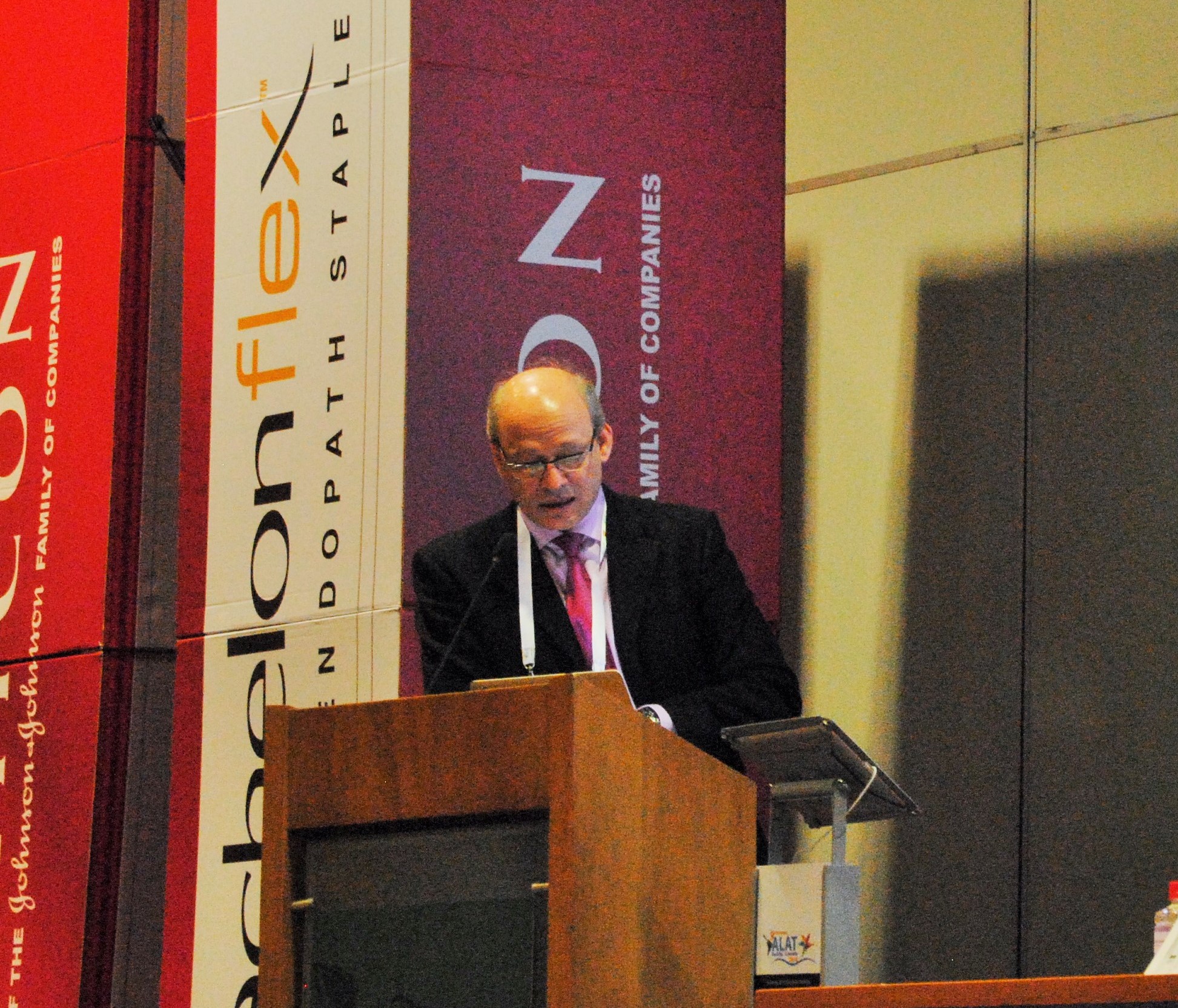
2. Lectures from the master surgeon himself; Dr. Diego Gonzalez Rivas: That’s where the second critical component comes in, in the form of the candid, direct and straight-forward lecture by Dr. Diego Gonzalez Rivas on Control of Inter-operative Bleeding. If you weren’t paying attention during this lecture, it’s obvious in the lab. This isn’t a computer course where you can dial in your answers, fast-forward thru lectures and print off a shiny new certificate. This isn’t a computer app, or a simulation that you can reset and re-start as soon as the surgery heads off course, to try again.. It’s real surgery.
3. Dr. Carlos Fernandez Crisosto
Lastly, if you didn’t attend VATS Peru, then you missed an opportunity to know and to talk to Dr. Carlos Fernandez Crisosto. VATS Peru is his brainchild, and the organization was created specifically to advance minimally invasive surgery in Peru. VATS Peru is separate from ALAT (the Latin American Society of Thoracic Surgeons), of which Dr. Fernandez is the current president. VATS Peru is also separate from the Peruvian Society of Thoracic Surgeons which has its own focus in the thoracic surgery specialty.
Dr. Carlos Fernandez Crisost0, Cardiothoracic and Vascular surgeon
Dr. Fernandez, a Tacna native, works at Daniel Alcides Carrion Essalud facility in the southernmost region of Peru. He is the sole cardiovascular and thoracic surgeon for the city of Tacna, and performs cardiac, vascular, and endovascular surgeries in addition to general thoracic surgery. While he is a trained cardiovascular surgeon, (in addition to general thoracic) thoracic surgery is what he enjoys most.
He trained in Argentina, and practiced in Cordoba, Argentina for 23 years before returning to Tacna in the last few years.
His average case volume is around 380 surgeries a year, and he reports that all of his thoracic surgeries are generally performed using the uniportal thoracoscopic approach. He also does transplant, which requires him to travel to Lima specifically to perform the procedure. The transplant program is small and performs 4 to 5 transplants per year.
In his practice he sees the usual oncology cases, and empyemas but he also sees a large number of patients with tuberculosis, as well as an assortment of hydatid cysts, and pectus cases. Trauma from accidents, as well as injuries from guns, and knives also comprises a large part of his practice.
Dr. Fernandez is pleased with the success of his course, since this is only the second time the course has been available here in Peru. It was a complex logistical arrangement to hold the course in Cusco this year, but with the help of his wife, a professional events planner, they were able to pull of the event with very few hiccups. Next year, they plan to hold the event in Lima, the capitol of Peru and a city famed for its gastronomic offerings.
If you missed this year’s VATS Peru, look for VATS Peru 2017 here at Thoracics.org next fall.
Dr. Diego Gonzalez Rivas headlines the ALAT sponsored event this September.
Cardiothoracic surgeon and the coordinator and director of VATS Peru, Dr. Carlos Fernandez Crisosto cordially extends an invitation for all interested thoracic surgeons to attend VATS Peru. This event is co-sponsored by ALAT being held at the Hospital Essalud Tacna in Tacna, Peru on the 21st and 22 of September. The 2 day course includes a wet-lab for a hands on approach at teaching uniportal VATS with Dr Gonzalez Rivas.
Thoracics.org has written for additional information – so I will update this post as information arrives. To register – click here.
Corrections: as many readers know, I do much of my writing on the fly, in airports, waiting rooms etc. The sometimes results in spelling and grammatical errors. As always my sincere apologies.
It’s the second day of the minimally invasive surgery course at Monaldi Hospital and there are a score of Italian physicians speaking in addition to the main events – Dr. Henrik Hansen and Dr. Diego Gonzalez Rivas.
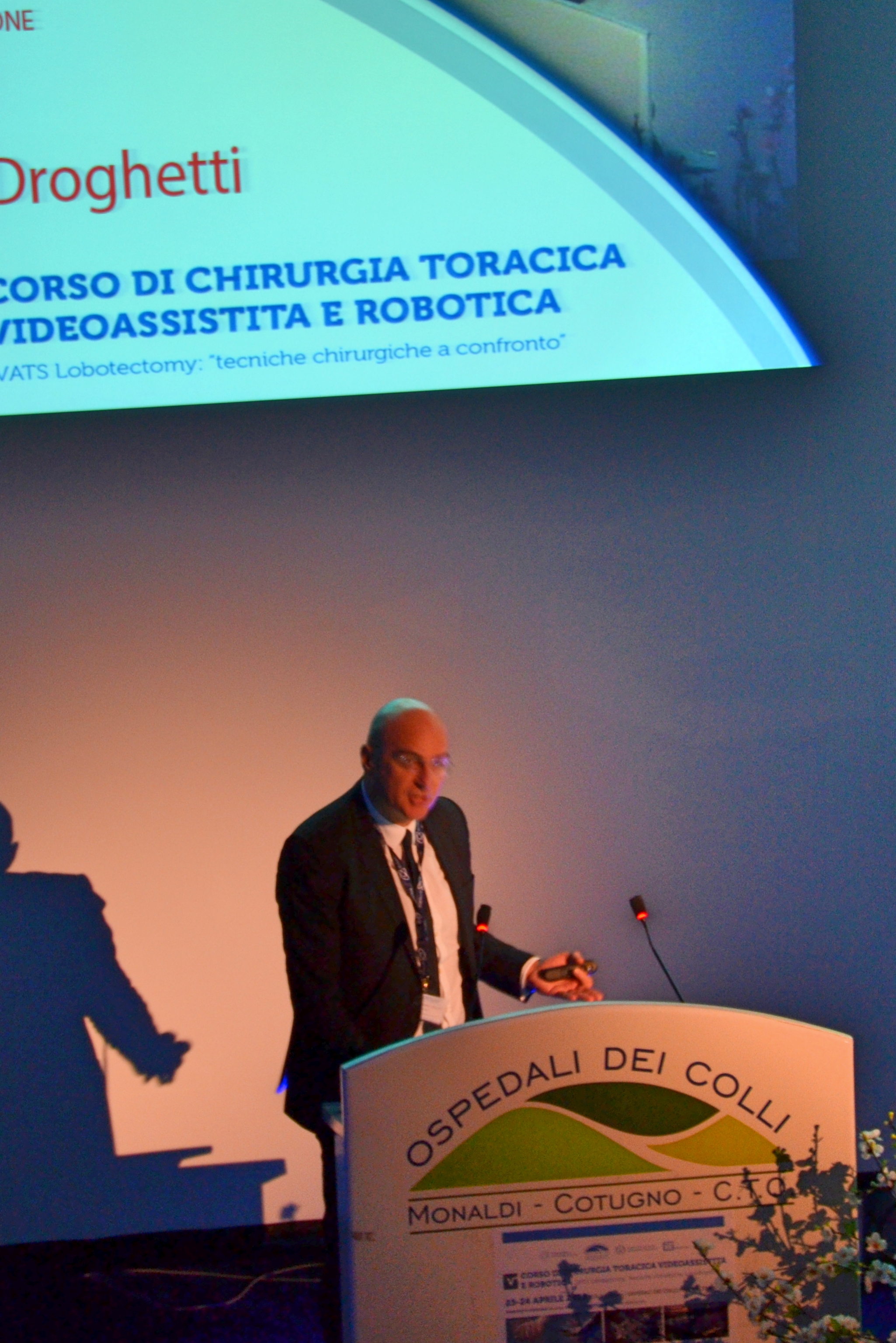
Dr. Andrea Droghetti
One of the surgeons addressing the group this morning is Dr. Andrea Droghetti, a thoracic surgeon from Carlo Poma Hospital in Mantova, Italy. Dr. Droghetti is here to present the latest information on the Italian VATS registry, Vatsgroup.it.
Now long-term readers know how we love a registry! We have talked a lot about the STS thoracic database and how it is woefully underutilized, we’ve talked to other surgeons who have been involved in creating their own national databases, and we even created our own.
As we discussed during a recent interview, data collection and publication are essential for research and advancement of the specialty – and that all starts with accurate data and statistics. But not all data collection tools are alike.
It is interesting, and encouraging to hear about the successful enrollment of 57 Italian facilities into a nationwide VATS registry to track VATS surgery and outcomes.
The database itself is pretty detailed and much more involved than the high altitude database or even STS. There are multiple risk stratification measures as well as quality of life indicators. The database is also designed to allow greater categorization – of pre-operative conditions, staging, procedures, and just about anything else you can think of.
the registry is extremely detailed
Sounds like a great way to improve the quality of the data being used for research. After all, plenty of surgeons in Italy are participating – and as we know, getting surgeons to participate is always difficult. Even the STS database is lagging with just over 215 surgeons participating.
That’s awesome.. Now if only we could get more global participation!
Unfortunately, these kinds of large-scale projects never go off without a hitch – and during the presentation, we noted several potential pitfalls. One the major ones that Dr. Droghetti addressed was:
– Getting surgeons/ hospitals to participate
Out of 57 sites that are eligible to participate, only 44 are actually submitting data, and the data volumes have been measly – at just over 2 cases per day. (There is certainly more than two cases being performed every day.)
It also makes you wonder about the ‘randomness’ of the cases being entered. Maybe it’s one very diligent site entering cases everyday, or maybe it’s different sites entering their best outcomes – so the potential for data skewing seems to be there.
But since it seems like such a great project, Thoracics.org asked Dr. Droghetti to talk to us some more about this project, (translational issues during the conference made parts of the presentation unclear) and answer some additional questions. He was nice enough to talk to Thoracics.org for a few minutes.
From our own experience, we identified several other potential problems for the registry: so we posed these problems to Dr. Droghetti for his input.
Time consuming / repetitive entries for single patient
Data has to be entered on two occasions for the registry. The first submission takes approximately 30 minutes and the second – the post-surgical follow up – takes around ten minutes. The nice part about the project is that the patients actually participate in the follow-up evaluation and enter their own answers for the quality of life answers.
Now the QoL stuff is pretty unique to this registry, and the two entries per patient – allows for real-time time entry instead of retrospective review (which can get pretty skewed) so these are also strengths of the project. But..
After our own adventure with data collection as well as our experiences with the STS (cardiac) database, that this also immediately identifies this study as relying on 3rd party data entry. That’s because there is no surgeon under the sun that is going to spend that kind of time entering data when he could be seeing consults, performing surgery etc..
Third party data entry
is a dirty word in my book since it requires surgeons to rely on others to enter data about their outcomes. It’s also a negative because in many cases, the data entry is being done by a person who is more computer literate than medically literate. This means that they can’t always extrapolate data correctly from charts because they often don’t understand the data in the first place. This leads to unnecessary errors which skew data.
Dr. Droghetti and his team are addressing this issue, by appointing a specific “team member” but if that team member is someone specifically hired to enter your data (and not your anesthesiologist or other invested person) – then it’s no different from the third-party data entry systems we’ve seen before with STS (so expect similar problems). Computerized data entry tends to be tedious – and that might also be leading to the low participation rates we are seeing. With the amount of data to be entered, 30 minutes of drop down boxes might actually translate to more than an hour (just take a look at the cardiology PCI registry).
Hopefully these issues won’t impede Dr. Droghetti and his colleagues in their efforts. We wish them luck and look forward to seeing more publications based on this data.
Cirugia de torax invites readers for an open discussion on the latest STS guidelines on multimodality treatment of esophageal cancer.
Guidelines for esophageal cancer?
Guidelines, guidelines, guidelines.. It seems like much of American medicine is now directed by guidelines, committees and government agencies. We have pay-for-performance, “Core Measures” and even more guidelines, recommendations and requirements that attempt to pre-script the care that we provide. This often leaves clinicians and surgeons feeling more like technicians following recipes for “cookbook medicine” to treat anonymous, “standardized” patients rather than highly skilled, extensively trained and experienced medical providers using clinical judgment, intellect and training to treat unique individuals.
Guideline fatigue, questionable “evidence” and mandated medicine
With that in mind, many healthcare providers are sick of reading and writing about “evidence-based practice recommendations and clinical guidelines”. Some of this frustration comes from the sometimes contradictory clinical evidence regarding these mandates, such as pre-operative beta blockade. While this medication is now mandated by the federal government, multiple studies* question the benefit of this treatment in patients undergoing noncardiac surgery.
As the debate continues to rage over this therapy, is it fair that surgeons must continue to risk their hospital’s performance scores, and surgical reimbursement for challenging the blanket administration of this medication to their patients?**
Not all guidelines created equally
The concept of clinical guidelines have its origins in the 1960’s. While differing political camps explain the emergence of these guidelines according to their individual bias (insurance cost-cutting versus autonomy etc.), it seems obvious that these guidelines were at least, initially, designed to improve the overall care of patients with similar diagnoses, symptoms or clinical scenarios.
But when it comes to these clinical guidelines – not all guidelines are created equally. In addition to criticism that many clinical guidelines are poorly supported by the existing literature, or based on poor quality studies, allegations of cronyism, obvious bias/ self-serving have plagued guideline committees particularly in the field of cardiology.
But what does this mean for thoracic surgery? We have our own organizational committees such as the Society for Thoracic Surgeons, (aka STS), our own recommendations, guidelines and ratings systems (national and international database). STS and thoracic surgery based clinical guidelines address the very lifeblood of our specialty and our clinical practice.
It behooves us as a professional specialty to read, review and know these guidelines so that we can determine when and if these guidelines serve our practices and our patients. If not, as representatives of thoracic surgery; it is our responsibility to participate and to voice our concerns and criticisms of these guidelines. We are the watchdogs, to prevent the over-representation of commercial interests or bias into our arena of patient care.
It is also crucial that we attempt to support the crafting of recommendations to support and adopt the best practices in thoracic surgery; after all, as practicing clinicians, we know thoracics better than any outside agencies, organizations or other specialties. With this philosophy in mind, Cirugia de Torax invites readers to become more familiar with the latest STS guidelines.
Society of Thoracic Surgeons guidelines
Thus far, the Society of Thoracic Surgeons has published eighteen guidelines on a wide variety of topics’ from antibiotic use, to cerebral protection of infants undergoing cardiac surgery, the use of TMR, to the newest guidelines on the treatment of esophageal cancer.
Cirugia de Torax would like to invite our American and International readers to participate in a review of the most recent guidelines in our next post. What do you think of trend towards guidelines in general? What about the guidelines for multi-modality treatment in esophageal cancer? Love them? Hate them? Any omissions or errors? Any changes or suggestions for future versions?
Deadline for submission of commentary, criticism or other remarks is January 15, 2015.
Notes:
* Link requires (free) subscription
** Surgeons can document a ‘variance’ on a case-by- case basis when omitting this and other prescribed core measures under a limited set of circumstances.
Article for Review
The Society of Thoracic Surgeons Practice Guidelines on the Role of Multimodality Treatment for Cancer of the Esophagus and Gastroesophageal Junction.
Little, Alex G. et al. (2014). The Annals of Thoracic Surgery , Volume 98 , Issue 5 , 1880 – 1885. pdf version.
Additional reference articles
1. Weisz G1, Cambrosio A, Keating P, Knaapen L, Schlich T, Tournay VJ. (2007). The emergence of clinical practice guidelines. Milbank Q. Dec;85(4):691-727.
2. The Society of Thoracic Surgeons Esophageal Cancer Guideline Series. Mitchell, John D. et al. The Annals of Thoracic Surgery , Volume 96 , Issue 1 , 7
3. The Society of Thoracic Surgeons Guidelines on the Diagnosis and Staging of Patients With Esophageal Cancer. Varghese, Thomas K. et al. The Annals of Thoracic Surgery , Volume 96 , Issue 1 , 346 – 356
Copies of all STS guidelines are available on-line here.
a new film showing the life-changing efforts of one thoracic surgeon.. It’s about time!
Dr. Diego Gonzalez Rivas
I am excited beyond words to hear that my long-time hero and champion of modern-day thoracic surgery, Dr. Diego Gonzalez Rivas, is featured in a new documentary film, “This is Life”. The film follows the life of a patient undergoing a single incision thoracoscopic lobectomy. The film is being released this December.
I eagerly await the film – and am happy to see thoracic surgery (and Dr. Diego Gonzalez Rivas) get their due. For too long, our humble specialty has been overlooked for the more ‘glamorous’ cardiac surgery. This oversight has led to a dire shortage of thoracic surgeons in many parts of the world.
Hopefully, this is only part of an ongoing effort to have thoracic surgery recognized as an independent and complex surgical specialty requiring extensive knowledge, advanced skills and training. It is not an ‘add-on’ for cardiac surgeons with insufficient cardiac consultations.
Dr. Gonzalez Rivas and single-port surgery in Shanghai, China
For those of you hoping to see – and learn from the best, Dr. Gonzalez Rivas will be spending much of the month of October in Shanghai, China at the “National Uniportal VATS Training Course & Continuing Medical Education Forum on General Thoracic Surgery” which runs from October 8th to November 8th, 2014 at Tongi University.
Alas! To my eternal regret, Cirugia de Torax will not be in attendance. However, I will have sources on the ground – and hope to post more information during the conference,
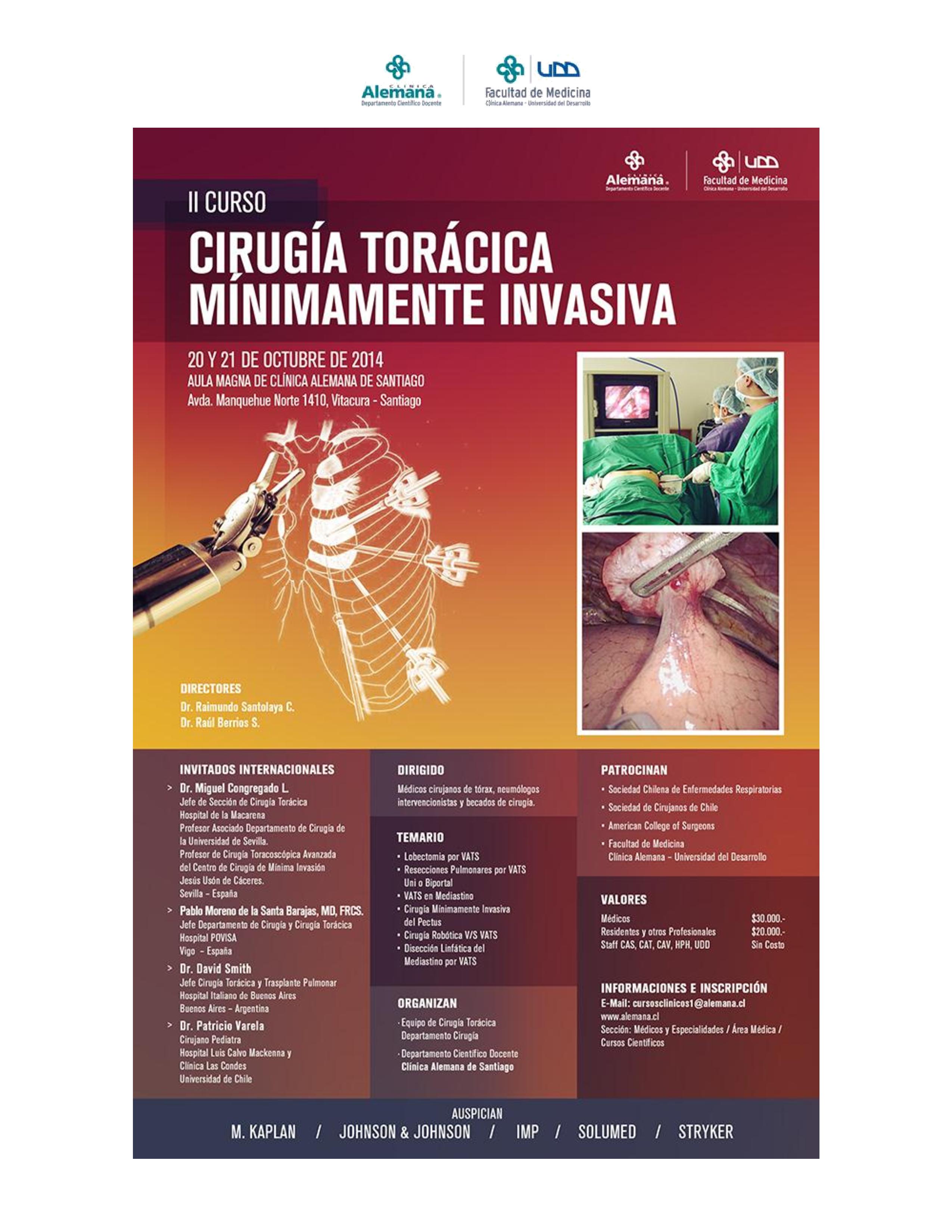
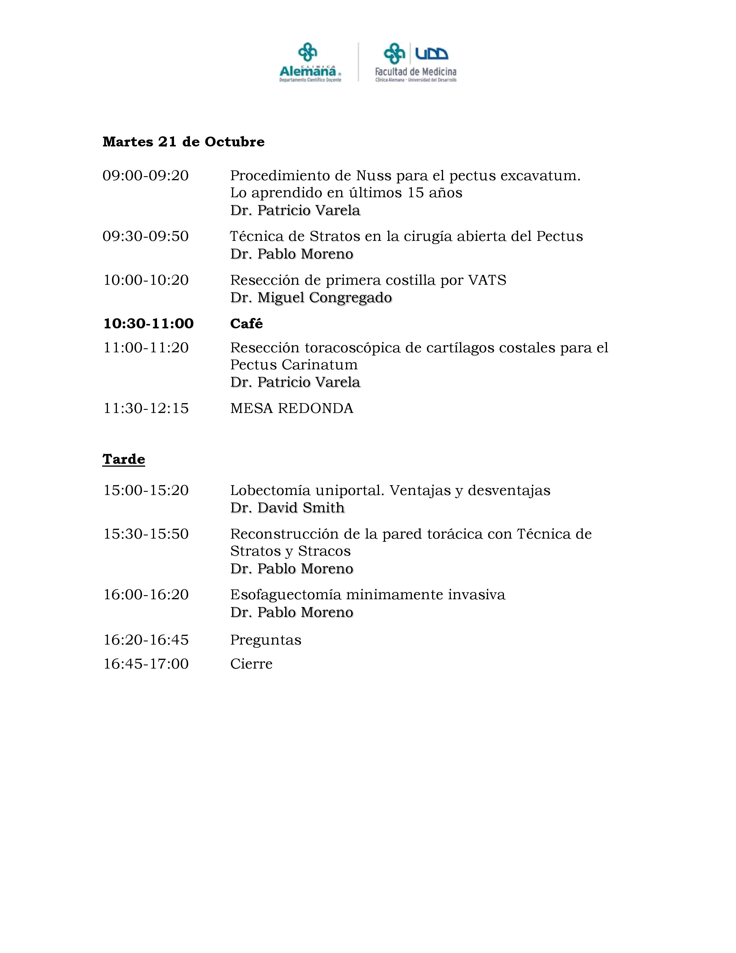
upcoming minimally invasive thoracoscopy course for my Spanish speaking readers at the Clinica Alemana in Santiago, Chile
Cirugia de Torax won’t be there this year – but Clinica Alemana, one of the highest ranked hospitals in Latin America is holding another course on Minimally invasive thoracic surgery this October.
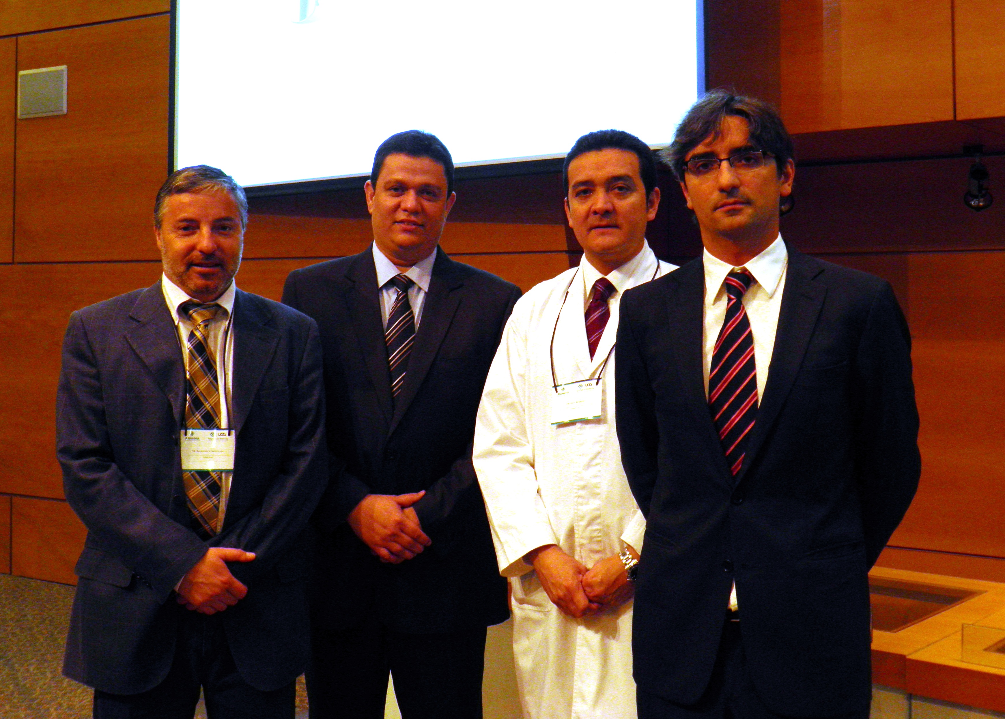
The facility, which is in Santiago, Chile is also home to Dr. Raimundo Santolaya, who was one of our first interviews here at Cirugia de Torax.
Featured speakers include Dr. Miguel Congregado (Seville, Spain), Dr. Pablo Moreno de Santa Barajas (Vigo, Spain), Dr. David Smith (Buenos Aires, Argentina) and Dr. Patricio Varela of the University of Chile.
Course content is sponsored by the Chilean Society of Respiratory Diseases, The Chilean Society of Surgeons, the American College of Surgeons and the Faculty of Medicine at Clinica Alemana – University of Desarrollo.
Interested surgeons should contact the clinic at this address: dmedico@alemana.cl or cursosclinicos1@alemana.cl or enroll on-line at www.alemana.cl
The event is jointly sponsored by M. Kaplan, Johnson & Johnson, IMP, Solumed and Stryker.
Talking about the roles of traditional VATS, single port surgery and robots in modern thoracic surgery.
The Ethicon (Johnson & Johnson) sponsored session was by far, the best of the conference – and an excellent overview of modern technologies in thoracic surgery.
starting with Dr. Ricardo Buitrago (purple tie), Dr. Diego Gonzalez Rivas (blue tie) and Dr. Mario Ghefter (pink tie) are changing the future of thoracic surgery
Dr. Diego Gonzalez Rivas
“Is uni-port surgery feasible for advanced cancers?” Short answer: Yes.
The first speaker, was Dr. Diego Gonzalez Rivas of Coruna, Spain. He is a world-renown thoracic surgeon and innovator of uni-port thoracoscopic surgery. He discussed the evolution of single port surgery as well as the most recent developments with this technique, including more advanced and technically challenging cases such as chest wall resections (2013), sleeve resections/ reconstructions (2013), pulmonary artery reconstructions (2013) and surgery on non-intubated, awake patients (2014).
Experience and Management of bleeding
The biggest challenges to surgeons learning this technique is management of bleeding. But as he explained in previous lectures, this can be overcome with a direct approach. (these lectures and YouTube videos, Dr. Gonzalez explains the best ways to manage intra-operative bleeding.) In the vast majority of cases – this did not require deviation or conversion from the uni-port technique.)
As surgeons gain proficiency with this technique which mirrors open surgery, the only contra-indications for surgical resection of cancerous tissue (by single port) are tumors of great size, and surgeon discomfort with the technique.
Dr. Mario Ghefter
My favorite lecture of the series was given by Dr. Mario Ghefter of Sao Paolo, Brazil. While his lecture was ostensibly about video-assisted thoracoscopy (VATS), it was more of a retrospective vision and discussion of the modern history of thoracic surgery as seen through the eyes of a 22 year veteran surgeon.
He talked about the beginnings of VATS surgery and the contributions from such legends as Cefolio and D’Amico, including the 2005 paper – and modern-day thoracic bible, “Troubleshooting video-assisted thoracoscopic lobectomy (Demmy, James, Swanson, McKenna and D’Amico).
Dr. Ghefter also talked about how improved imaging and diagnostic procedures such as PET-CT and EBUS have been able to provide additional diagnostic information pre-operatively that helps surgeons to plan their procedures and treatment strategies more effectively.
Dr. Mario Ghefter
As a counterpoint to both Dr. Gonzalez and Dr. Buitrago, Dr. Ghefter acquitted himself admirably. He reminded audience members that even the newer technologies have some drawbacks – both as procedures and for the surgeons themselves.
He also successfully argued (in my opinion) that while the popularity of procedures such as multiple port VATS and even open thoracotomies have dropped drastically as thoracic surgeons embrace newer technologies, there will always be a place and time for these more traditional procedures.
Dr. Mario Ghefter is the Director of Thoracic Surgery at Hospital do Servidor Público Estadual – Sāo Paulo and on staff at the Hospital Alemão Oswaldo Cruz.
Dr. Ricardo Buitrago
Native Colombian (and my former professor), Dr. Ricardo Buitrago is acknowledged as one of the foremost experts in robotic thoracic surgery in Latin America.
During his presentation, he discussed the principles and basics of use of robotic techniques in thoracic surgery. He reviewed the existing literature surrounding the use of robotic surgery, and comparisons of outcomes between thoracic surgery and traditional lobectomy.
He reviewed several recent robotic surgery cases and the use of robotics as a training tool for residents and fellows.
While he mentioned some of previously discussed limitations of robotic surgery (namely cost of equipment) he cited recent studies demonstrating significant cost savings due to decreased length of stay and a reduced incidence of surgical complications.
He also discussed recent studies (by pioneering surgeons such as Dr. Dylewski) demonstrated short operating times of around 90 minutes.
the latest from Dr. Diego Gonzalez Rivas and the masters of thoracic surgery.
Dr. Gonzalez Rivas and the Thoracic Surgery Unit in Coruna, Spain are hosting the “International Symposium on Uniportal VATS” this week (February 26th to 28th, 2014).
Dr. Gonzalez Rivas demonstrates uniportal VATS
While the in-person, on-site event is limited to just 100 attendees, the event will be offering real-time live streaming surgery for viewers worldwide.
With registrations from around the world, Dr. Gonzalez Rivas estimates that thousands of pairs of eyes will be watching; from Australia to Saudi Arabia, Hong Kong to Colombia, Brazil to Russia, and the United States.
If you’ve ever wanted to learn more about single port VATS, this is the time to find out.
For more information:
Livethoracic.com – link to the event and on-line registration. Registration is 500 Euros.
Article at Examiner.com with more details on this event.
Dr. Chin Hao Chen revisits one of the basic procedures in thoracic surgery: Chest tube placement
Even Hippocrates placed chest tubes or the history of tube thoracostomy
Chest tube placement has been performed since ancient Greek times. Early physicians, including Hippocrates himself, performed (and wrote about) the use of tube thoracostomy for the treatment of lung abscesses and empyema. Often this procedure is performed using a ‘blind approach’ based entirely on external anatomic features (intercostal spaces) and a fundamental knowledge of internal and chest wall anatomy. Over the years, surgeons have developed guidelines to this technique using palpation/ and other tactile information but none of these techniques challenged initial insertion technique.
With any blind procedure, there is a risk of inadvertent injury due to the lack of visualization, particularly in patients with previous thoracic procedures or infections (adhesions), or when performed by less experienced staff.
Direct visualization during this procedure (akin to VATS) may lessen this risk. However, little has been published on alternatives to the traditional technique.
VGTT: video-guided tube thoracostomy
Our latest post comes directly from Dr. Chih-Hao Chen at Mackay Memorial Hospital in Taiwan.
Dr. Chen presents a video clip demonstrating video-guided tube thoracostomy (VGTT), a technique used to avoid tube-related injury during the course of tube thoracostomy (versus blind insertion). This visualization technique is helpful particularly when performed by inexperienced staff, such as residents or in emergent situations.
A complete description of this technique was recently published in the Annals of Thoracic Surgery.
This paper describes the technique as well as discussing the clinical experience of Dr. Chen and his team in applying this technique to several patients.
Dr. Chin-Hao Chen is a thoracic surgeon at Mackay Memorial Hospital in Taiwan. Dr. Chen is a frequent and valued contributor here at Cirugia de Torax. He has provided several case studies as well as articles and videos on surgical techniques.
in the operating room with Dr. Diego Gonzalez Rivas for single port thoracoscopic (uniportal) surgery.
Hilar mass resection using single port thoracoscopy with Dr. Diego Gonzalez – Rivas
K. Eckland & Andres M. Neira, MD
Instituto Nacional de Cancerlogia
Bogota, Colombia
Surgeon(s): Dr. Diego Gonzalez Rivas and Dr. Ricardo Buitrago
Dr. Diego Gonzalez Rivas demonstrates single port thoracoscopy
Case History:
59-year-old female with past medical history significant for recurrent mediastinal mass previously resectioned via right VATS. Additional past medical history included prior right-sided nephrectomy.
Pre-operative labs:
CBC: WBC 7230 Neu 73% Hgb:14.1 Hct 37 platelets 365000
Pt 12.1 / INR1.1 PTT: 28.3
Diagnostics:
Pre-operative CT scan: chest
edited to preserve patient privacy
Procedure: Single port thoracoscopy with resection of mediastinal mass and lymph node sampling
After review of relevant patient history including radiographs, patient was positioned for a right-sided procedure. After being prepped, and draped, surgery procedure in sterile fashion. A linear incision was made in the anterior chest – mid clavicular line at approximately the fifth intercostal space. A 10mm port was briefly inserted and the chest cavity inspected. The port was then removed, and the incision was expanded by an additional centimeter to allow for the passage of multiple instruments; including camera, grasper and suction catheter.
Dr. Gonzalez Rivas and Dr. Ricardo Buitrago at National Cancer Institute
The chest cavity, pleura and lung were inspected. The medial mediastinal mass was then identified.
As previously indicated on pre-operative CT scan, the mass was located adjacent and adherent to the vessels of the hilum. This area was carefully dissected free, in a painstaking fashion. After freeing the mediastinal mass from the hilum, the remaining surfaces of the mass were resected. The mass was fixed to the artery pulmonary and infiltrating it) . The mass was removed en-bloc. Care was then taken to identify, and sample the adjacent lymph nodes which were located at stations (4, 7 and 10).
Following removal of the tumor and lymph nodes, the area was re-inspected, and the lung was re-inflated. A 28 french chest tube was inserted in the original incision, with suturing of the fascia, subcutaneous and skin layers.
closing the single port incision
Hemostasis was maintained during the procedure with minimal blood loss.
Patient was hemodynamically stable throughout the case, and maintained appropriate oxygen saturations. Following surgery, the patient was awakened, extubated and transferred to the surgical intensive care unit.
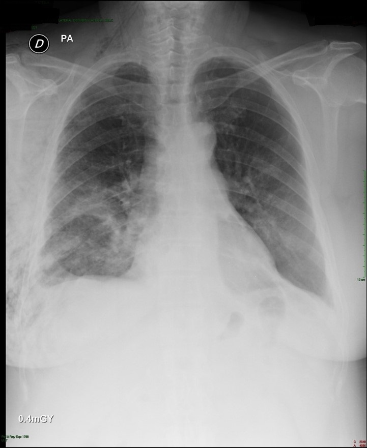
Post-operative: Post-operative chest x-ray confirmed appropriate chest tube placement and no significant bleeding or pneumothorax.
Immediate post-operative film (chest tube visible)
Patient did well post-operatively. Chest tube was discontinued on POD#2 and discharged home.
PA & LAT films on post-operative day 2
Discussion: Since the initial published reports of single-port thoracoscopy, this procedure has been applied to an increasing range of cases. Dr. Gonzalez and his team have published reports demonstrating the safety and utility of the single-port technique for multiple procedures including lobectomies, sleeve resections, segmentectomies, pneumonectomies and mediastinal mass resections. Dr. Hanao Chen (Taiwan) has reported several successful esophagectomies using this technical, as well as bilateral pleural drainage using a unilateral single-port approach.
Contrary to popular perception, the use of a single-port versus traditional VATS procedures (three or more) is actually associated with better visibility and accessibility for surgeons. Surgeons using this technical have also reported better ergonomics with less operating fatigue related to awkward body positioning while operating.
The learn curve for this surgical approach is less than anticipated due to the reasons cited above, and has been cited at 5 to 20 cases by Dr. Gonzalez, the creator of this approach.
The main limitations for surgeons using this technique is often related to anticipated (but potentially unrealized) fears regarding the need for urgent conversion to open thoracotomy. In reality, many of the complications that may lead to urgent conversion, such as major bleeding, are manageable thoracoscopically once surgeons are experienced and comfortable with this approach.
Dr. Gonzalez and his colleagues have reported a conversion rate of less than 1% in their practice. Subsequent reports by Dr. Gonzalez and his colleagues have documented these findings.
Other barriers to adoption of this technique are surgeon-based, and may be related to the individual surgeon’s willingness or reluctance to adopt new techniques and technology. Many of these surgeons would be surprised by how this technique mimics open surgery.
The successful adoption of this technique by numerous thoracic surgery fellows shows the feasibility and ease of learning single-port thoracoscopy by surgeons interested in adopting and advancing their surgical proficiency in minimally invasive surgery.
The benefits for utilizing this technique include decreased length of stay, decreased patient discomfort and greater patient satisfaction.
References/ Additional Readings
Bertolaccini, L., Rocco, G., Viti, A. & Terzi, A. (2013). Surgical technique: Geometrical characteristics of uniportal VATS. J. Thorac Dis. 2013, Apr 07. Article from thoracic surgeons at the National Cancer Institute in Naples, Italy explains how the geometric advantages of uniportal VATS improves visibility and spatial perception over traditional VATS, and mimics open surgery.
Calvin, S. H. Ng (2013). Uniportal VATS in Asia. J Thorac Dis 2013 Jun 20. Article discussing the spread of uniportal techniques in Taiwan, China and other parts of Asia.
Chen, Chin-Hao, Lin, Wei-Sha, Chang, Ho, Lee, Shih-Yi, Tzu-Ti, Hung & Tai, Chih-Yin (2013). Treatment of bilateral empyema thoracis using unilateral single-port thoracoscopic approach. Ann Thorac Cardiovasc Surg 2013.
Gonzalez Rivas, D., Fieira, E., Delgado, M., Mendez, L., Fernandez, R. & De la Torre, M. (2013). Surgical technique: Uniportal video-assisted thoracoscopic lobectomy. J. Thorac Dis. 2013 July 4.
Gonzalez Rivas, D., Delgado, M., Fieira, E., Mendez, L. Fernandez, R. & De la Torre, M. (2013). Surgical technique: Uniportal video-assisted thoracoscopic pneumonectomy. J. Thorac Dis. 2013 July 4.
Rocco, G. (2013). VATS and uniportal VATS: a glimpse into the future. J. Thorac Dis. 2013 July 04. After coming across several articles by Dr. Gaetano Rocco, and actively pursuing several other publications by this Italian thoracic surgeon, I have become increasingly convinced that Gaetano Rocco, along with Dr. Gonzalez Rivas is one of the world’s leading innovators in thoracic surgery. Hopefully, cirugia de torax will be able to catch up to Dr. Rocco at some point for an in-depth discussion.
Rocco G. Single port video-assisted thoracic surgery (uniportal) in the routine general thoracic surgical practice. Op Tech (Society of Thoracic and Cardiovascular Surgeons). 2009;14:326–335.
Rocco G, Khalil M, Jutley R. Uniportal video-assisted thoracoscopic surgery wedge lung biopsy in the diagnosis of interstitial lung diseases. J Thorac Cardiovasc Surg. 2005;129:947–948.
5 / Video-assisted thoracic surgery lobectomy: 3-year initial experience with 200 cases. Gonzalez D, De la Torre M, Paradela M, Fernandez R, Delgado M, Garcia J,Fieira E, Mendez L. Eur J Cardiothorac Surg. 2011 40(1):e21-8.
6 / Single-port Video-Assisted Thoracoscopic Anatomical Resection: Initial Experience. Diego Gonzalez , Ricardo Fernandez, Mercedes De La Torre, Maria Delgado, Marina Paradela, Lucia Mendez. Innovations.Vol 6.Number 3. May/jun 2011. Page 165.
the 2013 S.W.A.T conference, presented by Johnson & Johnson. Featured presenters Dr. Diego Gonzalez Rivas and Dr. Paula Ugalde discuss single port thoracoscopy and topics in minimally invasive surgery
Very pleased that despite the initial difficulties, we are able to provide information regarding the recent conference.
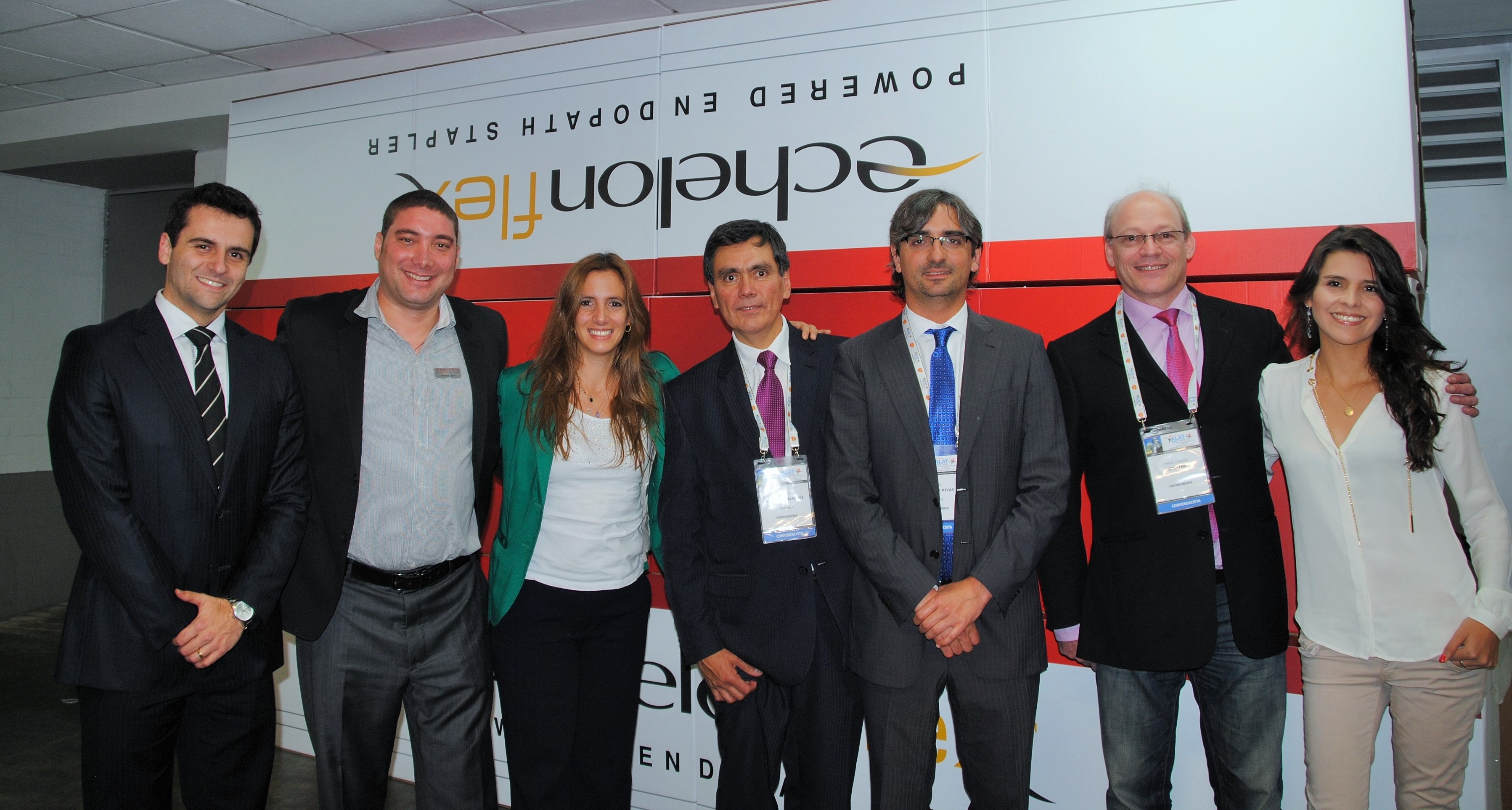
Talking about Single-port surgery in Bogotá, Colombia – 2013 S.W.A.T. Summit
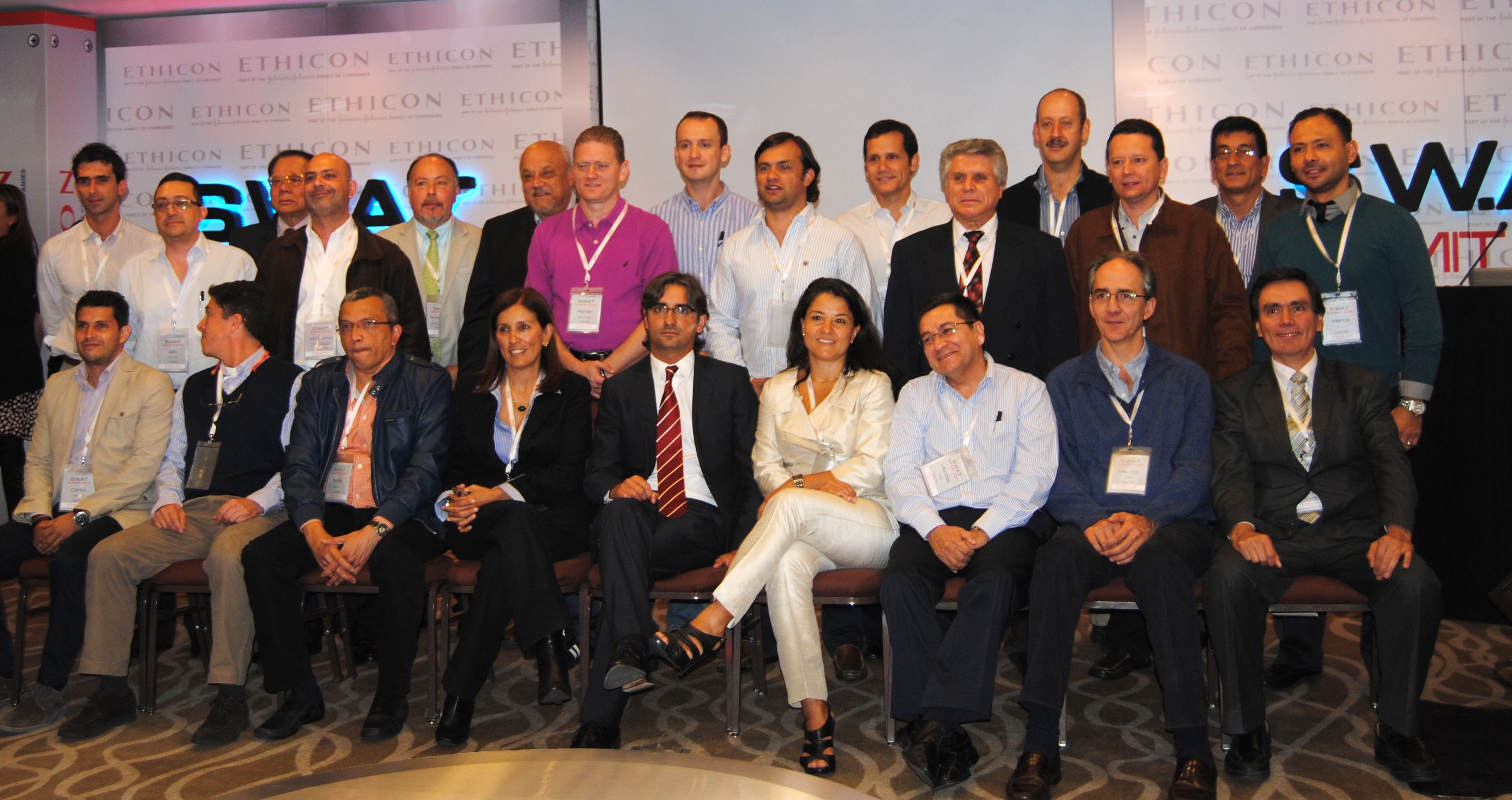
Dr. Diego Gonzalez Rivas and Dr. Paula Ugalde were the headliners at the recent Johnson and Johnson thoracic surgery summit on minimally invasive surgery. Both surgeons gave multiple presentations on several topics. They were joined at the lectern by several local Colombian surgeons including Dr. Stella Martinez Jaramillo (Bogotá), Dr. Luis Fernando Rueda (Barranquilla), Dr. Jose Maineri (Venezuela) Dr. Mario Lopez (Bogotá) and Dr. Pardo (Cartagena).
Thoracic surgeons at the 2013 S.W.A.T Summit in Bogota, Colombia. Drs. Gonzalez-Rivas and Dr. Paula Ugalde are center, front-row
Target audience missing from conference
The audience was made up of thirty Latin American surgeons from Colombia, Costa Rica and Venezuela. This surgeons were hand-picked for this invitation-only event. Unfortunately, while Johnson and Johnson organized and presented a lovely event; their apparent lack of knowledge about the local (Colombian) thoracic surgery community resulted in the exclusion of several key surgeons including Dr. Mauricio Velasquez, one of Colombia’s earliest adopters of single-port thoracoscopy. Also excluded were the junior members of the community, including Dr. Castano, Dr. Carlos Carvajal, and current thoracic surgery fellows. It was an otherwise outstandingand informative event.
The Gonzalez – Rivas dissector, photo courtesy of Scanlon International
As discussed in multiple publications, previous posts as well as during the conference itself, it is these younger members who are more likely to adopt newer surgical techniques versus older, more experienced surgeons. More seasoned surgeons may be hesitant to change their practices since they are more comfortable and accustomed to open surgical procedures.
Despite their absence, it was an engaging and interesting conference which engendered lively discussion among the surgeons present.
Of course, the highlight of the conference actually occurred the day before, when Dr. Gonzalez- Rivas demonstrated his technique during two separate cases at the National Cancer Institute in Bogotá, Colombia. (Case report).
Dr. Gonzalez-Rivas and Dr. Ricardo Buitrago performing single port thoracoscopy at the National Cancer Institute
Featured presenters:
Dr. Diego Gonzalez – Rivas is a world-renown thoracic surgeon jointly credited (along with Dr. Gaetano Rocco) with the development of single-port thoracoscopic (uni-port) surgery. He and his colleagues at the Minimally Invasive Surgery Unit in La Coruna, Spain give classes and lectures on this technique internationally. Recent publications include three papers in July alone detailing the application of this surgical approach, as well as several YouTube videos demonstrating use of this technique for a wide variety of cases.
Dr. Gonzalez Rivas
Dr. Paula Ugalde, a Chilean-borne thoracic surgeon (from Brazil) who gave several presentations on minimally-invasive surgery topics. She is currently affiliated with a facility in Quebec, Canada.
Dr. Paula Ugalde
Refuting the folklore
Part of the conference focused on refuting the ‘folklore’ of minimally-invasive procedures. Some of these falsehoods have plagued minimally-invasive surgery since the beginning of VATS (in 1991), such as the belief that VATS should not be applied in oncology cases. The presenters also discussed how uniportal VATS actually provides improved visibility and spatial perception over traditional VATS (Bertolaccini et al. 2013).
However, Gonzalez-Rivas, Ugalde and the other surgeons in attendance presented a wealth of data, and publications to demonstrate:
– VATS is safe and feasible for surgical resection in patients with cancer. (Like all surgeries, oncological principles like obtaining clear margins, and performing a thorough lymph node dissection need to be maintained).
– Thorough and complete lymph node dissection is possible using minimally invasive techniques like single-port surgery. Multiple studies have demonstrated that on average, surgeons using this technique obtain more nodes than surgeons using more traditional methods.
– Large surgeries like pneumonectomies and sleeve resections are reasonable and feasible to perform with single-port thoracoscopy. Using these techniques may reduce morbidity, pain and length of stay in these patients.
– Rates of conversion to open surgery are very low (rare occurrence). In single-port surgery, “conversion” usually means adding another port – not making a larger incision.
– Learning curve fallacies: the learning curve varies with each individual surgeon – but in general, surgeons proficient in traditional VATS and younger surgeons (the “X box generation”) will readily adapt to single-port surgery.
– Bleeding, even significant bleeding can be managed using single-port thoracoscopy. Dr. Gonzalez Rivas gave a separate presentation using several operative videos to demonstrate methods of controlling bleeding during single-port surgery – since this is a common concern among surgeons hesitant to apply these advanced surgical techniques.
Additional References / Readings about Single-Port Thoracoscopy
Scanlon single-port thoracoscopy kits – informational brochure about specially designed instruments endorsed by Dr. Gonzalez Rivas.
Dr. Diego Gonzalez Rivas – YouTube channel : Dr. Gonzalez Rivas maintains an active YouTube channel with multiple videos demonstrating his surgical technique during a variety of cases. Below is a full-length video demonstrating the uniportal technique.
Additional posts at Cirugia de Torax about Dr. Diego Gonzalez- Rivas
Upcoming conference in Florida – information about registering for September conference for hands-on course in single-port thoracoscopic surgery with Dr. Gonzalez-Rivas
Youtube video for web conference on Single-port thoracoscopic surgery
Bertolaccini, L., Rocco, G., Viti, A. & Terzi, A. (2013). Surgical technique: Geometrical characteristics of uniportal VATS. J. Thorac Dis. 2013, Apr 07. Article from thoracic surgeons at the National Cancer Institute in Naples, Italy explains how the geometric advantages of uniportal VATS improves visibility and spatial perception over traditional VATS, and mimics open surgery.
Calvin, S. H. Ng (2013). Uniportal VATS in Asia.J Thorac Dis 2013 Jun 20. Article discussing the spread of uniportal techniques in Taiwan, China and other parts of Asia.
Rocco, G. (2013). VATS and uniportal VATS: a glimpse into the future.J. Thorac Dis. 2013 July 04. After coming across several articles by Dr. Gaetano Rocco, and actively pursuing several other publications by this Italian thoracic surgeon, I have become increasingly convinced that Gaetano Rocco, along with Dr. Gonzalez Rivas is one of the world’s leading innovators in thoracic surgery. Hopefully, cirugia de torax will be able to catch up to Dr. Rocco at some point for an in-depth discussion.
While I advance criticism of this event – it was a fantastic conference. My only reservations were to the exclusivity of the event. While this was certainly related to the costs of providing facilities and services for this event – hopefully, the next J & J thoracic event will be open to more interested individuals including young surgeons and nurses.
writing about Dr. Diego Gonzalez Rivas and the other living legends in thoracic surgery and connecting people to the world of thoracic surgery
Readers at Cirugia de Torax have certainly noticed that there are numerous articles regarding the work of Dr. Diego Gonzalez Rivas. This week in particular, after a recent thoracic surgery conference and an afternoon in the operating room – there is a lot to say about the Spanish surgeon.
It’s also hard to escape that fact that I regard him in considerable awe and esteem for his numerous contributions to thoracic surgery and prolific publications. I imagine that this is similar to how many people felt about Drs. Cooley, Pearson or Debakey during their prime.
Making thoracic surgery accessible
But the difference is Dr. Diego Gonzalez Rivas himself. Despite the international fame and critical surgical acclaim, he remains friendly and approachable. He has also been extremely supportive of my work, at a time when not many people in thoracic surgery see the necessity or utility of a nurse-run website.
After all, the internet is filled with other options for readers; CTSnet.org, multiple societies like the Society of Thoracic Surgeons (STS), and massive compilations like journal-based sites (Annals of Thoracic Surgery, Journal of Thoracic Disease, Interactive Journal of Cardiothoracic Surgery).
But the difference between Cirugia de Torax and those sites is like the difference between Dr. Gonzalez Rivas and many of the original masters of surgery: Approach-ability and accessibility.
This site is specifically designed for a wider range of appeal, for both professionals in thoracic surgery, and for our consumers – the patients and their families. Research, innovation, news and development matters to all of us, not just the professionals in the hallowed halls of academia. But sometimes it doesn’t feel that way.
Serving practicing surgeons
For practice-based clinicians, and international surgeons publication in an academia-based journal requires a significant effort. These surgeons usually don’t have research assistants, residents and government grants to support their efforts, collect their data and clean up their grammar. Often English is a second or third language. But that doesn’t mean that they don’t make valuable contributions to their patients and the practice of thoracic surgery. This is their platform, to bring their efforts to their peers and the world.
Heady aspirations
That may sound like a lofty goal, but we have readers from over a 110 countries, with hundreds of subscribers along with over 6,000 people with Cirugia de Torax directly on their smart phone. Each month, we attract more hits and more readers.
Every day, at least 200 people read “Blebs, Bullae and Spontanous Pneumothorax”. Why? Because it’s a concise article that explains what blebs are, how a pneumothorax occurs and how it’s treated. Another hundred people usually go on to read the accompanying case report about blebectomy, for similar reasons. There are links for more information, CT scans and intra-operative photos included, so that people can find exactly what they need with a minimum of effort.
Avoiding ‘Google overload’
With the massive volume of information available on the internet, high-quality, easily understood, applicable information has actually become even more difficult for patients to find than ever before. Patients spend hours upon hours browsing through academic jargon, commercial websites and biased materials while attempting to sift through the reams of information for pertinent and easily understandable information. There is also a lot of great material out there – so we provide links to reputable sites, recommend well-written articles and discuss related research.
Connecting patients to surgeons
We also provide patients with more information about the people they are entrusting their bodies, their hopes and their lives to. It’s important that they know about the Dr. Benny Wekslers, the Dr. Hanao Chens, and the Dr. Diego Gonzalez Rivas out there.
Update: June 2019
After multiple reader requests from this site, we have launched a service to assist readers in pursuiting minimally invasive thoracic surgery, uniportal surgery, HITHOC and other state-of-the-art thoracic surgery procedures with the modern masters of thoracic surgery. We won’t talk a lot about this on the site, but we do want readers to know that we are here to help you. If you are wondering what surgery costs like with one of the world’s experts – it’s often surprisingly affordable.
If you are interested in knowing more, please head to our sister site, www.americanphysiciansnetwork.org or send an email to kristin@americanphysiciansnetwork.org.
Keeping it ‘real’
Looking over the shoulder of Dr. Gonzalez Rivas in the operating room
As much as I may admire the work and the accomplishments of Dr. Gonzalez-Rivas – it’s important not to place him on a pedestal. He and his colleagues are real, practicing surgeons who operating on regular people, not just heads of state and celebrities. So when we interview these surgeons and head to the OR, it’s time to forget about the accolades, the published papers and the fancy titles. It’s time to focus on the operations, the techniques, the patients and the outcomes because ‘master of thoracic surgery’ or rural surgeon – the operation and patient are all that really matters.
Reviewing “Ten years experience on 644 patients undergoing single-port (uniportal) video-assisted” by Gaetano Rocco et al. at the National Cancer Institute in Naples, Italy
In this month’s issue of the Annals of Thoracic Surgery, Dr. Gaetano Rocco and his colleagues at the National Cancer Institute, Pascale Foundation in Naples, Italy reported their findings on ten year’s worth of single-port surgery in their institution.
Who: 644 patients; (334 males, 310 females)
Indications:
Annals of thoracic surgery – Rocco et. al (2013)
What: Outcomes and experiences in single port thoracic surgery over a ten-year period. All procedures performed by a single surgeon at this institution, and single-port VATS accounted for 27.7% of all surgeries performed during this time period.
When: data collected on thoracic surgery patients from January 2000 – December 2010.
Technical Notes:
Pre-operative CT scan was used for incision placement planning. Incision was up to 2.5 cm (1 inch) in length depending on indications for surgery.
Conversion rate to 2 or 3 port VATS: 2.2% (14 patients)
Conversion to mini-thoracotomy: 1.5% (10 patients)
Patients underwent conversion due to incomplete lung collapse (22 patients) and bleeding (2 patients).
There were no re-operations or “take backs”. The four patients with malignant effusions who died within the 30 day post-op period were re-admitted to the ICU.
Post-operatively:
Otherwise, all patients were admitted to either the floor or the step-down unit following surgery.
Pain management: post-operative pain was managed with a non-narcotic regimen consisting of a 24 hour IV infusion pump of ketorolac (20mg) and tramadol (100mg*). After the first 24 hours, patients were managed with oral analgesics such as paracetamol (acetaminophen).
Limitations: in this study, uni-port VATS was not used for major resections, as seen in the work of Dr. Diego Gonzalez and others. This may be due to the fact that uni-port VATS was an emerging technique at the initiation of this study.
Strengths: This is one of the largest studies examining the use of single-port thoracic surgery – and showed low morbidity and mortality. (Arguably, the 30 day mortality in this study was related to the patients’ underlying cancers, rather than the surgical procedure itself.)
*Intravenous tramadol is not available in the United States.
Rocco G. Single port video-assisted thoracic surgery (uniportal) in the routine general thoracic surgical practice. Op Tech (Society of Thoracic and Cardiovascular Surgeons). 2009;14:326–335.
Rocco G, Khalil M, Jutley R. Uniportal video-assisted thoracoscopic surgery wedge lung biopsy in the diagnosis of interstitial lung diseases. J Thorac Cardiovasc Surg. 2005;129:947–948.
Discussing Dr. Joseph Coselli and ‘the cowboys of cardiac surgery’ along with some of our own heros of thoracic surgery here at Cirugia de Torax.
There’s a great article in this month’s Annals of Thoracic Surgery, by Dr. Joseph Coselli, from Texas Heart Institute and the Michael DeBakey Department of Surgery at Baylor. His article, entitled,” My heros have always been cowboys” is more than just a title torn from the song sheets of Willie Nelson. It’s a look back at both the pioneers of cardiac surgery and his own experiences as a cardiac surgeon. He also discusses the role of surgeons, and medical practitioners in American society in general and the promises we make to both society at large and our patients.
Here at Cirugia de Torax, I’d like to take a moment to look back at the surgeons that inspired and encouraged me in this and all of my endeavors. Some of these surgeons knew me, and some of them didn’t – but their encouragement and kindnesses have spurred a career and life that have brought immense personal and professional satisfaction.
Like Dr. Coselli, I too, took inspiration from the likes of Dr. Denton Cooley. But our stories diverge greatly from there. I never met Dr. Cooley and I probably never will. But it was a related story, from my former boss (and cardiothoracic surgeon), Dr. Richard Embrey that led to an email to Dr. Cooley himself. My boss had too trained under Dr. Cooley, Dr. Debakey and the Texas Heart Institute, the citadel of American heart surgery. Then, somehow, along the way – Dr. Embrey stopped to work at our little rural Virginia hospital. We were the remnants of a larger Duke cardiothoracic program but we were a country hospital all the same.
While I learned the ins and outs of surgery from Dr. Embrey (and Dr. Geoffrey Graeber at West Virginia University) on a day-to-day basis, I was also weaned on the folklore of cardiothoracic surgery – stories of the giants of history, like the ones mentioned in Dr. Coselli’s article, as well as local Duke legends who occasionally roamed the halls of our tiny ICU and our two cardiothoracic OR suites; Dr. Duane Davis, Dr. Shu S. Lin and Dr. Peter Smith. While never working side-by-side, Dr. D’Amico’s name was almost as familiar as my own. As the sole nurse practitioner in this facility, without residents or fellows, there was no buffer, and little social divide in our daily practice. Certainly, this changed me – and my perceptions. I asked the ‘stupid’ questions but received intelligent and insightful answers. I asked even more questions, and learned even more..
These opportunities fed my mind, and nurtured my ambitions. Not to be a physician or a doctor, but to learn as much as possible about my specialty; to be the best nurse possible in my field. It also nurtured a desire to share these experiences, and this knowledge with my peers, my patients and everyone else who ever had an interest.
It was that tiny little email, a gracious three-line reply from Dr. Cooley himself that made me realize that I didn’t have to rely on folklore and second-hand stories to hear more. That’s critical; because as we’ve seen (here at Cirugia de Torax) there are a quite of few of “Masters of thoracic surgery” or perhaps future giants that haven’t had their stories told. Dr. Coselli and his fellow writers haven’t written about them yet.. So I will.
Sometimes I interview famous (or semi-famous) surgeons here, but other times, I interview lesser-known but equally talented/ innovative or promising surgeons. All of them share similar traits; dedication and love for the profession, immense surgical talent and proficiency and sincere belief in the future of technology of surgery.
So, let’s hope that it won’t take forty more years for these surgeons to be recognized for their contributions to thoracic surgery in the way that Cooley, DeBakey and Crawford are heralded in cardiac surgery.
Awake epidural anesthesia for thoracoscopic pleurodesis: A prospective cohort study. a new publication from Dr. Mauricio Velasquez and his surgical team reviewing results from their 36 month study
On the heels of a recent announcement on CTSnet.org soliciting surgeon input on their experiences with non-general anesthesia for thoracic surgery procedures, Cirugia de torax is revisiting one of the surgeons we interviewed last year, Dr. Mauricio Velasquez at Fundacion Valle de Lili in Cali, Colombia.
Dr. Velasquez in the operating room with Lina Caicedo Quintero (nurse) Valle de Lili, Cali, Colombia
The trip to Cali was primarily to discuss Dr. Velasquez’s Thoracic Surgery Registry, and to observe him performing several single port surgery cases. However, during the trip, Dr. Velasquez also spoke about several other aspects of his current practice including some of his recent cases, and the thoracic surgery program at Fundacion Valle de Lili.
Dr. Mauricio Velasquez after another successful case
We also talked with his wife, (and lead author), the talented Dra. Cujiño, an anesthesiologist who subspecializes in thoracic anesthesia. Together, they have successfully performed several thoracic cases using thoracic epidural anesthesia on awake patients.
By chance, they published articles in both Revista Colombianas de anesthesia and Neumologia y cirugía de torax in the last few weeks.
Revista Colombianas de anesthesia
Patients receiving epidural anesthesia received a small dose of midazolam prior to insertion of epidural needle at the T3 – T4 intervertebral space. During the case, patients received bolus administration via epidural of 0.5% bupivacaine on a prn basis.
Short surgeries, single port approach
All patients, regardless of anesthesia type underwent single port thoracoscopic surgery for the talc pleurodesis procedure. Surgery times were brief, averaging 30 to 35 minutes for all cases (range 25 – 45 minutes) with the epidural patient cases being slightly shorter.
Dr. Mauricio Velasquez performing single port thorascopic surgery
Dramatic reduction in length of stay
In their study, patients receiving awake anesthesia had an average length of stay of four days compared with ten days for the general anesthesia group.
Decreased incidence of post-operative complications
There was a marked reduction in the incidence of post-operative respiratory complications (19 in general anesthesia group) versus 3 patients in the awake anesthesia group. Post-operative mortality was also decreased (six in general anesthesia) versus two deaths in the awake anesthesia group. However, the mortality statistics may also be impacted by the overall poor prognosis and median survival time of patients presenting with malignant effusions.
Post-operative pain
Study patients also self-reported less post-operative pain in the awake anesthesia group – with only one patient reporting severe pain versus seven patients in the general anesthesia group.
Conclusions
Cujiño, Velasquez and their team found awake thoracic epidural anesthesia (ATEA) was a safe and effective method for intra-operative anesthesia and was associated with a decreased post-operative pain, decreased length of stay (LOS) and decreased incidence of post-operative complications.
Notes
This study was funded by the authors with no relevant disclosures or outside financial support.
A discussion of Meimarakis’ recently published article, “Prolonged overall survival after pulmonary metastatectomy in patients with breast cancer.”
As reported in the Society of Thoracic Surgeons, and multiple other outlets, a newly published study by several surgeons in Germany shows that surgical removal of metastatic breast cancer that has spread to the lungs may improve overall patient survival. The study, by Meimarakis et al. was published in the April 2013 issue of the Annals of Thoracic Surgery.
pulmonary metastatectomy in metastatic breast cancer
The Meimarakis study included 81 patients over a twenty-five year period. The study looked at the overall survival time in breast cancer patients with a pulmonary metastasis. The study began in 1992, and data was collected retrospectively to 1982.
Poor median survival despite advances in chemotherapy
Current survival time in these patients ranges from 12 to 24 months. However, the authors note that in up to 23% of these patients, the sole metastatic lesion is in the lung or pleural space. In these patients with pulmonary metastasis alone, the majority survived less than 22 months after diagnosis, despite chemotherapy. The 10 year survival has been previously reported as a dismal 9% in this population in prior studies conducted as M. D. Anderson (Meimarakis, et. al, 2013).
Role of pulmonary metastatectomy in advanced breast cancer
Unlike pulmonary metastatectomy for colon cancer, metastatectomy has been used sparingly in this population and with no clear-cut criteria to distinguish which breast cancer patients would benefit from surgery, surgery in addition to chemotherapy, versus chemotherapy alone.
Aim of study
The authors, at Ludwig-Maximilian University in Munich, Germany attempt to address this deficiency by investigating surgical, pathological and demographic factors that impact survival in this patient population to help determine which candidates would benefit the most from surgical intervention.
The authors looked at a multitude of factors such as presence and type of hormone receptor, histological type, size of both primary and metastatic lesions, the number of metastatic lesions, surgical grade/ resectability and the laterality of these lesions. They also collected and compared additional markers such as CEA, LDH and CA 15-3.
These factors and their impact on survival were analyzed using statistical analysis, Kaplan-Meier estimators, log-rank tests as well as matched pair analysis of 2 year survival (metastectomy vs. standard therapy only). These factors included data from pathological specimens and tumor typing (Meimarakis, 2013).
What makes this study particularly interesting and noteworthy, is the operative inclusions. While patients with local residual disease, additional (non-lung) metastases or recurrent primary breast tumors were excluded, patients with contralateral lung lesions were not.
Selected patient demographics
Total number of patients: 81
Median age: 58.2 (range 28.2 to 76.3)
Breast cancers: Histological types
64.1% invasive ductal carcinoma, 17.2 % with ductal carcinoma in situ? and 18.7% other breast cancer.
Number and size of metastatic lesions:
61 (75.3%) lesions were less than 3 cm in size.
20 (24.7%) of lesions were 3 cm or greater.
The majority (51 (63%) of patients presented with a solitary lung lesion, whereas 30 (37. %) presented with two or more lesions.
Operative procedures
Meimarakis et al. performed a total of 92 operations. These included 71 patients who underwent one procedure, 9 patients for two procedures and 1 patient with three procedures.
All of the patients undergoing more than one procedure had contralateral surgery for newly occurring metastases. (The authors re-operated on patients within 4 to 6 weeks for synchronous metastatic lung lesions.) This is important to remember when reviewing the primary article since the terminology ‘re-do’ operations and repeat operations can be confusing. However, after clarifying with the primary author, there were no completion procedures (i.e. wedge converted to lobectomy based on final pathology) and no returns to the operating room for surgery due to complications. There was no return to the operating room for any procedures on the same side as the original procedure. Thus for clarification, no “re-do” procedures.
All patients underwent resection via anterolateral thoracotomy. However, patients with peripheral, previously unbiopsied nodules were initially approached via VATS with conversion to anterolateral thoracotomy for positive intraoperative pathology.
67 operations were wedge resection, with an additional 10 segmental resections. The remainder of procedures included 7 lobectomies, 7 pneumonectomies and 1 bilobectomy.
Median operating room time was 83 minutes, with a fairly lengthy hospitalization stay (median 9 days, with a range of 3 – 63 days.) Complication rate was 7.6% (3 patients with pneumonia, 4 patients with atelectasis).
Limitations of Study
The median follow-up was only 27.2 months. At the end of this period, 27 of the 81 patients (33.3%) had died. While the published study was lengthy and detailed (10 pages with multiple charts and graphs) much of this was related to discussion regarding receptor status, and existing literature. A clearer, more streamlined algorithmic approach or scoring system utilize to their findings would be more helpful to readers in determining the likelihood of successful outcomes with surgical resection, and for encouraging replication of their research.
Results
Despite the limited number of patients with multiple metastatic lung lesions in this study, the underlying rules of surgical resection remain consistent. Patients who did the best, with the longest overall survival time were patients with complete surgical resection (R0). While patients with a completely resection of a single metastasis lived longer than patients with complete resection of multiple metastases, the R0 patients with multiple metastases had greater median survival than all patients with incomplete resection, regardless of the degree of residual (R1, R2) disease (microscopic or gross disease).
Receptor positive patients with better outcomes
As seen in multiple studies, tumor types were a crucial factor in long-term outcomes; whether estrogen receptor positive (ER+), human growth factor receptor 2 positive (HER2+), progesterone receptor+ (PR+).
Median survival of all patients after metastatectomy was 82.4 months with the greatest median survival time in the 31 patients with + hormone receptor tumors (HR+) at 127.4 months (range 33.2 to 221.6 months). In comparison, the 8 patients with HER+ had a mean survival of 66 months and only 27 months median survival for the 14 triple negative patients)*.
These findings regarding longevity and tumor receptors are similar to those reported by Welter et. al (2008) and others, but the patients from this larger study demonstrated greater longevity, which gives weight to continued study in this area.
In Meimarakis’ work, the presence of pleural infiltration or lymphangiosis carcinomatosis denoted a reduced longevity (32.1 and 34.5 months). This may serve as a better marker of systemic disease for future classification and treatment of advanced breast cancer.
Implications: For breast cancer patients, the discovery of a metastatic lung lesion advances the stage of the disease, drastically changing current treatment options. Most breast cancer patients diagnosed with metastatic disease are not considered surgical candidates even if complete surgical resection is technically feasible.
Meimarakis’s study is one of the larger studies to date, using a large number of prospective patients versus retrospective chart review. This gives a more comprehensive look at a multitude of factors and patient demographics. It serves as an excellent framework for future study in this area.
But, more interesting to our readers is the low incidence of post-operative complications (7 operations; 3 patients with pneumonia, 4 patients with atelectasis).
None of the patients died post-operatively. There were no ‘take backs’ for post-operative complications such as bleeding, prolonged air leak or post-operative infections despite the fact that almost 10% (8 patients) underwent significantly larger procedures such as pneumonectomy or bilobectomy and that all patients underwent thoracotomies versus the smaller VATS procedures. There was no difference in outcomes in this set of patients by procedure (wedge versus pneumonectomy) though Meimarakis notes that “there is a trend to worse survival in case of pneumonectomy during R1/ R2 resection (considering the whole database [Munich Cancer Registry] i.e not only in this group of patients with breast cancer.”
As outcomes appeared independent of the surgical procedure itself; based solely on resectability and tumor type, even larger scale resections such as pneumonectomy may be worthy of consideration during preoperative surgical evaluation, particularly in patients with favorable tumor types with good potential for complete resection.
Future considerations
Using the work of Meimarakis and similar researchers, development of an algorithmic approach may be beneficial to thoracic surgeons and others who encounter pulmonary metastases from breast cancer outside of larger research facilities.
Related case reports: We previously reported a case of metastatic breast cancer that was discovered at the time of surgery, despite the use of multiple imaging and diagnostic modalities. However, in that case, the patient also had local metastases to bone (ribs), which were also resected.
*Please see original article for further detail on patient characteristics and outcomes.
While the data (statistics, patient outcomes) is from the original research of Meimarakis et al., the commentary has been written by writers at Cirugia de Torax and may not reflect the thoughts, considerations and experiences of the primary researchers.
Kycler, W. & Laski, P. (2012). Surgical approach to pulmonary metastases from breast cancer. Breast J. 2012 Jan-Feb;18(1):52-7. doi: 10.1111/j.1524-4741.2011.01176.x. Epub 2011 Nov 20. [no free full text available]. Retrospective data review of 33 patients who underwent pulmonary metastatectomy (1997 – 2002) at the Great Poland Cancer Center, in Poznan, Poland.
Welter S, Jacobs J, Krbek T, Tötsch M, Stamatis G. (2008). Pulmonary metastases of breast cancer. When is resection indicated?Eur J Cardiothorac Surg. 2008 Dec;34(6):1228-34. doi: 10.1016/j.ejcts.2008.07.063. Epub 2008 Sep 27 [free text available]. A review of 47 cases of metastatic breast cancer with pulmonary metastatectomy, Essen, Germany.
The Spanish-language lecture entitled, “El viaje de los pioneros: Dr. Diego Gonzalez Rivas” should be just as inspiring to readers/ and viewers as it is to Cirugia de Torax.
If you don’t speak Spanish – don’t despair! Dr. Gonzalez’ TED talk is now available with captions in multiple languages. (Click on the closed captioning icon for translation options.)
Sometimes, it’s lonely out front – and being innovative is difficult. It’s one thing to be Ivor Lewis, Pearson or McKeown but it’s another to be the first or sole surgeon to challenge edicts and procedures laid down by the giants of the specialty. But without the modern-day Dylewskis, Gonzalez Rivas, Chen, (and others) – technology within the specialty would remain static.
Changing the future of thoracic surgery
These surgeons take big risks with their careers and reputations by attempting to deviate from long-standing surgical traditions. But sometimes, it pays off – and when it does, it is wonderful to see these daring and forward thinkers receive the admiration and appreciation they deserve for their contributions to the field and to their patients.
Dr. Santolaya, Dr. Sales dos Santos, Dr.Berrios and Dr. Diego Gonzalez Rivas
Congratulations, Dr. Diego Gonzalez Rivas! Here’s to your continued success..
This article is one of several articles and content from Cirugia de Torax that has been used commercially without consent, permission or attribution/ proper citation.
the latest video from Dr. Diego Gonzalez Rivas demonstrating a sleeve lobectomy via single port surgery
On the heels of the recent conference in Hong Kong, one of our favorite surgeons (and presenter at the 1st Asian single port surgery conference), Dr. Diego Gonzalez Rivas has sent another link to one of his more recent cases – Single port lobectomy – Sleeve resection after chemotherapy.
the latest predictions on the impending shortage of surgeons in the United States
Unsurprisingly – rural area hospitals face additional challenges in attracting and retaining specialty surgeons in comparison to big cities/ metropolitan areas. However, as reported by Patrice Welding at Thoracic Surgery News in a report on the annual meeting of the Central Surgical Association, this may be viewed as a boon for the surgeons themselves as hospitals may devise new and enhanced incentives to attract surgeons to their facilities. The surgical specialties most likely to benefit from this strategy include (as previously reported), obstetrics and gynecology, orthopedic surgery, general surgery, otolaryngology, urology, neurosurgery, and thoracic surgery.
The article which quotes Dr. Thomas E. Williams, Jr. predicts that hospitals and institutions may break out into a ‘bidding war’ over surgeons.
While this is dire news for rural hospitals and the estimated 56 million patients served by these facilities, it comes as a relief for current thoracic surgery fellows and new thoracic surgeons who have faced an increasingly bleak economic landscape over the last few years.
Of course, more sanguine experts note that the impact of the impending shortage has been reported for several years – with little impact on the current job market for new graduates.
Dr. Thomas E. Williams Jr. is one of the main researchers on the impending shortage in the United States and published a book based on his findings in 2009, entitled, “The coming shortage of surgeons: why they are disappearing and what that means for our health“. (Praeger, ISBN #978-0313380709). His work has also be published in multiple journals, and presented in meetings and conferences across the country.
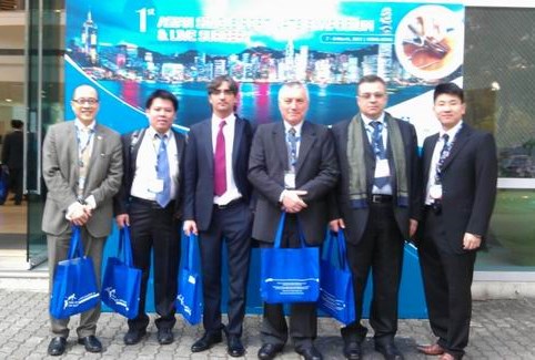
Interested in learning more about single port thoracoscopy, or talking to the inventors of this technique? This March – head to the 1st Asian single port surgery conference in Hong Kong.
It doesn’t look like Cirugia de Torax will be in attendance for this conference, but it’s another opportunity for practicing thoracic surgeons and thoracic surgery fellows to learn more about single port thoracoscopic surgery.
This March (7th – 8th), the Chinese University of Hong Kong, along with the Minimally Invasive Thoracic Surgery Unit (Coruna, Spain), and Duke University are presenting the 1st Asian Single Port Symposium and Live Surgery conference in Hong Kong.
This is your chance to meet the experts – and the inventors of this technique (such as Dr. Diego Gonzalez – Rivas, one of the new masters frequently featured here at Cirugia de Torax.)
what is the future of thoracic surgery education? A new American study asks the if it is time to separate the specialties of cardiac and thoracic surgery.
A new study by Cooke & Wisner performed at a large medical center in California (UC Davis) and published in the Annals of Thoracic Surgery provides additional weight to the idea that Thoracic Surgery has increasingly developed into it’s own subspecialty away from the traditional cardiothoracic surgery model (seen in the United States and several other countries.)
In an article published in Medical News Today, the authors of the study explained that the increased complexity of (noncardiac) thoracic surgery procedures for general thoracic conditions has led to increased referrals and utilization of general thoracic surgeons (versus cardiac or general surgeons). This shows a reversal in a previous trend away from specialists – with more patients now receiving “complex” thoracic surgery procedures from specialty trained, board-certified thoracic surgeons. Previously up to 75% of all thoracic surgery procedures were performed by general surgeons.
With lung cancer rates expected to climb dramatically in North America and Europe, particularly in women – along with esophageal cancer, and long waits already common, support and on-going discussion about the evolution of resident and fellow education is desperately needed.
Reference
Cooke, D. T. & Wisner, D. H. (2012). Who performs complex noncardiac thoracic Surgery in United States Academic Medical Centers? Ann Thorac Surg 2012;94:1060-1064. doi:10.1016/j.athoracsur.2012.04.018
a day in the operating room with one of Colombia’s New Masters of Thoracic Surgery
Cali, Colombia
Dr. Mauricio Velasquez is probably one of the most famous thoracic surgeons that you’ve never heard of. His thoracic surgery program at the internationally ranked Fundacion Valle del Lili in Cali, Colombia is one of just a handful of programs in the world to offer single port thoracic surgery. Dr. Velasquez has also single-handedly created a surgical registry for thoracic surgeons all over Colombia and recently gave a presentation on the registry at a national conference. This registry allows surgeons to track their surgical data and outcomes, in order to create specifically targeted programs for continued innovation and improvement in surgery (similar to the STS database for American surgeons).
Dr. Mauricio Velasquez after another successful case
Dr. Velasquez is also part of a team at Fundacion Valle del Lili which aims to add lung transplant to the repertoire of services available to the citizens of Cali and surrounding communities.
He is friendly, and enthusiastic about his work but humble and apparently unaware of his growing reputation as one of Colombia’s finest surgeons.
Education and training
After completing medical school at Universidad Pontificia Bolivariana in Medellin in 1997, he completed his general surgery residency at the Universidad del Valle in 2006, followed by his thoracic surgery fellowship at El Bosque in Bogotá.
The Colombia native has also trained with thoracic surgery greats such as Dr. Thomas D’Amico at Duke University in Durham, North Carolina, and single port surgery pioneer, Dr. Diego Gonzalez Rivas in Coruna, Spain. He is also planning to receive additional training in lung transplantation at the Cleveland Clinic, in Cleveland, Ohio this summer.
Single port surgery
Presently, Dr. Velasquez is just one of a very small handful of surgeons performing single port surgery. This surgery is an adaptation of a type of minimally invasive surgery called video-assisted thoracoscopy. This technique allows Dr. Velasquez to perform complex thoracic surgery techniques such as lobectomies and lung resections for lung cancer through a small 2 – 3 cm incision. Previously, surgeons performed these operations using either three small incisions or one large (10 to 20cm) incision called a thoracotomy.
By using a tiny single incision, much of the trauma, pain and lengthy hospitalization of a major lung surgery are avoided. Patients are able to recovery and return to their lives much sooner. The small incision size, and lack of rib spreading means less pain, less dependence on narcotics and a reduced incidence of post-operative pneumonia and other complications caused by prolonged immobilization and poor inspiratory effort.
However, this procedure is not just limited to the treatment of lung cancer, but can also be used to treat lung infections such as empyema, and large mediastinal masses or tumors like thymomas and thyroid cancers.
Dr. Velasquez in the operating room with Lina Caicedo Quintero (nurse)
Team approach
Part of his success in due in no small part to Dr. Velasquez’s surgical skill, another important asset to his surgical practice is his wife, Dr. Indira Cujiño, an anesthesiologist specializing in thoracic anesthesia. She trained for an additional year in Spain, in order to be able to provide specialized anesthesia for her husband’s patients, including in special circumstances, conscious sedation. This allows her husband to operate on critically ill patients who cannot tolerate general anesthesia. While Dr. Cujiño does not perform anesthesia for all of Dr. Velasquez’s cases, she is always available for the more complex cases or more critically ill patients.
In the operating room with Dr. Velasquez
I spent the day in the operating room with Dr. Velasquez for several cases and was immediate struck by the ease and adeptness of the single port approach. (While I’ve written quite a bit about the literature and surgeons using this technique, prior to this, I’ve had only limited exposure to the technique intra-operatively.) Visibility and maneuverability of surgical instruments was vastly superior to multi-port approaches. The technique also had the advantage that it added no time, or complexity to the procedure (unlike robotic surgery).
Dr. Velasquez performing single port thoracoscopy
Cases proceeded rapidly; with no complications.
close up view
Note to readers – some of the content, and information obtained during interviews, conversations etc. with Dr. Velasquez may be used on additional websites aimed at Colombia-based readers.
Dr. Chen discusses single port thoracoscopy – and specimen size.
Single port thoracoscopy for wedge resection – does size matter? Dr. Chih-Hao Chen, Thoracic Surgeon, Mackay Memorial Hospital, Taiwan Correspondence: musclenet2003@yahoo.com.tw
Case presentation and discussion
A 77 y/o woman was found to have a neoplasm in RML during routine health exam. (Full case presentation here.)
The incision was 2.5 cm. The specimen was removed successfully; patient experienced no complications and was discharged from the hospital without incident. However, when discussing this case with surgeon colleagues a specific question arose.
preparing to remove the specimen through the port
Does size matter? How big is too big to remove through a port incision?
An important issue is the theoretically smallest (and effective) incision size. The issue is very challenging for most surgeons. In the past, other surgeons have questioned me :
“The specimen to be removed is more than 12-15 cm. How can you remove the specimen from a 3.5 cm incision?”
BUT, the fact is “the smallest size depends mostly on the solid part of the neoplasm “.
The lung is soft in nature. In my experience, a 3.5 cm port is usually enough for any lobe. The only problem is that if the solid part is huge, pull-out of the specimen may be difficult.
Therefore, the solid part size is probably the smallest incision size we have to make, usually in the last step when dissection completed , if needed.
successful removal of lung specimen thru small portPort with chest tube in place
Using endo-bags to avoid larger incisions
There is an exception. I have never do this but I think it may be possible. We usually avoid opening the tumor (or cutting tumor tissue into half ) within the pleural space in order to prevent metastasis from spillage of cancerous tissue.
What if we can protect the tumor from metastasis while we cut it into smaller pieces? For example, into much smaller portions that will easily pass through the small port incisions?
My idea is to use double-layer or even triple layers of endo-bag to contain the specimen. If it is huge, with perfect protection, we cut it into smaller parts within the bag. Then the huge specimen can be removed through a very tiny incision. This is possible.
We still have to avoid “fragmentations” since this might interfere or confuse the pathologist for accurate cancer staging. Therefore, in theory, we can cut it into smaller parts but not into fragments. However this idea is worthy of consideration, and I welcome debate from my fellow surgeons and medical colleagues.
Robotic (thoracic) surgery comes to Clinica de Marly in Bogota, Colombia
A year and a half ago, I interviewed and spent some time with Dr. Ricardo Buitrago at the National Cancer Institute, and Clinica de Marly while doing research for a book about thoracic surgeons. At that time, Dr. Buitrago stated he was interested in starting a robotic surgery program – and was planning to study robot-assisted thoracic surgery with Dr. Mark Dylewski.
Dr. Ricardo Buitrago in the operating room, April 2011
Fast forward 1 year – when I received a quick little email from Dr. Buitrago telling me about his first robotic surgery at the Clinica de Marly. At that point, I sent Dr. Buitrago an email asking if I could come to Colombia and see his robotic surgery program to learn more about it. We had several phone conversations about it and I also outlined a research proposal to gather data on thoracic surgery patients and outcomes at high altitude, to which he enthusiastically offered to assist with. Thus began my current endeavor, in Bogota, studying with Dr. Buitrago.
Now – after completing a proctoring period with Dr. Dylewski, Dr. Buitrago has more than a dozen independent robotic surgeries under his belt. He has successfully used the robot for lobectomies, mediastinal mass resections and several other surgeries.
As part of my studies with Dr. Buitrago – I’ve made a video for other people who may be interested in robotic surgery with the DaVinci robot and what it entails.
Discovery of extensive pleural plaques during VATS
Usually with pleural plaques, you think of two possible diagnoses: metastatic cancer and tuberculosis.
But which is the more likely culprit?* That kind of depends on both your patient and your geographic location.
If this had been in my native Virginia – I’d “assume/ guess” metastatic cancer (since my patient population is usually older, high rate of smoking, other risk factors for cancer).
But luckily (who ever thought I’d be saying luckily) in my current location (Northern Mexico) in this patient (with multiple risk factors for infectious disease but no asbestos exposure) – tuberculosis is the more likely diagnosis.
* Prior to formal tissue pathology results, which confirmed tuberculosis in this patient.
** I apologize for the lack of formal references, but I was unable to find any comprehensive literature (available as free articles).
an interview with thoracic and tranplant surgeon, Dr. Raimundo Santolaya Cohen of Santiago, Chile
Dr. Raimundo Santolaya, Thoracic and Transplant Surgeon
After listening to Dr. Raimundo Santolaya Cohen at the National Conference, where he discussed the diagnosis and management of Pneumothoraces, I immediately contacted him following the lecture to arrange an interview. I was delighted to be able to spend several hours in the charismatic and elegant Chilean surgeon’s company while we talked about thoracic surgery, and the state of lung transplant in Chile.
While he reports that while lung transplant programs are fairly small in Chile (in comparison to American institutions) he performs several transplants every year.
In addition to his interest in pulmonary transplant, he is also keenly interested in minimally invasive surgery, including uni-port surgery. In fact, he is currently arranging for Dr. Diego Gonzalez, the Spanish thoracic surgeon to come to Chile in October to teach uni-port lobectomy.
Like most thoracic surgeons, he performs a wide range of thoracic procedures including lung surgeries, and mediastinal masses.
We also discussed the incidence of Hidatidosis/ hydatidosis, (echinococcus granulosus) which is endemic in Chile. Hidatidosis is an infection caused by a parasite transmitted by animals (commonly dogs), and is more commonly known as a tapeworm. Infection with this organism can affect multiple organs, but frequently affects the liver and the lungs, called hepatopulmonary hydatidosis. Infection is marked by the development of hydatid cysts which are filled with tapeworm larvae. In the lungs, these cysts can become quite large.
Dr. Raimundo Santolaya completed his thoracic surgery fellowship at the Universidad de Chile before traveling to Madrid, Spain to study lung transplant for an additional year.
Talking to the interesting and entertaining Dr. Marcelo Argote Green, of Mexico City at the LXXI Congreso for Sociedad Mexicana de Neumologia y Cirugia de Torax
Dr. Argote Green has been practicing in Mexico City for four years, since completing his fellowship in thoracic surgery at Brigham Women’s & Children’s Hospital in Boston, Massachusetts. He trained under the guidance of several of the most prominent American thoracic surgeons including the ever elusive* Dr. David Sugarbaker.
Dr. Argote maintains an active, and diverse practice as he is currently operating in several facilities within the supercity, including that National Institute of Medical Science. This gives him a wide range of exposure and experience to patients from all across the city, and across all demographic lines. As a surgeon at one of the countries more prominent public facilities, he also receives patients from around the country – particularly the more challenging or complex cases. He reports that this along with the high incidence of HIV and other autoimmune conditions such as scleroderma and lupus make his day-to-day practice different from the average small town physicians practice, or even the West Roxbury Veteran’s facility where he completed some of this training. “I saw maybe two or three cases with patients with this level of complex co-morbidities when I was training, but here I see it everyday.” He enjoys this aspect of his work which gives him a deeper level of experience than he might otherwise have at this stage in his career.
He has also embraced minimally invasive technologies such as VATS, and RATS, and currently performs uni-port lung biopsies, and VATS lobectomies. He also particularly enjoys treating patients with mediastinal masses, and uses a unilateral VATS approach for many of these tumor resections versus the traditional median sternotomy.
During our in-depth conversation, we also discussed some of the differences I had observed during my time in Mexico – particularly the inter-collegial relationships between pulmonologists and thoracic surgeons. He explained that this is due in part to a shared history, and that in Mexico – thoracic surgery was a outbranching of pulmonary medicine, as pulmonologists initially sought additional training in more invasive procedures, including surgery. While there is now more delineation between the specialties, there remains a sense of commonality often spoken of during this conference by the participants which is sometimes striking to outsiders like myself.
* Long-time readers know of our ongoing attempts to contact Dr. Sugarbaker for an interview.
I’m here at the National conference for pulmonologists and thoracic surgeons this year to hopefully interview (and possible recruit some surgeons to the research project).
It’s already been an eventful day, with several excellent presentations this morning as attendees continue to arrive to the official beginning of the conference tomorrow morning. Extensive discussions on the multi-drug resistant tuberculosis started today and will continue for the rest of the week in addition to offering aimed at multiple specialties including surgery, nursing, respiratory therapy, pulmonology and general internists.
One of the featured presenters is Dr. Richard Light, MD who almost seems like family to me at this point, since I’ve been reading much of his previous work while I write a new case presentation about dual port thoracoscopy. He’s one of the many people I hope to catch up with for a few minutes over the next few days.
Wish me luck! I’ll continue to post about updates over the next several days.
Developing electronic applications to ease the task of data collection for clinical research.
In clinical research; results are dependent on data. Data is only as good as the data collection tools used to gather it. Furthermore, the best and most accurate data comes from the surgeon or the surgical team themselves at the time of care (versus third-party lay person data collection via chart review). However, there are some limitations that are imposed when following these guidelines; such as the collection of 30-day follow-up information.
Data collection needs to be simple and relatively speedy. The ideal tools allow surgeons to collect the essential data at the time of discharge (when information is fresh), limit additional paper accumulation and are submitted at the time of completion. This necessitates the development of electronic applications.
At the time of this writing, I am currently working on the development of both smart phone and web-based applications for data collection for the altitude project. These electronic forms will allow data to be entered and submitted at the time of collection.
smart phones for data collection
A secondary application is being developed to address the patient re-admission or development of complications post-discharge in the first thirty days after discharge.
Downloadable pdfs and/or spreadsheets will also be available for surgeons who elect to use the ‘paper’ option.
Since the data has only limited patient identifiers, and all data is being submitted to a clinical investigator, (versus outside companies) patient privacy is protected.
When completed, further information will be posted here at Cirugia de Torax.
Calling all thoracic surgeons – particularly those in La Paz, Quito, Bogota and Mexico City.. We have an opportunity for collaborative research.
One of our newest endeavors is a research project on thoracic surgery at moderate altitude. We’ve completed our review of existing literature, and developed our patient parameters and data collection points.
The main base of our operations is Flagstaff, Arizona, which is located at 7000 ft. (2,000 meters) making it the highest altitude cardiothoracic program in the United States. The Flagstaff site has several strengths in that much of our patient population comes from the surrounding areas; which are located at both higher and lower elevations. However, one of the weaknesses in Flagstaff is our small patient population – as a single surgeon site devoted to both cardiac and thoracic surgery – our lung surgery volumes are fairly small.
Right now, I am doing some of the preliminary work with the hospital – meeting with staff to apply for IRB approval, and formalizing the data collection process. I am also interested in recruiting surgeons from other sites to participate in data collection. Dedicated thoracic surgeons with large thoracic surgery practices would be ideal – and all results will be published and presented by surgeon/ and site.
We are particularly interested in recruiting surgeons from the following areas:
1. La Paz, Bolivia
2. Quito, Ecuador
3. Bogotá, Colombia
4. Mexico City, DF – Mexico
These four cities combined house many millions of people living at moderate altitudes, and would help provide for a wider and more expansive collection of data on patients undergoing thoracic surgery. Demographic differences (such as pre-existing thoracic diseases, incidence of heavy cigarette smoking, etc.) of geographic locales will also allow for further points of comparison.
Please contact Cirugia de Torax if you are interested in participating. All participating surgeons and institutions will receive credit (in accordance to level of participation) in any and all subsequent professional and scientific publications.
Cirugia de Torax in Mexicali, Baja California to interview Dr. Carlos Cesar Ochoa Gaxiola.
I spent a very pleasant and interesting morning talking to the enthusiastic and charming young surgeon, Dr. Carlos Cesar Ochoa Gaxiola in Mexicali, Mexico. Dr. Ochoa is my favorite type of surgeon to interview. He loves what he does despite the challenges it sometimes presents (due to limited local resources such as PET/CT modalities*). His enjoyment of surgery and caring for his patients is obvious – and he readily invites me to round with him, and see his daily practice. Unfortunately, on this occasion, I am unable to do so.
Just a year and a half since completing his thoracic surgery residency, and Dr. Ochoa has made Mexicali, (the capital of Baja California) his home. As the only full-time thoracic surgeon* in this city of almost one million residents – Dr. Ochoa stays busy operating and seeing patients at both the general hospital and the ISSSTecali hospital system.
Since much of his practice is working in public facilities, Dr. Ochoa spends much of his time caring for the poor, and the underserved patients of Mexicali – who have little access to preventative health and wellness therapies. He reports that he performs a large volume of decortications and other procedures to treat empyemas and similar endemic diseases of poverty. This includes surgical treatments for tuberculosis, which remains a serious health problem in Mexico.
During our interview, we discussed his work with tuberculosis patients many of whom have multi-drug resistant tuberculosis. (The emergence of MDR and XDR strains of tuberculosis has become a rapidly spreading health threat not just in the middle east and Asia but in the United States and Mexico, particularly in border towns.) In adjoining Calexico, the University of San Diego has a tuberculosis project to help identify and aggressively treat these resistant strains. While this program has been successful in encouraging compliance and adherence to complicated (and expensive) long-term drug regimens, it also highlights the importance of thoracic surgery in the treatment of this disease. Dr. Ochoa reports that he frequently treats pulmonary complications of this [TB], and other chronic lung infections. He performs many of these operatives to prevent constrictive complications and to restore patients functional status/ prevent disability.
He also performs the entire spectrum of other thoracic surgery procedures including other types of pulmonary resections for the treatment of cancer(s), traumatic injuries to the chest, thymectomies and other mediastinal procedures, esophagectomies and tracheal surgeries. He embraces the use of minimally invasive procedures including dual port thoracoscopic procedures, and performs the majority of his cases by VATS.
He prefers the transhiatal approach for the majority of esophageal cases since it limits the development of catastrophic complications such as mediastinitis from anastomosis leak. He reports that he does not get do as many esophageal cases as he would like since the majority of the cases performed locally are done by general surgeons.
Dr. Ochoa is certified nationally as a cardiothoracic surgeon, though he explains that similar to the United States – the majority of programs are combined – and he subspecialized in Thoracic Surgery. He states that current board certifications in Mexico make no distinction between subspecialties. He has also received additional certification by the National Counsel of Thoracic Surgery, and is a member of multiple specialty organizations including: the LatinAmerican Association of Thoracics (ALAT), Sociedad Mexicana de Neumologico y Cirugia de Torax.
Dr. Ochoa attended medical school at the Universidad Autonoma de Baja California. He completed his general surgery residency (four years) at the Hospital General del Estado; in Hermosillo, Sonora. He then performed his thoracic surgery fellowship at the Instituto Nacional de Enfermedades Respiratorias (INER). This four-year program is one of the only Thoracic Surgery specialty training programs in Mexico. He had received additional training in bronchoscopy, and video assisted thoracoscopy (VATS).
He has presented multiple case reports at national conferences.
Publications: (note: I was unable to find live links for all of his publications).
* The nearest PET/ CT scanner in Baja California is located in Tijuana.
** There are two cardiothoracic surgeons who divide their time between Tijuana and Mexicali, who primarily perform cardiac surgery. Dr. Ochoa sometimes partners with these surgeons on more complex, and complicated cardiac and thoracic cases.
Extrapleural pneumonectomy for malignant mesothelioma
Extrapleural Pneumonectomy (EPP) is a radical operation in which the entire lung, and tissues of the lung space (pleura, diaphragm and pericardium) are removed. This is done as part of a cancer operation, often for an aggressive cancer called malignant mesothelioma. During cancer operations, surgeons have to remove all or as much cancer tissue as possible, including microscopic cells that are not visible to the surgeon at the time of surgery. Any tissue that is left behind may have cancer cells which will continue to grow, and spread. Due to the location of the cancer cells (in the lining), surgeons have to remove more tissue than if the cancer was centered in the lung itself. This includes replacing the diaphragm with a synthetic patch during surgery.
Mesothelioma is named after the cells it affects. These mesothelial cells make up the “linings” of the body cavities. For this reason, mesothelioma can affect other areas of the body, in the linings of the abdomen called the peritoneum, the pericardium (the lining around the heart), and most commonly, the pleura.
Inside the chest, coating the chest wall is a thin lining tissue called the pleura. The interior area of the rib cage and chest are thus called the pleural cavity. When mesothelial cancer cells invade this fragile tissue layer, it is called pleural mesothelioma, which is different from peritoneal mesothelioma. (Peritoneal mesothelioma affects the peritoneal cavity, or abdominal cavity.)
Not everyone is a candidate for this surgery. Due to the radical nature of this procedure, patients need to have good pulmonary function and overall functional status prior to surgery. (The patient is going to lose one whole lung during this procedure, so patients that are already oxygen dependent / bedridden or otherwise debilitated won’t be able to tolerate this procedure.) The best patients for this surgery (the patients who will have the best outcomes/ receive the most benefits from surgery) are patients with good functional status (able to perform normal activities of daily living) with earlier stages of the disease. In these patients – this surgery can extend their lives significantly. In patients with more advanced (stage III/ stage IV) disease, the surgery will make them live longer (months) but the quality of life may be worse post-operatively.
Prior to consideration for extrapleural pneumonectomy (or any other treatment) the doctors will want to definitively diagnose (prove the diagnosis through tissue biopsy) and do preliminary staging. (Final staging occurs after the operation when further tissue / lymph node biopsies are evaluated by the pathologist).
Preliminary staging and pre-operative evaluation is the process to try to figure out how much cancer is present (has it spread?) and whether the patient can tolerate a large operation. Mediastinoscopy; a surgical procedure to look at mediastinal lymph nodes (lymph nodes behind the sternum or breast bone), PET scans and blood for tumor markers help determine how much cancer is present. The tumor markers also help the oncologists figure out which chemotherapy drugs will work the best.
Pre-operative testing is looking at lung function, to see how well the patient will do with only one lung. Cardiac testing may be done as well since surgery can be stressful to the heart.
If the disease is controllable with surgery, and the surgeon thinks the patient can withstand surgery – the surgeon will consult with an oncologist about the timing of surgery and adjuvant treatments (chemotherapy/ radiation).
Even with radical surgery, the prognosis for malignant pleural mesothelioma is poor, but improves with combination therapies (surgery with chemotherapy/ radiation.) Currently, surgeons are investigating the use of cytoreductive hyperthermic chemotherapy (HIPEC/ Hithoc) for treatment of pleural based mesothelioma. (Previous studies by Dr. Paul Sugarbaker has shown this treatment to be effective with advanced abdominal cancers including malignant peritoneal mesothelioma.) We will discuss HIPEC further on a future post.
There are numerous studies looking at extrapleural pneumonectomy for treatment of malignant pleural mesothelioma. The links below is just a small sampling.
1. Extrapleural pneumonectomy for malignant pleural mesothelioma (2005) – Argote- Greene, Chang, and Sugarbaker. (Note: this article was co-authored by Dr. David Sugarbaker, Department of Thoracic surgery, Brigham Womens & Children’s, not Dr. Paul Sugarbaker, developer of HIPEC. I am going to attempt to contact Dr. Davis Sugarbaker for commentary for the site since he is the expert on this topic, so hopefully I’ll be able to update the site with his post in the future.
2. Radical surgery for malignant pleural mesothelioma – Japanese study comparing results for EPP versus pleurectomy / decortication. The main points to take away from this study is that stage of disease has a huge impact on prognosis, and outcomes after surgery. (The patients with stage I and II that underwent EPP did fairly well.)
3. Review of 83 cases of EPP – (2009) French study which unintentionally highlights the potential complications of surgery of this magnitude(almost 40% had major complications and had a re-operative rate of almost fifteen percent.)
Discussion of article by Bilimoria and the importance of high volume esophageal surgery programs for successful post-operative outcomes.
Here’s another study highlighting the importance of having esophagectomies (esophageal surgery) at high volume centers. It’s a topic we’ve talked about before, and as it’s something I feel very strongly about – something readers will probably see mentioned again. It also helps answer the question – “Do I need to travel to X for surgery or can I have it at the local hospital?” This was the main questions the researchers were looking at for this study in terms of costs, logistics and burdens on patient and family.
This article by Bilimoria et. al (2010), published in the Annals of Surgery, was actually comparing outcomes for multiple surgery types at small community hospital versus large specialty center, not just thoracic surgeries but the research findings are similar to what we have reported previously. The irony of this study is that the researchers were expressly trying to prove the opposite, that small hospitals are safe for high complexity, high morbidity/ mortality operations – as a way to cut costs, and save money by preventing additional patient shifting to larger institutions that may be at a considerable distance for patients. They demonstrated limited success in their results for other surgeries – but the need for high volume esophagectomy programs for successful surgical outcomes remains unchanged. (Some of this may be due to the fact that many of these esophagectomies at smaller hospitals are performed by general, not thoracic surgeons.)
The answer for patients with esophageal cancer is: Yes – you do need to go to the esophageal cancer center (not your local community hospital). This is regardless of classification of low or high risk (which is based on age, and a Charlson score – which is a score used to add up other risk factors). This is something I have had to address with patients in my own personal practice as both a referring provider (at a smaller facility) and as a receiving provider (when I was at a larger esophageal surgery center.)
I’ve reposted the abstract below, so you can read for yourself. (The article itself is several pages long). [Italics are mine..]
Bilimoria, et. al. (2010). Risk-based Selective Referral for Cancer Surgery: A Potential Strategy to Improve Perioperative Outcomes Annals of Surgery. 2010;251(4):708-716.
Abstract
Background: Studies have demonstrated volume-outcome relationships for numerous operations, providing an impetus for regionalization; however, volume-based regionalization may not be feasible or necessary. Our objective was to determine if low-risk patients undergoing surgery at Community Hospitals have perioperative mortality rates comparable with Specialized Centers.
Methods: From the National Cancer Data Base, 940,718 patients from ~1430 hospitals were identified who underwent resection for 1 of 15 cancers (2003–2005). Patients were stratified by preoperative risk according to age and comorbidities. Separately for each cancer, regression modeling stratified by high- and low-risk groups was used to compare 60-day mortality at Specialized Centers (National Cancer Institute-designated and/or highest-volume quintile institutions), Other Academic Institutions (lower-volume, non-National Cancer Institute), and Community Hospitals.
Results: Low-risk patients had statistically similar perioperative mortality rates at Specialized Centers and Community Hospitals for 13 of 15 operations. High-risk patients had significantly lower perioperative mortality rates at Specialized Centers compared with Community Hospitals for 9 of 15 cancers. Regardless of risk group, perioperative mortality rates were significantly lower for pancreatectomy and esophagectomy at Specialized Centers. Risk-based referral compared with volume-based regionalization of most patients would require fewer patients to change to Specialized Centers.
Conclusions: Perioperative mortality for low-risk patients was comparable at Specialized Centers and Community Hospitals for all cancers except esophageal and pancreatic, thus questioning volume-based regionalization of all patients. Rather, only high-risk patients may need to change hospitals. Mortality rates could be reduced if factors at Specialized Centers resulting in better outcomes for high-risk patients can be identified and transferred to other hospitals.
The majority of general thoracic surgical operations in the United States are performed by surgeons not specializing in thoracic surgery. [despite the fact that] Both general thoracic surgeons and cardiac surgeons achieve better outcomes than general surgeons.” Schipper et. al (2009).
Research has shown that speciality specific training contributes greatly to surgical outcomes, yet large numbers of surgeons persist in operating outside their area of expertise.
In fact, in the United States, the majority of thoracic surgery procedures are not performed by board-certified thoracic surgeons. Unfortunately, the majority of patients are uninformed about the different training and subspecialties among surgeons, and it appears that general surgeons are not hastening to inform them. While most patients are sophisticated enough to realize and understand that a general surgeon is not the best candidate to remove a large brain tumor, this does not apply to a lung tumor.
Why does this happen? As Wood & Farjah (2009) explain: (italics are mine)
“Thoracic surgeons are well aware of the apparent moral hazard that occurs in a community when a patient is referred to the local general surgeon for lung cancer resection but to the general thoracic surgeon if the patient is higher risk, is a “VIP” (health professional or relative, community or business leader), or if the patient demands specialist care. If high-risk or “important” patients benefit from operations done by thoracic surgeons, it seems likely that other patients will as well. This tacit understanding of the benefits of specialty care is obvious and is supported by research from Schipper and others, yet appears to be undermined by local factors that have yet to be confronted by hospitals, payers, patient advocacy groups, or policy makers.
Physicians referring patients requiring thoracic operations may prefer to direct a patient to a nonspecialist due to local politics and economics, potentially benefiting directly or indirectly if the patient is cared for within the same hospital or same medical group. Although many hospital credentials committees require specialty board certification to provide specialty care, this is often overlooked because of local traditions, reluctance to restrict or offend current medical staff, and concern about potential financial implications if lack of hospital “specialists” results in redirection of certain patients to a competing hospital.”
“National specialty societies representing surgeons are generally silent on the issue in an effort to avoid disenfranchising one or more of their constituencies. These well-intended but incongruous local incentives could be overcome by policy decisions by health care systems, payers, agencies evaluating quality, and government policy makers.”
Does local politics, local traditions and financial incentives to the referring physician seem like a good reason to refer a patient to an unqualified surgeon – when conclusive, and comprehensive data shows otherwise?
the development and application of single-port thoracoscopic surgery, (or the lack there of.)
Right now, single incision scopic surgery (laparoscopic, generally*) is in all the literature. This is a minimally invasinve technique using only one port (or incision) for access to the surgical area (usually the abdomen).
I’ve seen it performed by several general surgeons as part of my travels for BogotaSurgery.org and read the literature surrounding it, but hadn’t heard much about it’s close kin, single port thoracoscopic surgery, though I’d seen it performed during a trip to Cartagena early last year. At the time, I immediately noticed the difference in technique in the operating room (it’s not something you miss) but the surgeon performing the procedure just sort of shrugged, and went back to work, a “Yeah, well.. I do this all the time sort of thing.”
Since that trip, I’ve talked to several thoracic surgeons about this technique, and they all agreed; that due to limited visibility and maneverability, it was a procedure with “limited applications”. But it didn’t sound like any of them had attempted it, or knew much on the subject.
Since ‘limited applications’ describes many surgical techniques, I decided to go to the literature, and see what has been published on the topic.
Hmm.. Not much.
An article from two Spanish surgeons dating to 2009. It’s a well written article with a decent amount of subjects (24) for the treatment of spontaneous pneumothorax. They mention one of the adaptations required is use of the Coviden multi-station system to hold instruments – this is a silly piece of equipment that costs about a thousand dollars. I know that in general surgery, several surgeons have adapted a sterile surgical glove for the same purpose. Since use of this costly but specialized piece of rubber also requires an even bigger incision – I hope these surgeons have since moved on to the sterile glove technique. In this study, length of stay and amount of post-operative pain were not greatly reduced, which was a little surprising.
Jutley, Khalil and Rocco published a paper in 2005 in the European Journaol of Cardiothoracic Surgery on the same technique for spontaneous pneumothorax with 16 patients having uniport surgery (versus 19 in the standard three port group) with positive findings of reduced pain, and less residual neuralgias.
An Italian group reported similar positive findings (compared to Jutley, et. al) in 2008 on a similar sized group/ population (28 patients uniport versus 23 ‘traditional’ 3 port). They also reported a minimum of problems with the uniport technique.
So, three published studies (there are probably more, but this is what I could find over at Pubmed) with minimum of fuss or problems… So, why isn’t there more single incision thoracoscopic surgery? Where are the surgeons performing this technique? Maybe I’m just not talking to enough thoracic surgeons, or the right thoracic surgeons..
I’ll get back to you on this.
* This laparoscopic technique goes by the anacronym: SILS for single-incision laparoscopic surgery. It is also called uni-port (uniportal) laparoscopy and it has both it’s champions and detractors.
How to maximize your chances before lung surgery to speed healing, post-operative recovery and reduce the incidence of complications.
As most of my patients from my native Virginia can attest; pre and post-operative surgical optimization is a critical component to a successful lung surgery. In most cases, lung surgery is performed on the very patients who are more likely to encounter pulmonary (lung) problems; either from underlying chronic diseases such as emphysema, or asthma or from the nature of the surgery itself.
Plainly speaking: the people who need lung surgery the most, are the people with bad lungs which makes surgery itself more risky.
During surgery, the surgeon has to operate using something called ‘unilung ventilation’. This means that while the surgeon is trying to get the tumor out – you, the patient, have to be able to tolerate using only one lung (so he can operate on the other.)
Pre-surgical optimization is akin to training for a marathon; it’s the process of enhancing a patient’s wellness prior to undergoing a surgical procedure. For diabetics, this means controlling blood sugars prior to surgery to prevent and reduce the risk of infection, and obtaining current vaccinations (flu and pneumonia) six weeks prior to surgery. For smokers, ideally it means stopping smoking 4 to 6 weeks prior to surgery.(1) It also means Pulmonary Rehabilitation.
Pulmonary Rehabilitation is a training program, available at most hospitals and rehabilitation centers that maximizes and builds lung capacity. Numerous studies have show the benefits of pre-surgical pulmonary rehabilitation programs for lung patients. Not only does pulmonary rehabilitation speed recovery, reduce the incidence of post-operative pneumonia,(2) and reduce the need for supplemental oxygen, it also may determine the aggressiveness of your treatment altogether.
In very simple terms, when talking about lung cancer; remember: “Better out than in.” This means patients that are able to have surgical resection (surgical removal) of their lung cancers do better, and live longer than patients who receive other forms of treatment such as chemotherapy or radiation.
If you are fortunate enough to have your lung cancer discovered at a point where it is possible to consider surgical excision – then we need you to take the next step, so you are eligible for the best surgery possible.
We need you to enhance your lung function through a supervised walking and lung exercise program so the surgeon can take as much lung as needed. In patients with marginal lung function,(3) the only option is for wedge resection of the tumor itself. This is a little pie slice taken out of the lung, with the tumor in it. This is better than chemotherapy or radiation, and is sometimes used with both – but it’s not the best cancer operation because there are often little, tiny, microscopic tumor cells left behind in the remaining lung tissue.
The best cancer operation is called a lobectomy, where the entire lobe containing the tumor is removed. (People have five lobes, so your lung function needs to be good enough for you to survive with only four.(4) This is the best chance to prevent a recurrence, because all of the surrounding tissue where tumors spread by direct extension is removed as well. Doctors also take all the surrounding lymph nodes, where cancer usually spreads to first. This is the best chance for five year survival, and by definition, cure. But since doctors are taking more lung, patients need to have better lung function , and this is where Pulmonary Rehab. comes in. In six weeks of dedicated pulmonary rehab – many patients who initially would not qualify for lobectomy, or for surgery at all – can improve their lung function to the point that surgery is possible.
Post-operatively, it is important to continue the principles of Pulmonary rehab with rapid extubation (from the ventilator), early ambulation (walking the hallways of the hospitals (5) and frequent ‘pulmonary toileting’ ie. coughing, deep breathing and incentive spirometry.
All of these things are important, where ever you have your surgery, but it’s particularly important here in Bogota due to the increased altitude.
One last thing for today:
a. Make sure to have post-pulmonary rehab Pulmonary Function Testing (PFTs, or spirometry) to measure your improvement to bring to your surgeon,
b. walk daily before surgery (training for a marathon, remember)
c. bring home (and use religiously!) the incentive spirometer provided by rehab.
ALL of the things mentioned here today, are things YOU can do to help yourself.
Footnotes:
1. Even after a diagnosis of lung cancer, stopping smoking 4 to 6 weeks before surgery will promote healing and speed recovery. Long term, it reduces the risk of developing new cancers.
2. Which can be fatal.
3. Lung function that permits only a small portion (or wedge section) to be removed
4. A gross measure of lung function is stair climbing; if you can climb three flights of stairs without stopping, you can probably tolerate a lobectomy.
5. This is why chest tube drainage systems have handles. (so get up and walk!)
and the snowball effect of atrial fibrillation after surgery. Discussion includes beta blockers and vitamin C as methods to reduce the incidence of post-operative atrial fibrillation with discussion of the literature supporting its use.
In previous posts, we’ve talked about prevention and management of respiratory complications of lung surgery. However, one of the more common complications of lung surgery, is atrial fibrillation, or an abnormal heart rate and rhythm. Most of the time, atrial fibrillation after surgery is temporary – but that does not make it a benign problem.
Developing atrial fibrillation is problematic for patients because increases length of stay (while we attempt to treat it) and increases the risk of other problems (such as stroke – particularly if we can’t get the heart rhythm to return to normal).
‘The Cootie Factor’ Length of stay is important for more than cost and convenience. One of the things I try to explain to my patients – is that hospitals are full of sick people, and in general, my surgery patients are not sick– they’ve had surgery..
But surgery increases their chance and susceptibility to contracting infections from other patients, and visitors. I call this ‘the cootie factor’. (Everyone laughs when you say cooties – but everyone knows exactly what you mean.) So the reason I am rushing my patients out the door is more than just for patient convenience and the comforts of home – it’s to prevent infection, and other serious complications that come from being hospitalized, in close quarters, with people who have may have some very bad cooties indeed (MRSA, resistant Klebsiella, VRE, Tuberculosis and other nasties.)
But besides, length of stay – atrial fibrillation, or a very rapid quivering of the atrial of the heart (250+ times per minute) increases the chance of clots forming within the atrial of the heart, and then being ejected by the ventricles straight up into central circulation – towards the brain – causing an embolic stroke.. Now that’s pretty nasty too..
Atrial fibrillation risk reduction
But there are some easy things we can do to reduce the chance of this happening..
One of the easiest ways to prevent / reduce the incidence of post-operative atrial fibrillation – to slow down the heart rate. We KNOW that just by slowing down the heart by 10 – 15 beats per minute, we can often prevent abnormal heart rhythms.
Most of the time we do this by pre-operative beta blockade, which is a fancy term for using a certain class of drugs, beta blockers (such as metoprolol, carvedilol, atenolol) to slow the heart rate, just a little bit before, during and after surgery.
In fact, this is so important – national/ and international criteria uses heart rate (and whether patients received these medications prior to surgery) as part of the ‘grading’ criteria for rating surgery/ surgeons/ and surgery programs. It’s part of both NSQIPs and the Surgical Apgar Scale – both of which are important tools for preventing intra-operative and post-operative problems..
The good thing is, most of these drugs are cheap (on the $4 plan), very safe, and easily tolerated by patients. Also, most patients only need to be on these medications for a few days before and after surgery – not forever.
Now, if you do develop atrial fibrillation (a. fib) after surgery – we will have to give you stronger (more expensive, more side effects) drugs such as amiodarone, or even digoxin (old, but effective) to try to control or convert your heart rhythm back to normal.
If you heart rhythm does not go back to normal in a day or two – we will have to start you on a blood thinner like warfarin to prevent the blood clots we talked about previously. (Then you may have to have another procedure – cardioversion, and more medicines, if it continues, so you can start to see why it’s so important to try to prevent it in the first place).
Research has also looked at statin drugs to prevent atrial fibrillation after surgery – results haven’t been encouraging, but if you are already on cholesterol medications prior to surgery, there are plenty of other reasons for us to continue statins during and after surgery.. (Now, since the literature is mixed on whether statins help prevent a. fib – I wouldn’t start them on patients having lung surgery, but that’s a different matter.)
Now Dr. Shu S. Lin, and some of the other cardiac surgeons did some studies down at Duke looking at pre-operative vitamin C (along with quite a few others) and the results have been interesting.. That doesn’t mean patients should go crazy with the supplements.. anything, even Vitamin C can harm you, if taken willy-nilly (though the risk with vitamin C is usually minimal).
In fact, the evidence was strong enough (and risk of adverse effects was low enough) that we always prescribed it to our pre-operative patients for both heart and lung surgery. (Heart patients are at high risk of atrial fibrillation too.) We prescribed 500mg twice a day for a week before surgery, until discharge – which is similar to several studies. I’ve included some of these studies before – please note most of them focus on atrial fibrillation after heart surgery.
Contrary to popular belief, performing a VATS procedure (versus open surgery) does not eliminate the risk of post-operative atrial fibrillation.
Now Dr. Onaitis, D’Amico and Harpole published some interesting results last year (and of course, as Duke Thoracic surgeons, I am partial) – but I can’t repost here since it’s limited access articles..
Discussion of treatment goals, and patient centered care for Malignant pleural effusions. This is the first in a series of articles on lung cancer, and lung surgery topics. Originally posted at our sister site.
Not all conditions are curable, and not all treatments are curative. Some treatments are based on improving quality of life, and alleviating symptoms. This is a hallmark of patient centered care – doing what we can to make the patient feel better even when we can’t ‘fix’ or cure the underlying disease. No where is this more evident than in the treatment of malignant effusions.
By definition, a Malignant Effusion is the development of fluid in the fluids related to an underlying (and sometimes previously undiagnosed) malignancy. Malignant effusions can be seen with several different kinds of cancers, most commonly lung and breast cancers. The development of a malignant effusion is a poor prognostic sign as it is an indicator of metastasis to the pleural tissue/ space.
The development of a malignant effusion usually presents with symptoms of shortness of breath, and difficulty breathing. While the treatment of the underlying cancer may vary, the primary goal of treatment of an effusion is palliative (or symptom relief). The best way to relieve symptoms is by removing the fluid.
This can be done several ways – but each has its own drawbacks.
Thoracentesis:
The fluid can be drawn out with a needle (thoracentesis) either bedside or under fluroscopy. This procedure is quick, and can be performed on an out-patient basis, in a doctor’s office, or in radiology.
The potential drawbacks with this treatment strategy are two-fold:
1. There is a chance that during the procedure, the needle will ‘poke’ or ‘pop’ the lung, causing a pneumothorax (or collapse of the lung). This then requires a chest tube to be placed so the lung can re-expand while it heals. However, if the procedure is performed uneventfully, (like it usually does) the patient can go home the same day.
2. The other complication – is rapid re – accumulation – since you haven’t treated the underlying cause, but have only removed the fluid. This also happens when the cause of the effusion (nonmalignant) is from congestive heart failure. This means the fluid (and symptoms of shortness of breath) may return quickly, requiring the patient to return to the hospital – which is hard of the patient and their family.
Video- Assisted Thoracoscopy: (VATs)
Malignant effusions can also be treated by VATS – this is a good option if we are uncertain of the etiology (or the reason) for the effusion. While all fluid removed is routinely sent for cytopathology (when removed during surgery, thoracentesis or chest tube placement) – but cytopathology can be notoriously inaccurate with false negative reports, because the diagnosis is dependent on the pathologist actually seeing cancer cells in the fluid. However, during the VATs procedure – the surgeon can take tissue samples, and photos along with fluid for diagnostic testing. This is important because I have had cases in the operating room (VATS) where the surgeon actually sees the tumor(s)** with the camera but the fluid comes back as negative.
** in these cases, we send biopsies of the tumor tissue – which is much more accurate and definitive.
But a VATS procedure requires an operation, chest tube placement and several days in the hospital.
Chest tube placement:
Another option is chest tube placement – which also requires several days in the hospital..
During both chest tube placement and VATS, a procedure called pleurodesis can be performed to try to prevent the fluid from re-accumulating.
But what if we know it’s a malignant effusion? What are the other options for treatment?
Catheter based treatments: (aka PleurX style catheter, or Heimlich valve)
(note: catheter means a small tube – a foley catheter is the type used to drain urine, but other types are used for many things – even an IV is a catheter.)
One of the options used in our practice was pleur X (brand) catheter placement. This catheter was a small flexible tube that could be placed under local anesthesia – either in the office or the operating room – as an ambulatory procedure. After some patient teaching, including a short video, most family members felt comfortable emptying the catheter every two or three days at home, to prevent fluid re -accumulation (and allowing the patient to continue normal activities, at home.)
PleurX catheter placement is preferred in many cases due to ease of use, and patient convenience. The Heimlich valve is messier – as it tends to leak, and harder for patients to hide under clothing.
Sometimes a visiting nurse would go out and empty the catheter, and in several cases, patients would come to the office, where I would do the same thing – it was a nice way to relieve the patient’s symptoms without requiring hospitalization, and several studies have shown that repeated drainage often caused spontaneous pleurodesis (fluid no longer accumulated.) We would then take the catheter out in the office.. Now, like any procedure, there is a chance for problems with this therapy as well, infection, catheter can clog, etc..
But here’s another study, showing that even frail patients benefit from home-based therapy – which is important when we go back and consider our original treatment goals:
-Improving quality of life
-Relieving symptoms
In the article, the authors used talc with the catheters and then applied a Heimlich valve, which is another technique very similar to pleurX catheter placement. (Sterile talc is used for the pleurodesis procedure – which we will talk about in more detail in the future.)