Featured

International VATS 2018: Better than before – Extreme Fast track thoracic surgery

Some of the videos are silly, cheesy even…  But ugly track suits aside…

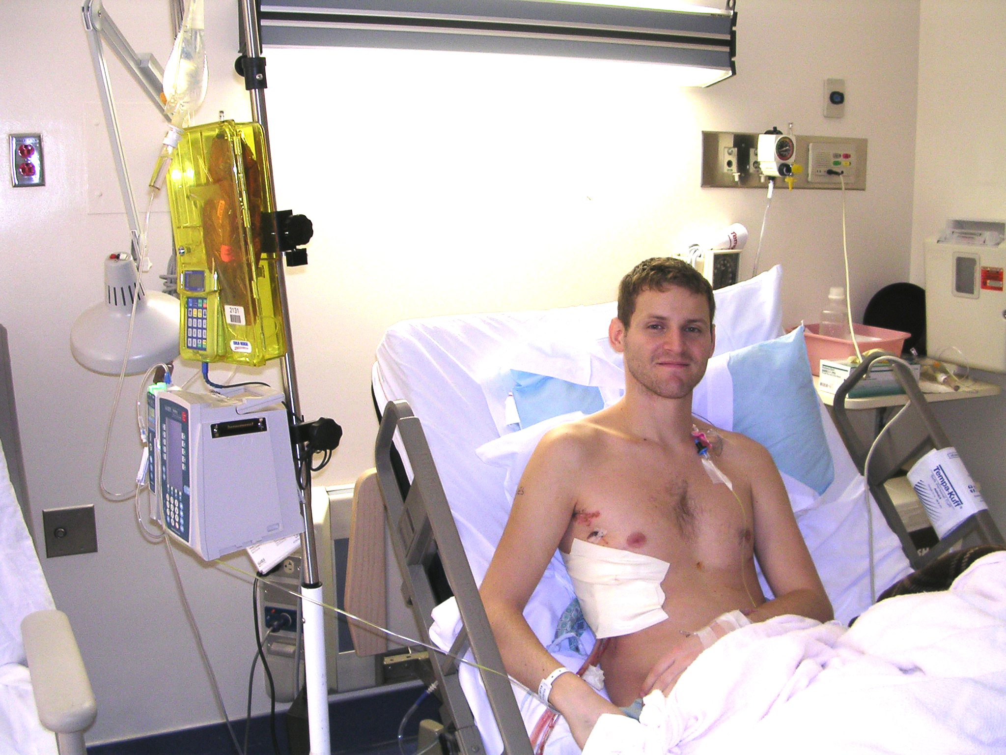
The results are, in arguably, wonderful.  Patients eating, drinking, walking, and relaxing just an hour after major lung surgery.

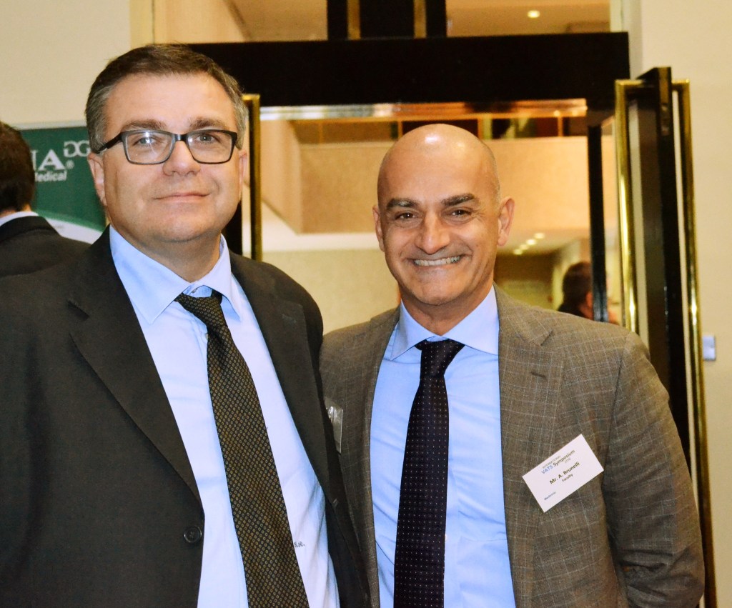
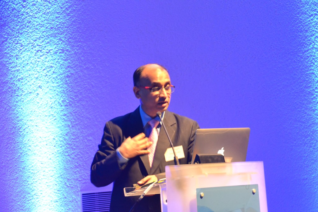
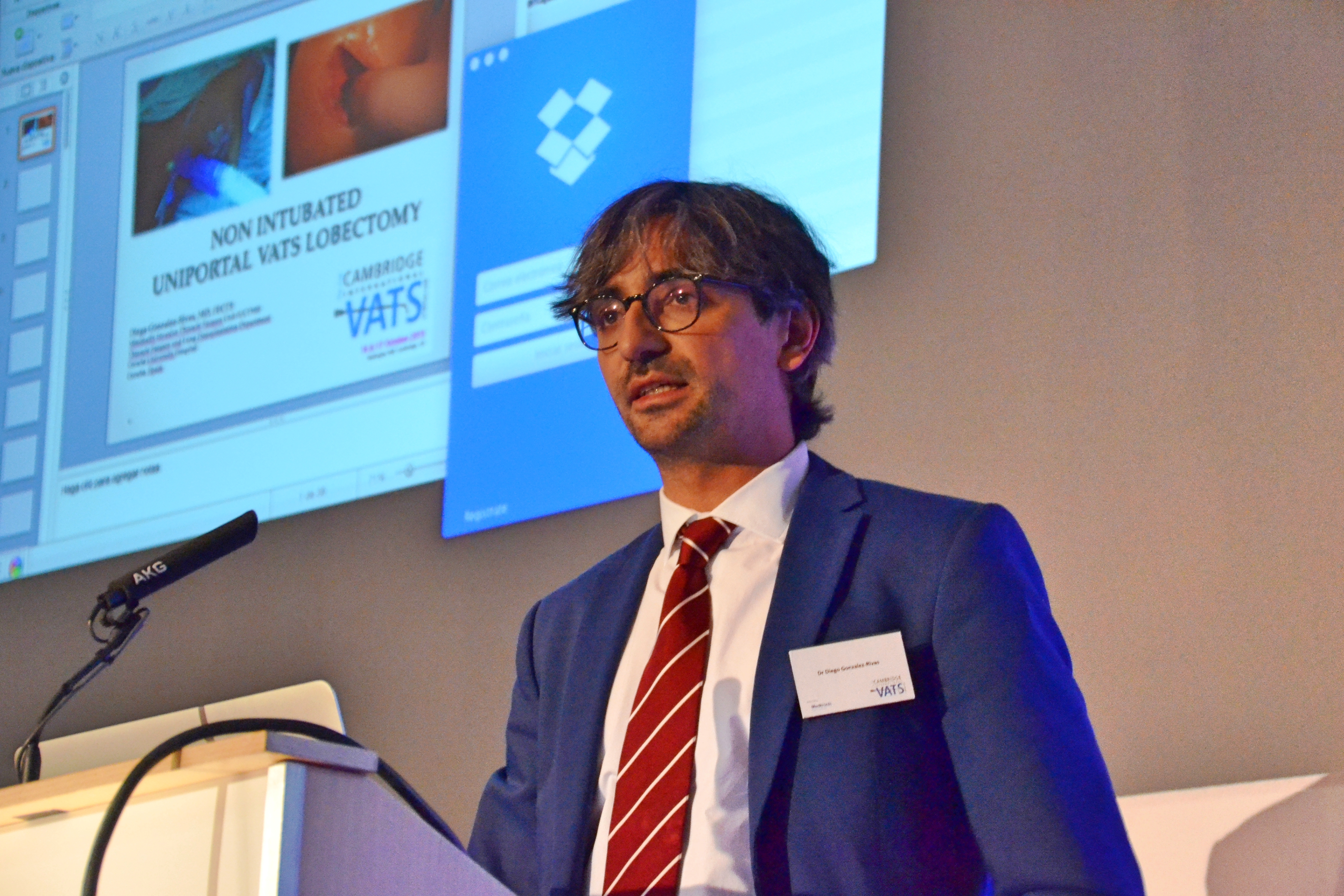
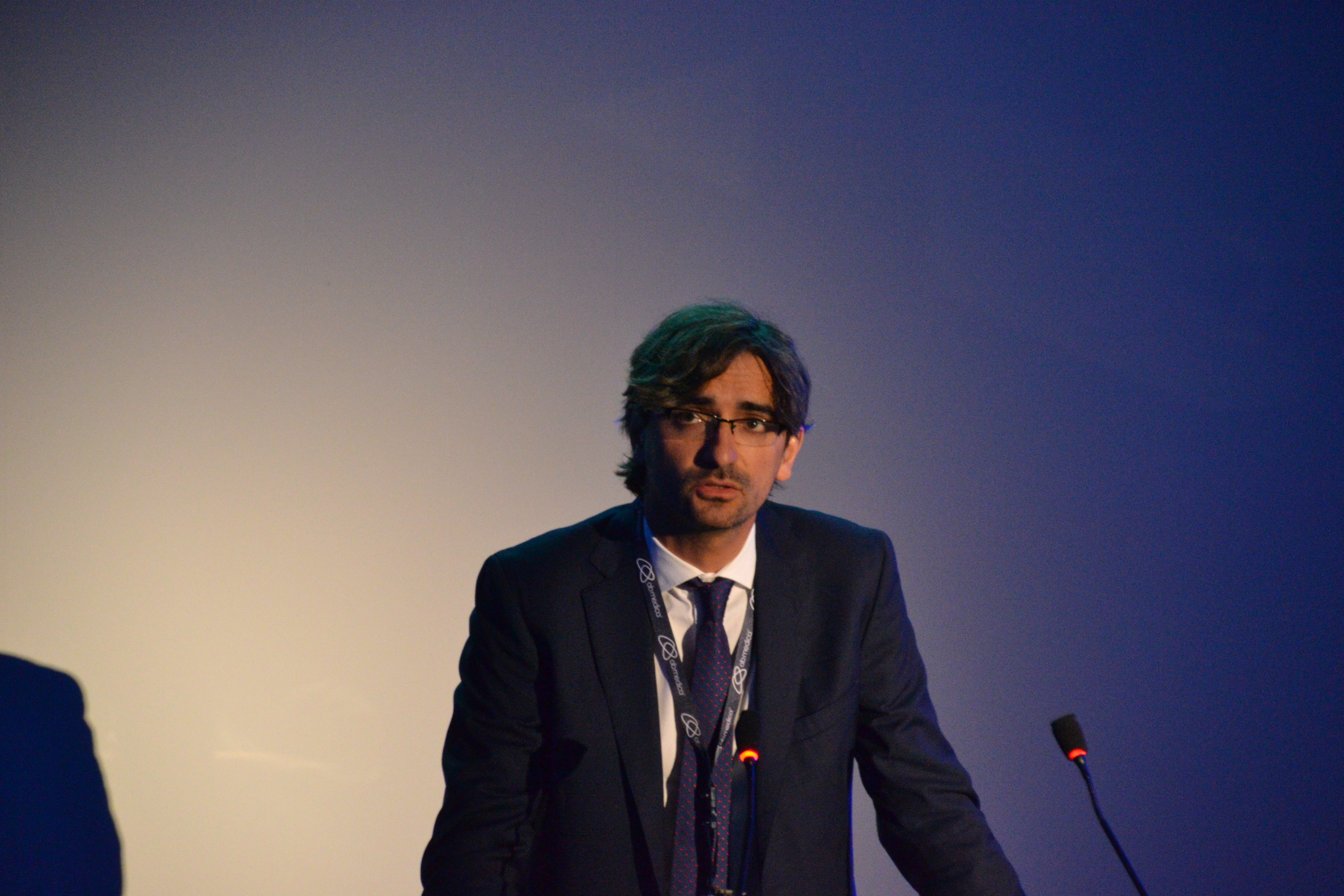
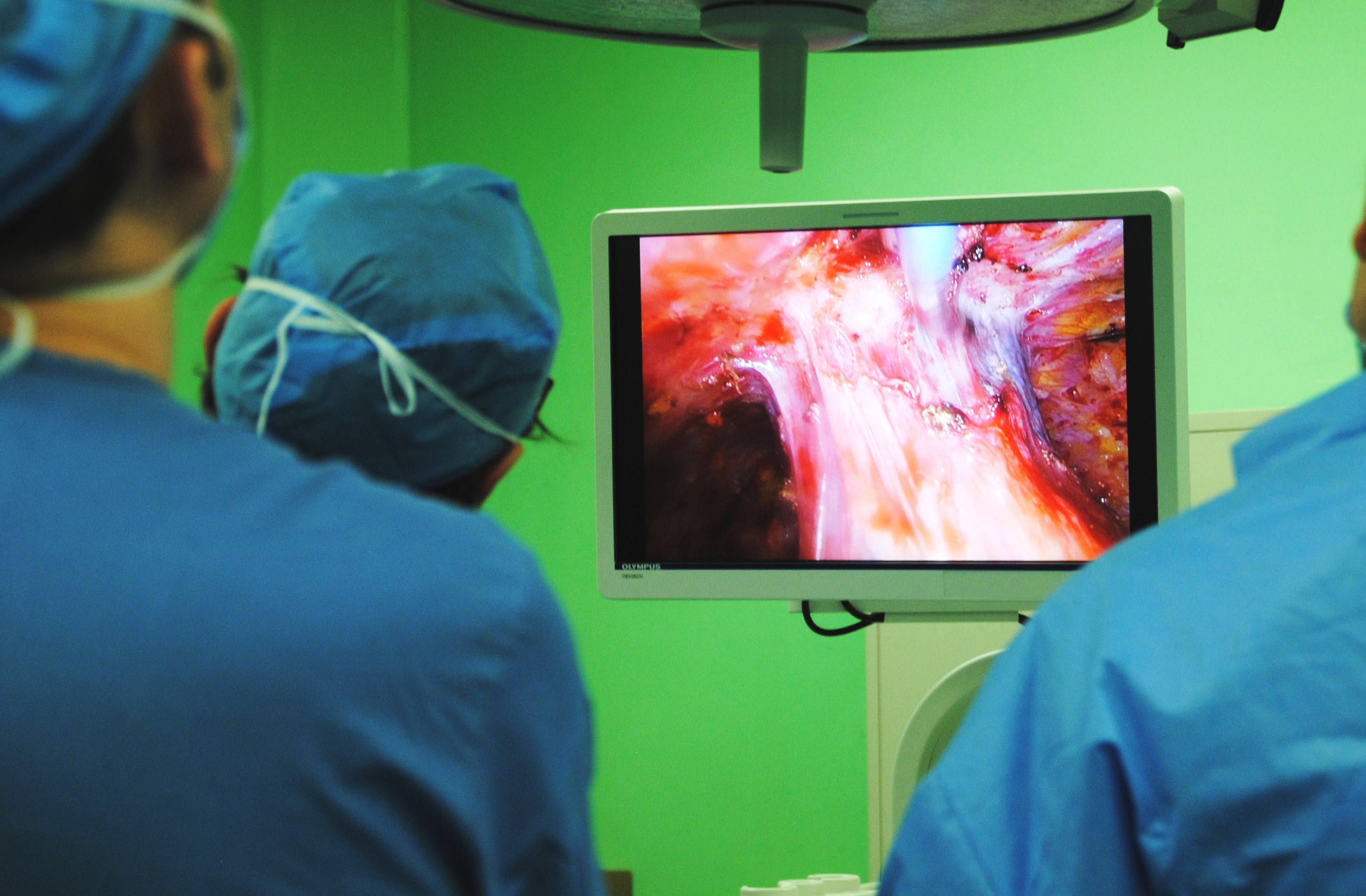
Dr. Joao Carlos Das Neves Pereira is a Brazilian thoracic surgeon, who has been the primary author and leader on several articles, and programs for what he is calling ‘extreme fast track thoracic surgery’.   He was also one of the featured speakers at International VATS 2018.  “Better than before” is his model.  “Patient empowerment’ is his ideology.

His presentation was easily the best in show, so to speak.. What’s more remarkable about his results are – that this isn’t new.  He’s been doing it for more than a decade.  In fact, he published an article on his experience in the European Journal of cardio-thoracic surgery was back in 2009.  And now he is responsible for the implementation of a multi-disciplinary program at two hospitals on two continents – one on Paris, France and the second in Sao Paulo, Brazil.

So what does he do?  How does he do it?  And why aren’t the rest of us doing it?

What he does:  “Feed & walk”

Change the existing surgical traditions:

  • no prolonged fasting
  • no cold operating rooms
  • no IV opioids

He does this with a multifaceted program that starts several weeks before surgery; with a comprehensive nutrition, smoking cessation and and an exercise regimen.

The night before:

  • Aromatherapy with lavender / Orange to promote sleep.
  • Avoid benzodiazepines

On the morning of surgery

  • patients are able to drink liquids within two hours of surgery, preventing dehydration and eliminating the need for IV fluids (no starving!)

During surgery

  • Normothermia
  • Multi-modality approach for anxiety/ nausea/ vomiting / pain
  • Opioid-free

Patient analgesia and anesthetic is treated with a combination of approaches including hypnosis, pre-emptive oral medications, BIS for awake anesthesia, minimally invasive airways.  Patients are only given very short acting medications such as ketamine, or propofol combined with local anesthesia.  By avoiding narcotics, there is a reduction in both sedation, and GI complications post-operatively.

Patients who are able to readily wake up after surgery and who haven’t had narcotics that adversely affect bowel function) are able to eat and drink immediately after surgery.

Post-operative

  • Immediate extubation (once the specimen is out of the chest)
  • Immediate feeding
  • Immediate exercise
  • “Hands free” care:  No IV lines, oral medications only, patient controlled and opioid free.

Patients are encouraged to wear their own clothing before going to the exercise room, the outside garden or walking the halls.  Post-operative pain management consists of oral medications only, and is augmented by physical therapy, acupuncture, aromatherapy and massage.  Friends and family are instructed in the proper massage techniques so that they are able to participate in the patients care (also shortage of massage therapists).  Patient recovery is enhanced by conviviality: patients don’t spend time in the rooms, alone or in bed.  Patients are welcome to spend time in open spaces, aromatherapy areas, exercise rooms, a japanese style garden, an indoor garden and a tea room.  Patients are encouraged to socialize and spend time with other patients.

While some of these ideas are novel, there is no magic surgical technique, and no miracle drug to account for these results – which are arguably better the most of ours.  But it’s not just aromatherapy, it’s a philosophy of care.

More importantly, what Dr. Das Neves Pereira and his colleagues have; that many of us find difficult to replicate – is patient buy-in.   We can call it “Patient empowerment” but it’s the part that many of us continue to struggle with.

But Dr. Das Neves Pereira’s lecture leaves us with more questions as well as answers..

Would this work for your practice?  And why aren’t the rest of us already doing it?  Will the patients accept it?

For the answer to this – we have to look at our own practices, in the here and now, in late 2018.    A recent issue of the thoracic journal of disease did just that, devoting an entire issue to ERAS (enhanced recovery and fast track programs) while providing blueprints for anesthesiologists, nursing and physical therapists.  But for many of us, the pat and simple answer is something like this:

“While most of my patients wouldn’t mind some aromatherapy or a massage after surgery, the unfortunate truth is that few would participate in a pre-operative program stressing diet and exercise.  Even fewer patients would sign on for a program that restricts narcotics.  Many of us already know this about our patient populations because we try routinely to incorporate more holistic practices into our treatment in a daily basis.  While holistic premises and alternative treatments make billions of dollars in the United States (under the guise of prevention) it’s still a culture that is highly dependent on fast, and immediate remedies and a strong belief that very little post-operative pain is acceptable or tolerable.  For every one patient that would embrace the philosophies of extreme rehabilitation, there would be another 200 screaming at the nurses for IV dilaudid.”

Much of the research actually confirms this view:

British researchers, Rogers et al. (2018) had a similar experience, noting in their recent publication that benefits of enhanced recovery protocols were dependent on compliance (and adherence) to protocols – particularly in regards to pre-operative dietary modification, and early post-operative ambulation.  Refai et al. (2018) have attempted to address these issues with a comprehensive patient education component. However, their publication does not address whether these interventions increased compliance and reduced patient stress or anxiety.

Does this mean that we are skeptical of extreme rehabno, not at all!  Interest, participation and development in fast track thoracic surgery programs continues to grow despite these obstacles.

In fact, the tightening of many federal and state restrictions on narcotics due to the American opioid crisis may make this the best time in modern American medical history to bring this ideas and approaches to our patients (Bruera & Del Fabbio, 2018, Herzid, 2018).   It also means that many of us have some preliminary hurdles and preconceived notions  (on all sides)  to overcome to engage our patients, nurses, therapists and fellow medical professionals to get their buy-in on the idea.  We might be over a decade behind – but it’s not too late to start today.

References:

Das-Neves-Pereira, et al. 2009).  Fast track rehabilitation for lung cancer lobectomy: a five year experience.  European Journal of Cardio-thoracic surgery, 36 (2009) 838-392. primary reference article.

Additional references:

Bruera, E. & Del Fabbio, E. (2018). Pain management in the era of the opioid crisis.  Am Soc Clin Oncol Educ Book 2018 May 23 (38): 807-812.

D’Andrilli, A. & Rendina, E. (2018). Enhanced recovery after surgery and fast-track in video-assisted thoracic surgery lobectomy: preoperative optimisation and care-plans.  Journal of visualized surgery, 2018:4 (4).

Herzid, S. (2018). Annals for hospitalists Inpatient Notes: Managing acute pain in the hospital in the face of the opioid crisis.  Annals of internal medicine 169(6): H02-H03.

Rogers, et. al (2018).  The impact of enhanced recovery after surgery (ERAS) protocol compliance on morbidity from resection for primary lung cancer.  Journal of thoracic and cardiovascular surgery.  155(4) April 2018: 1843 -1852.

European Society of Anaesthesiology. “Hypnosis/local anesthesia combination during surgery helps patients, reduces hospital stays, study finds.” ScienceDaily. ScienceDaily, 21 June 2011.

From the Journal of thoracic disease – special issue:  Supplemental issue #4 2018

Ardo et. al. (2018). Enhanced recovery pathways in thoracic surgery.

Bertani et al. (2018). A comprehensive protocol for physiokinesis therapy and enhanced recovery after surgery in patients undergoing video assisted thoracoscopic surgery: lobectomy.

Picconi, et. al. (2018). Enhanced recovery pathways in thoracic surgery from Italian VATS group: perioperative analgesic protocols. 

Refai et. al. (2018). Enhanced recovery after thoracic surgery: patient information and care-plans.

Featured

Act differently or watch thoracic surgery die

Dr. Eric Lim challenges thoracic surgeons to remain relevant with a call to action at the 3rd VATS International conference in London, England

“Act different or watch thoracic surgery die”

With that dramatic shot across the bow, the dynamic and forthright Mr. (Dr.) Eric Lim of Royal Brompton Hospital opened the third VATS International conference.   In a lecture entitled,  “The Why of advancing minimally invasive surgery,”  Dr. Lim put out a call to action to thoracic surgeons around the world, in an effort to remain relevant.

In an increasingly competitive world of thoracic oncology,  nonsurgical options like stereotactic radiotherapy, and the developing MRI proton beam therapy  are gaining traction for the treatment of early stage lung cancers.   These nonsurgical treatments are gaining publicity and popularity due to the efforts of radiation oncologists.

dsc_0076

The PCI of Lung Cancer Treatment

Reminding thoracic surgeons of the plight of their cardiac counterparts, Dr. Lim taunted the audience that having superior long-term outcomes does not guarantee success in a consumer-driven market.   Public and medical perception is shaped not only by clinical research findings, but by the inherent bias introduced by the authors of these publications.  As he explained, this bias, along with a public desire for simplicity, has driven the overwhelming success of percutaneous angioplasty (PCI)  and declining rates of cardiac surgery despite well-documented research studies and clear evidence demonstrating the overwhelming superiority of coronary artery bypass grafting (CABG) for long term survival.  Thoracic surgeons must not fall into the trap of complacency and arrogant belief in surgical superiority  that has plagued cardiac surgery if we want the specialty to survive.

Dr. Lim has identified three behaviors of thoracic surgeons that are harming the specialty:

  1. Refusal to look at the evidence –  thoracic surgeons must be willing to continuously review, understand and accept new clinical evidence and publications.  Evolving and emerging treatments have changed many of the cornerstones of thoracic surgery, and core concepts of 1980’s thoracic oncology management need to give way to the increasingly body of knowledge favoring VATS resections,  neo-adjuvant treatments, and improved outcomes.

An important caveat to this – is the need for Surgeon led research, and clinical trials to help eliminate the medical bias that has crept into much of the existing literature.  Surgeons need to stop allowing other specialties to control the narrative.  This is what allowed cardiologists to introduce concepts such as “non-inferiority” when research studies failed to show the benefit of cardiology interventions.

These research trials need to compare surgical interventions with non-surgical treatments.  Our inter-specialty debates over which surgical technique need to take a backseat to studies designed to compare relevant outcomes like long-term survival and cancer recurrence if we want to demonstrate surgical superiority over medical treatments.  “We need to stop arguing about which surgical technique and favor surgery over other therapies,” Dr. Lim explains.

  1. Refusal to engage with industry – industry drives and funds innovation. If we want to continue to develop wireless technologies, robotics and other innovations for use in minimally invasive surgery, thoracic surgeons must be willing to engage and participate with industry.

3.  Refusal to evolve – this is a fundamental problem plaguing thoracic surgery and addressing this issue is the underlying theme of many of the presentations at this year’s course. Dr. Lim has also addressed this refusal to evolve previously.  Surgeons need to evolve, and be willing and able to change their surgical practices based on evidence and clinical guidelines.  The failure to adopt VATS as the primary surgical approach in thoracic surgery in North America, and Europe despite decades of evidence and  clear clinical guidelines favoring this approach is a symptom of this failure to evolve.

The future of thoracic surgical oncology

How will thoracic surgery survive?  We already know that surgical excision offers the best long-term outcomes for our patients.  But as we have seen, having ‘right’ on your side isn’t enough.

Make surgery the most attractive option

For surgery to succeed, thoracic surgeons need to focus on making surgery safer and more acceptable to our patients.  Many patients prefer surgical removal on a philosophical level, but this preference is being eroded by promises of “easy” with SABER and newer chemotherapy regimens.

One of the benefits of surgery versus many of the newer treatments is that surgery is a single treatment versus multiple episodes of care.  If we can make that single encounter better for our patients, with shorter hospital stays, less pain/ less trauma and less risk, then surgery will remain the first and preferred treatment option for lung cancer.


Featured

 Dr. Hung, Dr. Chen and nonintubated and awake thoracic surgery

Dr. Ming-Hui Hung & Dr. Jin-Shing Chen at National Taiwan University Hospital talk about their work in nonintubated and awake thoracic surgery.

After attending multiple recent thoracic surgery conferences, where the topic of nonintubated thoracic surgery sparked murmurs and outspoken criticism, thoracics.org conducted a brief review of the literature to attempt to discern if this criticism and skepticism was warranted.  As part of this review, we reached out to several of the leaders in the field, including Dr. Ming-Hui Hung, a well-respected Taiwanese anesthesiologist and widely acknowledged expert on this topic.

Thoracics.org asked for Dr. Hung’s commentary as well as his response to several specific questions on nonintubated thoracic surgery.  Here is his response in it’s entirety (re-formatted to fit the Question and Answer format posed by our correspondence).

Question:  Would you tell me more about your initial research in this area.   What lessons have you learned (overall) in patient selection for non-intubated thoracic surgery?  What additional tips or advice would you offer interested thoracic surgeons/ members of anesthesia?

As we had discussed in our publications, we are facing more and more aging and frail patients with minor thoracic procedures. As surgical approach evolves toward a minimally invasive thoracoscopic technique, we expect that there would be a need for less invasive anesthetic management (i.e. nonintubated VATS) as well. Traditional intubated one-lung ventilation does offer a safe and quiet surgical environment for surgery; however, we still suffer occasionally to have patients complicated with intubation-related adverse effects, not to mention the consuming procedures for successful one-lung ventilation. Actually, there was a short-stature elderly lady complicated with pneumo-mediastinum because of tracheobronchial laceration after a double-lumen tracheal intubation. We was driven by this case we suffered to find a solution and whereas we developed our nonintubated techniques since 2009. As you noted, now nonintubated VATS is a routine part of our armamentarium for thoracic surgery.

To summarize, there are important steps that we learned from our experiences:

  1. Thoracic epidural anesthesia (TEA) vs internal intercostal nerve blocks (INB)

In the beginning, we applied TEA. It does provide satisfying analgesia but it is time-consuming and carries more risks for neurological complications. Once again, we had a nonintubated case coincidentally complicated with acute transverse myelitis after surgery. Although we excluded the epidural procedure per se, to be the direct cause of the regretful complication, we were still bothered by a legal suit against us. Then we learned that internal INB is equally effective as a thoracic epidural catheter. It saves time and risk free to do it as we do it under a direct vision by scope, and no touching on any spinal structures. Now INB is our routine part of nonintubated VATS. TEA is considered for those doing a bilateral VATS. We think this is important because it makes nonintubated VATS more safe and even more less invasive, for which our patients would accept this approach more. We Taiwanese are mostly reluctant to have someone doing anything on our spines, as we usually call them the “dragon bones”, the most important part of our bodies.

  1. Intrathoracic vagal block

Since cough reflex is a visceral part of autonomic nerve, which is not blocked by TEA or INB, unpredictable cough reflex during surgery could quietly bothering and even dangerous. We soon learned that we could block the cough reflex via intrathoracic vagal nerves. It really works. It alleviates the tension upon surgeons who working on a spontaneously breathing lung and enable them more manipularity of lung parenchyma and hilar structures. Surgeons are still needed to be as gentle as possible for that excessive traction still can trigger cough reflex from the dependent side where vagal nerve function is intact.

  1. Sedation and titration of its depth

We know there is an “awake, or not awake” issue on nonintubated VATS. We prefer to sedate our patients just because our patients do not want to be awake during surgery. Lateral decubitus position is not a confortable position. Most of our patients undergo surgery because of lung cancer or potential lung cancer. It usually takes 1-2 hours to have a diagnosis first and complete the definite treatment upon the final pathological result. We believe no one would like to be anxiously awake for the result with an open chest in an awkward position. In addition, the initial phase of iatrogenic pneumothorax would cause the patient dyspneic and tachypneic for a while, giving patient sedated with supplemental opioid is useful to alleviate the respiratory disturbances and accelerate the operated lung to collapse. By applying bispectral index EEG monitor, we can observe the BIS index increasing during the initial phase of open pneumothorax, it could be caused by inadequate analgesia, or just because of a dyspneic response. We may give the patients some more anesthetic and it usually recovered after effective vagal block. Carefully observe the respiratory pattern (from the video, or using an noninvasive end-tidal capnography) is of importance. Anesthesiologists should keep vigilant on the respiratory pattern and airway patency of the nonintubated patients, including a plan B for intubation conversion.

  1. Patient selection

We operate on spontaneous breathing lungs (most of the time, the operated lung collapses well because of positive pressure introduced into the chest cavity). The remaining opposite lung is sufficient to maintain satisfactory oxygen saturation, despite unavoidable hypoventilation. However, a vigorous diaphragm would jeopardize the balance. For surgery, it causes excessive movement of the operated lung and makes hilar manipulation dangerous or even impossible. For respiration, COrebreathing (an to-and-fro phenomenon between the dependent and the non-dependent lung) would further exacerbate the breathing pattern and decrease the alveolar oxygen fraction of the nondependent lung, leading to oxygenation desaturation. It is the most common scenario of our difficult cases and we changed to intubation conversion in some of them, especially in major resections (i.e. lobectomy) for lung cancer. We learned that obese patients tend to be an abdominal breather because of an elevated diaphragm and they are usually associated with excessive diaphragmatic movement during nonintubated surgery. Other contraindications for nonintubated VATS are also listed on the literature. We suggest that are mostly at the discretions of the caring surgeon and anesthesiologist as their good clinical practice routines.

Question:  How have your findings of your work been received internationally?  At several recent conferences, there has been a lukewarm or even critical response towards nonintubated thoracic surgery.  Is this a frequent response? 

A typical unfriendly tone from other colleagues is “just because it can be done, should it be done?” We have the same feelings as you experienced in those meeting. Nonetheless, our findings are relevant and robust that nonintubated VATS is feasible and safe in selected patients with a variety of thoracic procedures. They were published in well-known surgical journals in cardiothoracic field, including Annals of Surgery, Journal of Thoracic and Cardiovascular Surgery, Annals of Thoracic Surgery and the European Journal of Cardio-Thoracic Surgery. Still, there are surgeons and anesthesiologists enthusiastic about less invasive alternative for their caring patients visiting our hospital for nonintubated VATS, including Korea, China, Switzerland internationally and other hospitals nationally.

We believe it is human nature being anxious and doubtful to do something you do not get familiar with, especially when intubated one-lung ventilation is nearly an unbreakable only golden standard for thoracic surgery for decades, and almost all thoracic surgeons in current generations would request a fully collapsed lung to operate upon. But at this time, we are approaching a 1000 nonintubated VATS case volume, and all thoracic anesthesiologists and thoracic surgeons in our hospital are dealing with nonintubated VATS if this is appropriate for their patients. We think it is quiet a milestone in our program.

Five years ago, I asked one of my colleagues, a nursing anesthetist [emphasis mine] whether she would choose nonintubated technique if she needs a VATS procedure.

She said, “Well, I need to think about it. You better give me an double lumen even though I know how big it is.”

One year later, her answer to the same question is a “Yes, please, no tube.”

Question: Are there any other obstacles for researchers in this area?  Do you have other on-going research programs at your facility?

Obviously, nonintubated patients recover from surgery fast. They can shift to the gurney on their own from the surgical table. They experience less pain and less PONV in PACU, which enables them to recover oral intake sooner with oral analgesics and early ambulation, not to mention those common adverse effects after double lumen intubations, such as a sore throat and a change of voice quality. Currently, we are drafting our manuscripts about nonintubated VATS pulmonary resection in patients with compromised lung function. Meanwhile, a randomized trial is under investigation to compare the recovery differences of nonintubated VATS vs. intubated VATS.   There are also several more nonintubated trials in Clinicaltrial.org in different countries.

Question: Do you know of any programs that have adopted your techniques and protocols?

To our knowledge, Dr. Jianxing He from the First Affiliated Hospital of Guangzhou Medical University, Guangzhou, China, is also an enthusiastic advocator and pioneer in nonintubated VATS. He is also leading journals such as Journal of Thoracic Disease andAnnals of Translational Medicine as an open forum to accelerate the impact of nonintubated VATS. He is going to publish a state-of-the-art monograph dedicated to nonintubated VATS in the near future. We believe you can get useful information regarding nonintubated VATS in China and different perspectives from him.

As always, we keep doing our best to satisfy our patients’ need during their curing and recovery processes, just because “our patients, first.”

Ming-Hui Hung, MD, MS

Anesthesiologist, Department of Anesthesiology

Jin-Shing Chen, MD, PhD

Professor, Department of Surgery

National Taiwan University Hospital

Thoracics.org would like to thank Dr. Hung and his colleagues for their continued work in this area.  Thoracics.org would also like to thank Dr. Hung for his willingness and frank candor in addressing some of the other issues in this area.

Additional References and Resources

Ke-Cheng Chen1,3, Ya-Jung Cheng2, Ming-Hui Hung2, Yu-Ding Tseng3, Jin-Shing Chen (2012).  Nonintubated thoracoscopic lung resection: a 3-year experience with 285 cases in a single institution.  Journal of Thoracic Disease, Aug 2012, 4(4).

Hung MH, Hsu HH, Cheng YJ, Chen JS. (2014).  Nonintubated thoracoscopic surgery: state of the art and future directions. J Thorac Dis. 2014 Jan;6(1):2-9. doi: 10.3978/j.issn.2072-1439.2014.01.16. Review. (Best read in pdf form).

Liu YJ, Hung MH, Hsu HH, Chen JS, Cheng YJ. (2015).  Effects on respiration of nonintubated anesthesia in thoracoscopic surgery under spontaneous ventilation.  Ann Transl Med. 2015 May;3(8):107. doi: 10.3978/j.issn.2305-5839.2015.04.15. Review

Fast track thoracic surgery: nonintubated minimally invasive surgery for complex procedures.  October 4, 2015.

Readers: Thoracics.org has highlighted a key phrase in Dr. Hung’s response that also, unintentionally but directly addresses one of the criticisms recently advanced by a noted American thoracic surgeon who challenged Dr. Martinez as to whether he would ever delegate the care of a nonintubated patient to a nurse anesthetist.  When Dr. Martinez hesitated in his response, the surgeon claimed victory, stating, “See?  That would never work in American hospitals, [where certified nurse anesthetists oversee the majority of cases]”.  This was his rationale for dismissing this technique, even when it might make otherwise inoperable patients eligible for life-saving surgery.  That dismissal of both his American colleagues and the needs of the more fragile subset of our thoracic surgery population demonstrates some of the limitations in our so-called “masters” or “giants” of thoracic surgery.  While great, and influential surgeons, they are not infallible.  Their experiences carry wisdom, but their opinions shouldn’t carry more weight than any other published study.

Thoracics.org is committed to giving a voice and forum to all specialties and members of the thoracic surgery community.       

Featured

Fast track thoracic surgery: Non-intubated minimally invasive surgery for complex procedures

Single port thoracoscopic surgery and awake anesthesia: the micro-invasive thoracic surgery? The current research and use of these state of the art techniques to bring minimally invasive surgery to complex surgery and high risk patients.

At a recent high-profile American thoracic surgery conference, one of the speakers presented data from his program showing the safe and effective use of regional and conscious sedation techniques to perform complex thoracic surgery procedures on non-intubated patients.

Instead of being greeted with enthusiasm or professional interest, the great majority of well-known giants in thoracic surgery dismissed the idea with a few, repeated sarcastic, albeit joking remarks about the inconvenience of having conscious patients in the operating room.  This attitude seemed perplexing given the results of Pompeo et; al.’s (2014) survey of the European Society of Thoracic Surgeons, in which a large number of respondents (59%) reported using nonintubated thoracic surgery (NITS) procedures.  These mixed attitudes led thoracics.org to perform an in-depth literature search to determine the state of non-intubated thoracic surgery.

What is the current status of non-intubated thoracic surgery (and the literature surrounding it)?

Is it a wild, unsustainable idea promoted by a few dynamic but misguided surgeons?  Is it a well-researched and promising developing technique that is being rejected by surgeons who may lack vision? Or does it fall into that gray area where we suspect that this technique has real value and benefits for a special subset of patients but there isn’t quite enough high level clinical evidence to demonstrate that to the surgical community?

Is non-intubated thoracic surgery destined to fall to the same fate of VATS – a game-changing technique that emerged in the early 1990’s, has been clinically demonstrated to be superior to open surgery with an overwhelming preponderance of evidence, but still being discussed by many surgeons as the ‘new kid on the block’**?  Will people still debate the merits of non-intubated surgery ad infinitude thirty years from now, even when clinical guidelines have made it the standard of care (like VATS and oncology surgery)?   Will there be the same reluctance to set firm standards for training in these techniques?

“Not a new concept”

photo courtesy of the US Army

As it turns out – non-intubated thoracic surgery is not a new idea or concept.  It was developed early in the 20th century and was used successfully for many years for even the most complex thoracic cases such as esophagectomies until the development of double lumen intubation in the 1950’s made the use of single lung ventilation possible (Gonzalez-Rivas et. al. 2015, Pompeo 2015, Kiss & Castillo 2015).  Since its rediscovery in the last several years, many of the problems that plagued this technique during its inception over a century ago have been addressed through better understanding of human physiology.  Now, this seemingly fringe technique has been shown to be a feasible approach for treating the very margins of the thoracic surgery population (the extreme elderly, patients with advanced respiratory disease or other serious medical co-morbidities) that are often deemed inoperable using current techniques.

The dreaded complication (spontaneous pneumothorax) of early use of this technique by pioneers in thoracic surgery has now become one of the main advantages.  Surgically created pneumothorax results in almost perfect deflation of the operative lung, achieving better results than even the most experienced of anesthesiologists using traditional single lung ventilation.  Surprisingly to many observers, instead of creating a ventilatory emergency, this process is readily tolerated by most patients, even those with poor baseline pulmonary function (David, Pompeo, Fabbi & Dauri, 2015).

Current research

The majority of the current series of research on this topic are being performed by a small group of surgeons which includes Dr. Diego Gonzalez Rivas (Spain), Dr. Eugenio Pompeo and the Awake Thoracic Surgery Research Group in Italy and Dr. Ming-Hui Hung and colleagues (Taiwan).  Pompeo’s group (Drs Benedetto Cristino, Augusto Orlandi, Umberto Tarantino, Tiziana Frittelli (General Director of the Policlinico Tor Vergata), Leonardo Palombi, Paola Rogliani, Roberto Massa, Mario Dauri) has been especially prolific in 2015 after several of their works were published in a special issue of Annals of Translational Medicine.

In multiple studies, these researchers have reported successful thoracic surgery outcomes in non-intubated patients, thus eliminating the majority of risks related to general anesthesia as well as uni-lung ventilation via mechanical ventilation and intubation.  In several of these studies, the authors were also able to successfully perform these surgeries in fully awake patients (versus consciously sedated), making surgery possible for even the frailest of candidates.  These studies included a small number of comparisons between traditional and non-intubated surgeries.  While the numbers of patients enrolled have been small, and there are few randomized studies, the results have been encouraging.

Chen et. al

Chen et. al’s 2012 study has been one of the largest studies to date, with 285 cases.  In this study, patients underwent lobectomies, wedge resections and segmentectomies with 4.9% requiring conversion with tracheal intubation.  Lung resection was undertaken with traditional (3 port) VATS or a needlescopic approach.

The authors report the biggest problem they encountered was increased bronchial tone and airway hyper-reactivity during manipulation of the pulmonary hilum during lobectomies and segmentectomies.  This was effectively treated without significant alteration in hemodynamics via intrathoracic vagal blockage which eliminated the cough reflex in these patients.

The authors caution judicious patient selection to prevent emergent conversion (intubation) particularly while surgeons are initially attempting this technique.  Chen et al. also believe that non-intubated thoracic surgery is best suited for petite or small-sized female patients because the small tracheal size of these patients predisposes them to a higher rate of complications and potential tracheal injury with traditional surgery and intubation.

Awake anesthesia and lung volume reduction surgery

Pompeo et. al’s review of the literature surrounding of the use of this technique in patients with severe emphysema undergoing nonresectional lung volume reduction surgery (LVRS by awake anesthesia) showed significant treatment advantages for patients undergoing lung volume reduction surgery without intubation or administration of general anesthesia.

With an average mortality of 5% and a morbidity of 59% for traditional lung volume reduction surgery as reported during the National Emphysema Treatment Trial, findings from Tacconi et al.’s 2009 study of 66 LVRS awake patients (matched with 66 patients undergoing traditional surgery) appears promising.  The authors report a reduced incidence of prolonged air leaks (18%) versus 40% in the traditional surgical group as well as a decreased length of stay.  In this study, 3 patients required conversion to general anesthesia – one patient due to an elevated paCo2 of 83% and the remaining two patients for anxiety attacks.

Rate of intubation/ respiratory failure/ mortality in Tacconi et al.

Mortality in both non-intubated and the traditional surgical group was the same, with one patient from each group.  In both cases, the patients had developed massive airleaks following surgery.   In the non-intubated group, the patient developed acute lung injury requiring intubation of POD#12 and died POD#38.

In the traditional surgical group, 4 patients were unable to be extubated at the end of the case, with one patient requiring an additional day of mechanical ventilation.   Another patient was reintubated on POD#3 for respiratory failure and died on POD#67.

Pompeo et. al, over the course of over eleven years, have also investigated the use of non-intubated (and awake) thoracic surgery for a wide variety of cases including urgent /emergent cases, wedge resections, decortications, talc pleurodesis as well as nonintubated anesthesia combined with single (uniportal) thoracoscopic approaches (aka “microinvasive thoracic surgery”).

Anesthesia for non-intubated thoracic surgery

The role of anesthesiologists in caring for patients undergoing non-intubated or awake thoracic surgery is more challenging than general anesthesia.  While thoracic anesthesia already requires specialized skills for initiating, managing and maintaining uni-lung ventilation, the switch to non-intubated patients with either localized anesthesia or conscious sedation adds a new set of complexity to managing these often frail patients.  Kiss & Castillo (2015) in their review of the literature, provide an excellent overview of the pros and cons of non-intubated anesthesia as well as guidelines for patient selection and eligibility criteria for use of this technique.  Special populations who may benefit from this technique include patients with severe respiratory disease (and a high risk of ventilator dependency with intubation), patients with severe but stable dyspnea, or multiple cardiovascular and respiratory co-morbidities.

Kiss et. al also reviews the contraindications to use of this technique including: phrenic nerve paralysis on the non-operative side, patients at risk for difficult intubation, or patients who are unwilling to undergo awake thoracic surgery.  Wang & Ge (2014) expand on these complications to include ASA status 4 or higher, bleeding disorders, decompensated heart failure, extreme obesity, unfavorable airway or spinal anatomy as well as specific respiratory conditions including bronchiestasis, asthma, sleep apnea, clinically significant sputum production and strict contralateral lung isolation.

Wang & Ge also give specific anesthesia dosing guidelines for  conscious sedation, local anesthesia and regional blocks in additional to monitoring parameters.

Alterations in oxygenation and ventilation

David et. al. (2015) describe the pathophysiology and alterations in oxygenation and ventilation in surgical pneumothorax including hypercapnia, hypoxia and the associated hypoxic pulmonary vasoconstriction that occurs along with the development of  intrapulmonary shunt as the deflated (and unventilated lung) maintains perfusion.  The authors also explain how this effect can be either exacerbated or minimized thru the choice of anesthetic agents,  and the administration of supplemental oxygen, which further demonstrates the importance of involving the thoracic anesthesia team in preparation for non-intubated cases.

The relationship between ventilation and perfusion. (A) Relationship between ventilation (roundes) and perfusion (rectangles) in different lung zones, in upright (A1) and lateral position (A2); (B) relationship between ventilation (roundes) and perfusion (rectangles) in lateral position with surgical pneumothorax, during spontaneous inspiration (B1) and exhalation phase (B2). Black arrows show paradoxical ventilation and mediastinal shift. (Illustration and caption from David et. al, 2015)

This “permissive hypercapnia” has been reported in multiple articles as having minimal to no clinical effects and is easily treated with supplemental oxygen by nasal cannula or facemask.

Editor’s note: In advance of this article, Dr. Pompeo, Dr. Gonzalez Rivas and Dr. Min-Hui Hung were contacted for their additional comments and insights on non-intubated thoracic surgery. This and subsequent articles may be augmented, as applicable with their replies.

Conclusion

Should we really abandon pursuit of better patient outcomes, faster mobility, recovery and reduced length of stay in lieu of the security to tell off-color jokes with our patients safely under general anesthesia?  Should we abandon all hope in treating patients previously deemed inoperable due to our own fears and hesitations to embrace newer techniques and procedures?

Or as Mineo et al, suggests, should we enlist our colleagues to design and devise several large scale studies at multiple institutions so that we can move to the next level of investigation and answer the question: “Should my patient be awake for this?”

References

Ke-Cheng Chen1,3, Ya-Jung Cheng2, Ming-Hui Hung2, Yu-Ding Tseng3, Jin-Shing Chen (2012).  Nonintubated thoracoscopic lung resection: a 3-year experience with 285 cases in a single institution.  Journal of Thoracic Disease, Aug 2012, 4(4).

David P, Pompeo E, Fabbi E, Dauri M. (2015). Surgical pneumothorax under spontaneous ventilation-effect on oxygenation and ventilation. Ann Transl Med. 2015 May;3(8):106. doi: 10.3978/j.issn.2305-5839.2015.03.53. Review.

Gonzalez-Rivas D1, Bonome C2, Fieira E3, Aymerich H4, Fernandez R5, Delgado M3, Mendez L3, de la Torre M5. (2015).  Non-intubated video-assisted thoracoscopic lung resections: the future of thoracic surgery?  Eur J Cardiothorac Surg. 2015 Apr 19. pii: ezv136.  article requires subscription, no free full-text available.  Nice discussion of the risks of traditional anesthesia and mechanical ventilation in thoracic patients.

Gonzalez-Rivas D1, Fernandez R2, de la Torre M2, Rodriguez JL3, Fontan L4, Molina F4. (2014).  Single-port thoracoscopic lobectomy in a nonintubated patient: the least invasive procedure for major lung resection? Interact Cardiovasc Thorac Surg. 2014 Oct;19(4):552-5. doi: 10.1093/icvts/ivu209. Epub 2014 Jul 7.

Kiss G, Castillo M. (2015).  Nonintubated anesthesia in thoracic surgery: general issues.  Ann Transl Med. 2015 May;3(8):110. doi: 10.3978/j.issn.2305-5839.2015.04.21. Review.

Hung MH, Hsu HH, Cheng YJ, Chen JS. (2014).  Nonintubated thoracoscopic surgery: state of the art and future directions. J Thorac Dis. 2014 Jan;6(1):2-9. doi: 10.3978/j.issn.2072-1439.2014.01.16. Review. (Best read in pdf form).

Liu YJ, Hung MH, Hsu HH, Chen JS, Cheng YJ. (2015).  Effects on respiration of nonintubated anesthesia in thoracoscopic surgery under spontaneous ventilation.  Ann Transl Med. 2015 May;3(8):107. doi: 10.3978/j.issn.2305-5839.2015.04.15. Review

Mineo TC, Tacconi F. (2014). Nonintubated thoracic surgery: a lead role or just a walk on part? Chin J Cancer Res. 2014 Oct;26(5):507-10. doi: 10.3978/j.issn.1000-9604.2014.08.11. No abstract available.  Very enjoyable, almost conversational article with the authors sharing their experiences with non-intubated thoracic surgery while calling for larger clinical research studies on the topic.

Noda, M., Okada, Y., Maeda, S., Sado, T., Sakurada, A., Hoshikawa, Y. et al. (2012).   Is there a benefit of awake thoracoscopic surgery in patients with secondary spontaneous pneumothorax?. J Thorac Cardiovasc Surg. 2012; 143: 613–616

Pompeo E, Cristino B, Rogliani P, Dauri M; Awake Thoracic Surgery Research Group (ATSRG). (2015).  Urgent awake thoracoscopic treatment of retained haemothorax associated with respiratory failure.  Ann Transl Med. 2015 May;3(8):112. doi: 10.3978/j.issn.2305-5839.2015.04.13.  Authors review their experiences treating emergent and trauma patients with awake, nonintubated VATS.

Pompeo E, Sorge R, Akopov A, Congregado M, Grodzki T; ESTS Non-intubated Thoracic Surgery Working Group. (2015).  Non-intubated thoracic surgery-A survey from the European Society of Thoracic Surgeons. Ann Transl Med. 2015 Mar;3(3):37. doi: 10.3978/j.issn.2305-5839.2015.01.34.

Pompeo, E. (2015).  Non-intubated thoracic surgery: nostalgic or reasonable?  Annals of Translational Medicine, 2015; 3(8): 99.  Review of the historical development on non-intubated thoracic surgery  and techniques in regional anesthesia for complicated thoracic surgery procedures including esophagectomies in the era predating the development of double lumen intubated and unilung ventilation.  A timely reminder that some of the greatest developments in medicine and surgery are ‘rediscoveries’ of our predecessors.

Pompeo E. (2014).  State of the art and perspectives in non-intubated thoracic surgeryAnn Transl Med. 2014 Nov;2(11):106. doi: 10.3978/j.issn.2305-5839.2014.10.01.  Nicely written article.  Has link to video presentation for purchase.

Pompeo, E. (2014).  Non-intubated vodeo-assisted thoracic surgery under epidural anesthesia – encouraging early results encourage randomized trials.  Chinese Journal of Cancer Research 2014, 26(4); 364-367.

Pompeo E1, Rogliani P1, Palombi L1, Orlandi A1, Cristino B1, Dauri M1; Awake Thoracic Surgery Research Group (ATSRG). (2015).  The complex care of severe emphysema: role of awake lung volume reduction surgery.  Ann Transl Med. 2015 May;3(8):108. doi: 10.3978/j.issn.2305-5839.2015.04.17. Related table:  Lung Volume Reduction Surgery Criteria – Awake Anesthesia

Pompeo E, Dauri M; Awake Thoracic Surgery Research Group (2014).  Is there any benefit in using awake anesthesia with thoracic epidural in thoracoscopic talc pleurodesis? J Thorac Cardiovasc Surg. 2013 Aug;146(2):495-7.e1. doi: 10.1016/j.jtcvs.2013.03.038. Epub 2013 Apr 17. No abstract available.

Pompeo E; Awake Thoracic Surgery Research Group (2012). To be awake, or not to be awake, that is the questionJ Thorac Cardiovasc Surg. 2012 Jul;144(1):281-2; author reply 282. doi: 10.1016/j.jtcvs.2012.01.083. No abstract available.  Comment on article by Noda et. al.

Pompeo E, Mineo D, Rogliani P, Sabato AF, Mineo TC (2004).  Feasibility and results of awake thoracoscopic resection of solitary pulmonary nodules.  Ann Thorac Surg. 2004 Nov;78(5):1761-8.  One of the earlier modern publications on awake thoracic surgery.

Tacconi F, Pompeo E, Mineo TC. (2009). Duration of air leak is reduced after awake nonresectional lung volume reduction surgery. Eur J Cardiothorac Surg 2009;35:822-8; discussion 828.

Wang B, Ge S. (2014).  Nonintubated anesthesia for thoracic surgery. J Thorac Dis. 2014 Dec;6(12):1868-74. doi: 10.3978/j.issn.2072-1439.2014.11.39. Review.   Related table: the advantages and disadvantages of nonintubated anesthesia for thoracic surgery.

Note: This is not an exhaustive list of literature available on this topic but a select listing of the most recent and relevant citations (and are available as free full text).

**Long time readers of thoracics.org may have noticed that we no long cover or report on ‘debates’ or discussions as to whether VATS can be used in oncology cases, or whether an adequate lymph node dissection can be performed using VATS.  The literature clearly demonstrates that it can – and clinical guidelines reflect this, making the discussion one-sided, tedious, out-dated and repetitious.

Featured

Interview with the Brazilian Society of Thoracic Surgery

Thoracics.org talks to the Brazilian Society of Thoracic Surgery and result isn’t what you might expect.

A very different article here at Thoracics.org!  For starters, I’m the interviewee – which doesn’t happen very often.  This interview was a joint collaboration after meeting and talking about issues in thoracic surgery with several Brazilian surgeons including Dr. Sergio Tadeu Pereira, at the ALAT conference in Medellin last July.

at ALAT with Bolivian surgeon, Dr. Cristian Anuz

Translation of interview from the December 2014 Journal of the Brazilian Society of Thoracic Surgery*:

The positive outcome of a thoracic surgery depends on several aspects, among them is the teamwork, the harmony between the various professionals involved in making decisions and actions. All experts have an instrumental part in restoring the health and maintenance the patient’s life. Each with its due importance, towards a single goal. The SBCT ratifies such thinking, and this issue of the Journal conducted an interview with K. Eckland, an acute care nurse practitioner in thoracic surgery, writer, and also the founder of Thoracics.org” – a blog about thoracic surgery with an international focus.  She has written several books on surgery in Latin America, including a community sociological examination thoracic surgery in Bogotá, Colombia.

In this conversation, K. Eckland talks about the future of thoracic surgery worldwide and recognizes the contribution of Brazilian surgeons for growth in the art.

Journal SBCT: For us at SBCT is a great pleasure to have their participation in our newspaper. How do you evaluate the specialty in Brazil?
K. Eckland: First, I would like to thank the editors this paper for the invitation to forward my message to Brazilian doctors. More importantly, I would like to serve as call to action to all the experts and future thoracic surgeons. When I look at Brazil, I see the future of thoracic surgery. While, in my own country, our thoracic surgeons are aging with an average age of 60 years, Brazil is full of young, dynamic and innovative surgeons.

Journal SBCT: This predisposition to new techniques of many the Brazilian thoracic surgeons implies an increase in research?

K. Eckland: The high fluency in minimally invasive techniques (in Brazil) combined with some of the largest academic and clinical settings worldwide, outside China, affords unique opportunities in research, development and discovery. Brazil is already home to many of the modern masters of thoracic surgery, names that resonate worldwide.

These surgeons have brought Brazil to the forefront, but it is up to the newest crop of thoracic surgeons to maintain Brazil’s forward momentum for the future.  However, this is hampered by a lack of awareness of the contributions of many Brazilian and other Latin American surgeons.

As a foreigner, writing about developments within the international surgical community, I have noted a large dearth in published research from much of Latin America including Brazil.  What research I do find, is often not widely dispersed or readily available to the rest of the world.  It has taken several years and many thousands of dollars for Cirugia de Torax to acquire and publish information about your many triumphs.  However, this is not the most efficient way for research to be disseminated.

Journal SBCT: In addition to increased investment in research, what more should be done in its assessment to mitigate this  gap in publications and contributions (to the specialty of thoracic surgery)?

K. Eckland: It’s possible to change this story from one of limited international exposure to greater recognition.  But for that to happen, several things need to occur. Firstly, the momentum must be Brazilian thoracic community to participate  and publish research on a large-scale.

Surgeons in São Paulo, for example, have unique opportunities to publish practice-changing work. The Department of Thoracic Surgery, University of São Paulo tracks more thoracic cases in a year than many American institutions have access in a decade. This gives greater impact to studies from this institution than anything that their (North) American colleagues could expect do.

Second, Brazilian surgeons need push for further publication in international journals, and in the international literature language, in English.

Lastly, surgeons need to look outside their corner of the globe and present their findings internationally and outside Latin America on a greater scale.  More groups of Brazilian surgeons should attend international conferences to gain knowledge,and take the opportunity to spread their own knowledge and research findings. Surgeons should not depend on the United States and Europe to take the lead in surgical innovation or research.

There is no reason why these findings will not occur at home, but research needs to be part of your daily practice. It should be more than reading the occasional surgical journal.  It should be a part of active problem solving and solution-seeking.

Journal SBCT: The wide practical experience associated with the host new techniques can be considered as a basis for the growth of the specialty and development more innovative research in Brazil?

K. Eckland:

For this to happen, each surgeon needs ask yourself**:

–  How can I improve my practice? – How can I improve the lives of my patients?

– What can I do to identify and document the phenomena I’m seeing?  – What we are doing now that we need to change? How can we implement these changes? How does this apply to people outside my immediate environment?

Once a potential search area is identified, other questions to ask include:

How I can improve my specialty? How can I represent my country to the world? Sometimes the answers
involve the development of new technologies, sometimes a reframing of the information we already know,
to apply the new clinical scenarios. Other times, we simply need to identify the phenomena and document it to serve as guidance to other professionals. That is what drives the research, and this combined insight with professional curiosity are essential for growth within the specialty. By embracing these concepts, we can begin a new era of thoracic surgery in Brazil and worldwide.

* Corrections to the English translation have been made for ease of reading.  This is an excerpt from a larger interview.

** This is how we identify research to discuss and publish here at Thoracics.org/ Cirugiadetorax.org

For the original article, click here.

XIX Congresso Brasileiro de Cirurgia Torácica

ikone

If you are interested in learning more about the latest research and developments in thoracic surgery in Brazil, the annual conference is this May.

Featured

Welcome to Shanghai Pulmonary Hospital

Shanghai Pulmonary Hospital, largest thoracic surgery center in the world

Shanghai Pulmonary Hospital – Shanghai, China

Shanghai Pulmonary Hospital is a dream come true for a thoracic surgery aficionado like myself.  Twelve operating rooms, a 30 ICU beds, 30 to 40 operations per day and over 40 staff surgeons means that there is always something interesting going on down the hall.

Am case presentations is like a review of Robbins’ pathology.  Bilateral nodules, ground glass opacity nodules, giant-sized tumors, mediastinal masses of all types and atypical presentations abound.   By tradition, all tuberculosis cases must come to the medical complex at Shanghai Pulmonary Hospital because they have a designated tuberculosis hospital on site.  Even with other facilities competing for some of the pathology, there is plenty to go around, and I am able to see a couple of lung abscesses as well as several varieties of cavitary lesions.  I am sure that there is still a wealth of untapped pathology for me to explore, but I suspect that more interesting infectious cases and occupational diseases are probably confined to the more distant provinces.

I briefly talk to one of the surgeons here, who is from Kashdar region, on the far western side of China.  Kashdar is located in one of the more mountainous regions of China, which was part of the famous Silk Road trade route explored by the likes of Marco Polo more than half a millennia ago.  We discuss the region and compare it to its American counterpart of West Virginia.  It’s not a perfect analogy but there are enough similarities to help me for a picture of life, and industry there.  That is where the mines are concentrated, and that is where I might find the black lung disease, the silicosis and similar type diseases, though the surgeon I speak with reports that the rates of occupational disease for this occupation to be quite low.  Given the dangerousness of underground mining, I wonder if many of the miners worry about living long enough to see a chronic disease like black lung.  I don’t know enough about China to ask a lot of the other interesting questions that are swarming in my mind, but I wonder about mesotheliomas and other diseases related to all the heavy industry that forms the backbone of the booming Chinese economy.  I wonder about the prevalence of empyemas given the pollution levels and the closeness in which many people are forced to live.  It seems like it would be a daily surgical feast, but I don’t know a polite way to ask directions to the hospital with the pus-filled buffet.

My hosts also tell me regretfully that they also only see a limited amount of esophageal cancer because many people are misled by the name of the facility, and are unaware that esophagectomies are performed here.  One of the surgeons looks so woe-begotten as he tells me this that I know he has the same love of that surgery  as I do – that feeling of joy when surgical planning, pre-operative optimization, surgical skill and aggressive post-operative care come together flawlessly for an uncomplicated post-operative course in a complex case.   It’s not just satisfaction with a job well-done but pure joy at seeing our patients walk out of the hospital and back to the regular lives.

I am here at part of the Uniportal VATS conference with Dr. Diego Gonzalez, but it’s also an opportunity to literally wander the operating rooms at will,  listen to case discussions and interview surgeons as I encounter them.  I always joke about feeling like a kid in a candy store, now I really am.  If I hear a particularly interesting case during am rounds, I am welcome to come into the operating room, watch the case, listen to the discussions and talk to the surgeons.

Attendees of the uni-portal VATS conference

As one of the largest general thoracic surgery departments in the world*, it would be impossible for me to know and present all  staff surgeons in the few days that we are here for the Uni-portal Surgery conference.  Instead I would like to highlight a just a few of the interesting and talented surgeons at this busy facility.

Dr. Jiang Gening – Chief of Thoracic Surgery

Dr. Jiang is the head of one of the world’s largest thoracic surgery services, but it doesn’t seem to faze him.  Then again, he’s been here at Shanghai Pulmonary Hospital (SPH) since he came here to train in 1982.  At that time, the thoracic surgery department was just a fraction of the size it is today.

As resident, staff surgeon, and then now Chief for the last ten years, Dr. Jiang has seen numerous changes, many of which have occurred in just the last few years.  Volumes have dramatically increased, resulting in annual hospital construction to expand the operating rooms.  A  16 bed thoracic surgery department has grown to over 250 beds.

Dr. HaiFeng Wang with Dr. Jiang Gening (right)

Dr. Jiang has a strong vision of where his hospital ranks in the world, and where he would like it to be.  He sees a strong future for this facility as an international leader in thoracic surgery and clinical research, and he has been working aggressively towards these aims.

Dr. Jiang has trained with Dr. Patterson (Bowman Grey, St. Louis) and other leaders in thoracic surgery in Boston and Los Angeles, and he encourages his surgeons to do the same.  He strongly supports surgical development among his staff such as bringing leading surgeons like Dr. Diego Gonzalez Rivas to train staff in the most up-to-date procedures.

As a surgeon himself, he enjoys the more complex cases, the larger surgeries for the challenges they bring.  When I mention, ‘chest wall resection,’ he smiles and nods before Dr. Wang can translate.

He is also very interested in expanding the lung transplant program but acknowledges that his facility has  difficulties in obtaining donors now that China has discontinued the policy of using incarcerated people for organ donation.  Organ procurement has been complicated by the traditionally low rates of voluntary donation in Chinese societies.  As Dr. Jiang explains, Chinese culture and many Chinese families has a hard time recognizing and reconciling with the concept “brain death” in the absence of physical death.  This means that Dr. Jiang and his program are focusing on donation after cardiac death and ex-vivo implantation.  But this too is problematic – the scarcity of organs means that despite being in a metropolitan area of almost 30 million, surgeons may have to travel to Beijing or other locations for available organs.  Often on arrival, these organs are not in suitable condition for transplantation.  Another problem is the reluctance of Chinese insurance companies and third-party payers to cover the cost of ex-vivo support. Dr. Jiang acknowledges that his facility has several large hurdles to overcome if Shanghai Pulmonary Hospital is to become the transplant center for Singapore, Korea and the rest of Asia, as he envisions.

I am hoping to find my way into Dr. Jiang’s operating room.  I have been advised by the Taiwanese surgeons that Dr. Jiang is widely-known and admired for his ‘nimble fingers’ so I want a chance to see him in action.

Dr. HaiFeng Wang

Dr. Haifeng Wang, thoracic surgeon

Dr. HaiFeng Wang is a very nice 41 year old surgeon who assisted in translating during the interview with Dr. Jiang.  It was strange, as soon as I started talking to Dr. Wang, it was like we recognized each other.  I immediately knew that we would see eye to eye.  And so it was, as he presented his daily cases, and we discussed the findings, the planned surgeons and related research.   So it seemed natural for me to spend the day with Dr. Wang in the operating room.

Dr. Wang in the operating room

Like Dr. Jiang, Dr. Wang is from Shanghai.  After completing a combined medical school and internship program, (with training in urology), Dr. Wang decided to switch to thoracic surgery (a decision that cirugia de torax wholeheartedly supports).

After receiving the World Health Organization fellowship, he traveled to Australia to train with Dr. Peter Clarke at Austin Hospital in Victoria.  He focused his studies on the surgical control of cancer.  More recently, in 2013, he received the Graham fellowship (from AATS) to study with Dr. Mathisen in Boston, Massachusetts.  He also spent one month with Dr. McKenna in Los Angeles and another month with Dr. Patterson in St. Louis.

His areas of interest include minimally invasive surgery, tracheal surgery, lung transplantation and the diagnosis and treatment of ground glass opacities.

HIs first surgery of the day is an asymptomatic middle-aged patient with an incidental finding[1] of a large bronchiogenic cyst in the right middle lobe.

On the CT scan, it looks like an egg-yolk with solid material within a fluid-based cyst.  The initial suspicion is a possible aspergilloma but this presumptive diagnosis is eliminated during surgery based on tumor appearance.

egg yolk appearance

Frozen section is requested intra-operatively but the results of that only deepen the mystery when a diagnosis of a possible sarcoma is suggested by the pathologist.  The resection is completed quickly, but the mysterious aspect of the case has me intrigued as we wait for the final pathology.

Update:  Final pathology completed 3/17/2015.  The report says pulmonary lymphangioma, a rare disease in the lung,  and the pathologist says that the cyst itself is actually the lymphangioma, not only the small nodules we see inside the cyst wall.

The second case is a young patient with a GGO (ground glass opacity) in the right upper lobe.  A needle biopsy confirms that the mass is a malignancy, an adenocarcinoma.  This surgery is also smooth and uneventful.

There is a third case still scheduled, (and interesting too!) but unfortunately, it’s time for me to race back to the hotel, do some writing before I go to sleep to get ready to do this all over again.

I’ll be here in China for three weeks, so this isn’t the last you will hear about Thoracic surgery in China or Shanghai Pulmonary Hospital.

[1] In China, a CT scan is a fairly affordable diagnostic tool ($40) for most middle class Chinese citizens.  Healthcare among certain classes, is also consumerized to a degree that the United States is only beginning to approach.  This means that many Chinese residents have CT scans with the same gravitas that a many of us may approach a new hairstyle, or similar type purchase.

* noncardiac.  There may be larger combined CTS departments.

Note:  this article has been edited for corrections due to translational and other inaccuracies.

Featured

Dr. Ahmet F. Işık talks about pleural mesothelioma, HITHOC, and thoracic surgery in Gaziantep, Turkey

updates on the on-going HITHOC project, war surgery, foreign body obstructions and bronchoscopy for infants

DSC_0033

Gaziantep, Southeastern Anatolia 

antep

It’s been over a year since I first read Dr. Isik’s work on treating pleural mesothelioma.  Since that time, Dr. Işik has continued his research into HITHOC and has now enrolled over 79 patients into the hyperthermic treatment group including one of the patients I met during my visit.  (There are 29 surviving patients in the study, 13 in the mesothelioma group, the remainder are secondary pleural cancers.).

(If you are a patient seeking treatment, or would like more information about Dr. Isik (or Dr. Gonzalez Rivas, Dr. Sihoe or any of the other modern Masters of thoracic surgery), we  are happy to assist you.  Contact me at kristin@americanphysiciansnetwork.org

First impressions are deceiving

I don’t know what I expected Gaziantep to look like as one of the world’s oldest cities, but from the moment the airplane begins its descent into a beige dust cloud, to the desolate brush and dirt of the airport outside the city, it isn’t what I expected.  Much of the antiquity of the biblical city of Antiochia has been replaced by a bustling modern city.  Historic ruins and ancient Roman roads marking this as part of the original Silk Road are conspicuous, only by their scarcity.

modern Gaziatep is featureless at first glance
modern Gaziantep is featureless at first glance

There are a handful of museums and monuments to the area’s rich history, but like the new name of Gaziantep (replacing Antep after the first world war), Turkey’s sixth largest city is modern; a collection of traffic and squat square buildings of post-modern architecture.

Kale
Kale

The city is also a mosaic of people.  There are groups of foreign journalists in the lobby of our hotel, and convoys of United Nations vehicles cruising the streets.  Crowds of Syrian children play in the park, calling out in Arabic to their parents resting on the benches nearby.  There is a smattering of Americans and English speakers interspersed, many are college students and other foreign aid workers on humanitarian missions to help alleviate the strain caused by large numbers of people displaced by the Syrian civil war.

Gaziantep is famed for their copper work

But like a mosaic, there is always more to see, the closer you look.  For me, as I look closer, I just want to see more.  I feel the same about Dr. Elbeyli’s thoracic surgery department.

The closer you look, the more you see. photo courtesy of wiki-commons
The closer you look, the more you see.
photo courtesy of wiki-commons

The border (and the largest Syrian city of Aleppo) lies just to the south – and the impact of the Islāmic militants is felt throughout the region.  No where is this more evident than at the local university hospital, where I meet Dr. Ahmet Işık and the Chief of Thoracic Surgery, Dr. Levent Elbeyli.

with Dr. Elbeyli (left) and Dr. Isik

Dr. Ahmet Feridun Işık

I like Dr. Işık immediately.  He is friendly and appears genuinely interested by my visit.  He’s from Giresun in the Black Sea region of northern  Anatolia of Turkey.  He attended medical school at Ankara University and completed his thoracic surgery training in Ankara before going to Adiyaman State Hospital in the bordering Turkish province of Adiyaman in southeastern Turkey.

He was an associate professor of thoracic surgery at Yuzuncu Yil University in the far eastern province of Van, Turkey before coming to Gaziantep in 2005.  He became a full professor at the University of Gaziantep in 2013.  In additional to authoring and contributing to his own publications, he also served as a reviewer for the Edorium series of open access journals.

It helps that his English is miles better than my non-existent Turkish.  (Reading about the Turkish language in phrase books is one thing, pronouncing words correctly is another.)

He doesn’t seem to mind my questions tumbling out one after another.  I’d like to be the cool, sophisticated visitor, but I’ve been waiting so long to ask some of these questions – and frankly, I am just excited to be there.

Dead-ends in medicine

There are a lot of “dead ends” in medicine – treatments that at first appear promising, but then end up being either impractical or ineffective.  In fact, for the first ten years of HIPEC, most surgeons dismissed it as a ‘dead-end’ treatment; the surgery was too radical and mortality too high.  But researchers kept trying experimental protocols; tweaking medications (less toxic) and procedures – and finding the right patients (not too frail prior to surgery) – and the literature shifted; from a largely useless ‘last ditch’ salvage procedure to a large, but potentially life-saving treatment. HITHOC is HIPEC in another color…

So I fire away –

Since our last post about Dr. Işık – he has performed several more cases of HITHOC on patients with pleural mesothelioma, pleural based cancers and advanced lung cancers.  He now has 79 patients in the HITHOC treatment group.  He has been receiving patients from all over Turkey, including Istanbul to be evaluated for eligibility for this procedure.  While the majority of patients are referred by their oncologists, others come to Gaziantep after reading about Dr. Işık on the internet.

None of the original patients (from 2009) are still alive, but their survival still exceeded all expectations, with 13 patients (of 14 HITHOC patients) living 24 to 36 months after the procedure.  (I don’t mean to be vague – but I was asking some of these questions in the operating room and I forgot to stuff my little notebook in my scrub pocket.)

While much of the literature surrounding the procedure cites renal failure as one of the major complications of the procedure, Dr. Işık has had one case of renal failure requiring dialysis.  Any other instances of elevated creatinine were mild and transient.  He doesn’t use any chemical renal prophylaxis but he does use fluid rehydration to limit nephrotoxicity.

He reports that while many surgeons consider sarcomas to be a contraindication to this procedure, he has had good outcomes with these patients.

He does state that diaphragmatic involvement in mesothelioma is an absolute contraindication because while the diaphragm can be resected / patched etc, it is almost impossible to guarantee or absolutely prevent the seeding of microscopic cancer cells from the diaphragm to the abdominal cavity – which increases the risk of disseminated disease.

He still uses Cisplatin – since that is what the original HITHOC researchers were using, but he uses a slightly higher dose of 300mg.  He’d like to do some prospective studies utilizing HITHOC (these have all been retrospective in nature – comparing today’s patients with past patients that received PDD and pleurodesis for similar conditions).  Prospective studies would allow him to better match his patients and to compare treatments head to head.  It would also allow him to compare different techniques or chemotherapeutic agents.

Unfortunately, as he explained, many of these types of studies of ineligible for government funding in Turkey because the government doesn’t want to pay for experimental / unproven treatments for patients even if there are few or no alternatives for treatment.  He is hoping to appeal this regulation so that he can continue his research since there is such a high rate of mesothelioma, that disproportionately affects rural Turkish patients.

 The University of Gaziantep Hospital

The University of Gaziantep Hospital

The University Hospital is one of several hospitals in Gaziantep.  The academic institution has over 900 beds and 20 operating rooms spread out over three floors.  There is a large 24 bed surgical ICU which includes 4 dedicated thoracic surgery beds.

Thoracic surgery may not be the advertised superstar of the hospital but it is the backbone of patient care.  There are three full-time professors of surgery; Dr. Ahmet Isik, Dr. Levent Elbeyli and Dr. Bulent Tunçözgür, along with an associate professor, Dr. Maruf Sanli, several thoracic surgery fellows and research assistants.  Together the thoracic surgery team performs over 1000 cases a year.

Dr. Levent Elbeyli is the driving force for thoracic surgery.  A Gaziantep native, he founded the department in 1992, and has seen it grow from a few scattered beds to a full-fledged program with a full-time clinic, 2 dedicated operating rooms, 4 ICU beds and 15 to 20 cases a week.

Dr. Levent Elbeyli (in loupes) in the operating room

For the thoracic nurse, the department of Thoracic Surgery is a dream come true; tracheal cases, surgical resections, esophagectomies, thoracic trauma – all of the bread and butter that makes our hearts go pitter-pat.  But then there is also plenty of pediatric cases, pectus repair, foreign body removal (oro-esophageal) and on-going surgical research.  They do a large amount of pediatric and infant bronchoscopies (for foreign body obstructions, tracheal malformations etc).

There is the slightly exotic hydatid cysts and the more mundane (but my personal favorite) empyema thoracis to be treated.  Cancers to be staged, and chest wall resections to undertake.  I feel almost overwhelmed in my own petite version of a candy store; everywhere I turn I see opportunities to learn, case reports to write and new things to see.

Dr. Levent Elbeyli operates as Dr. Isik observes.

My non-medical readers might be slightly repulsed by my glee – but it is this intellectual interest that keeps me captivated, engaged and enamored with thoracic surgery and caring for thoracic surgery patients.   And then there is the HITHOC program.  With a large volume of mesothelioma and pleural based cancers due to endemic environmental asbestos in rural regions of Turkey, there is an opportunity to bring hope and alleviate suffering on a larger level.  (Dr. Isik sees more cases here in his clinic in one year than I have seen in my entire career).

What’s not to love about that?

Article updates:

Since our original visit to Dr. Isik, he has continued his work on HITHOC for malignant pleural mesothelioma and other cancers.  You can read his latest paper, “Can hyperthermic intrathoracic perfusion chemotherapy added to lung sparing surgery be the solution for malignant pleural mesothelioma?

In this study, Dr. Isik and hs team looked at 73 patients with malignant pleural mesothelioma (MPM) who were in three different treatment groups.  Group 1 received surgery only (extrapleural pneumonectomy).  Group 2 received palliative treatment only.  Group 3 received lung sparing surgery with hyperthermic chemotherapy (HITHOC).  Lung sparing surgery included pleural decortication.

While the treatment groups are small, the results show a clear survival benefit to the patients receiving HITHOC.   Surprisingly, the palliative group lived longer than the surgery alone group.

Survival based on treatment modality:

Surgery only:  5 months average surgery.  15% survival at 2 years

Palliative treatment only: 6 months average survival   17.6% at 2 years

HITHOC group:  27 months average survival    56.5% at 2 years

Selected Bibliography for Dr. Işık  

Işık AF, Sanlı M, Yılmaz M, Meteroğlu F, Dikensoy O, Sevinç A, Camcı C, Tunçözgür B, Elbeyli L (2013). Intrapleural hyperthermic perfusion chemotherapy in subjects with metastatic pleural malignancies. Respir Med. 2013 May;107(5):762-7. doi: 10.1016/j.rmed.2013.01.010. Epub 2013 Feb 23. The article that brought me to Turkey, and part of our series of articles on the evolving research behind HITHOC.

Isik AF, Tuncozgur B, Elbeyli L, Akar E. (2007).  Congenital chest wall deformities: a modified surgical technique.  Acta Chir Belg. 2007 Jun;107(3):313-6.

Isik AF, Ozturk G, Ugras S, Karaayvaz M. (2005).  Enzymatic dissection for palliative treatment of esophageal carcinoma: an experimental study.  Interact Cardiovasc Thorac Surg. 2005 Apr;4(2):140-2. Epub 2005 Feb 16.

Er M, Işik AF, Kurnaz M, Cobanoğlu U, Sağay S, Yalçinkaya I. (2003).  Clinical results of four hundred and twenty-four cases with chest trauma. Ulus Travma Acil Cerrahi Derg. 2003 Oct;9(4):267-74. Turkish.

Sanli M, Arslan E, Isik AF, Tuncozgur B, Elbeyli L. (2013). Carinal sleeve pneumonectomy for lung cancer. Acta Chir Belg. 2013 Jul-Aug;113(4):258-62.

Maruf Şanlı, MD, Ahmet Feridun Isik, MD, Sabri Zincirkeser, MD, Osman Elbek, MD, Ahmet Mete, MD, Bulent Tuncozgur, MD and Levent Elbeyli, MD (2008). Reliability of positron emission tomography–computed tomography in identification of mediastinal lymph node status in patients with non–small cell lung cancer. The Journal of Thoracic and Cardiovascular Surgery, Volume 138, Issue 5, Pages 1200–1205, November 2009.

Sanlı M, Isik AF, Tuncozgur B, Elbeyli L. (2009).  A new method in thoracoscopic inferior mediastinal lymph node biopsy: a case report.  J Med Case Rep. 2009 Nov 3;3:96. doi: 10.1186/1752-1947-3-96.

Sanli M, Isik AF, Zincirkeser S, Elbek O, Mete A, Tuncozgur B, Elbeyli L. (2009).  The reliability of mediastinoscopic frozen sections in deciding on oncological surgery in bronchogenic carcinoma. J Thorac Cardiovasc Surg. 2009 Nov;138(5):1200-5. doi: 10.1016/j.jtcvs.2009.03.035. Epub 2009 Jun 18.

Sanli M, Işik AF, Tunçözgür B, Arslan E, Elbeyli L. (2009).  Resection via median sternotomy in patients with lung cancer invading the main pulmonary artery.  Acta Chir Belg. 2009 Jul-Aug;109(4):484-8.

Sanli M, Isik AF, Tuncozgur B, Elbeyli L.  (2010).  Successful repair in a child with traumatic complex bronchial rupture.  Pediatr Int. 2010 Feb;52(1):e26-8. doi: 10.1111/j.1442-200X.2009.03000.x

Sanli M, Işik AF, Tunçözgür B, Meteroğlu F, Elbeyli L. (2009).  Diagnosis that should be remembered during evaluation of trauma patients: diaphragmatic rupture].  Ulus Travma Acil Cerrahi Derg. 2009 Jan;15(1):71-6. Turkish.

Migliore et al. present final results of pilot study Pleurectomy/ Decortication with HITHOC versus VATS pleurodesis. P.S. – here comes the MARS 2 trial results..

We are here with the latest HITHOC pilot study updates from Dr. Migliore et al. If you remember, Dr. Migliore and his colleagues in Sicily have been investigating and researching the effectiveness of HITHOC for well over a decade. In fact, we’ve talked about this study before, when we presented preliminary findings.  But now the authors are presenting the results of a six-year pilot study with long-term follow-up in a paper that was published in the scientific journal, Cell. (Original paper here).

Why this is important

In the general population, patients diagnosed with malignant pleural mesothelioma (MPM) have a mean survival of 9 to 12 months.  

The mean age at time of death was 70, meaning this disease affects relatively young patients after a long incubation period.  It affects men at a ratio of almost 4 men (3.6)  to one woman. (Presumably, this is related to occupational/ industrial exposures and trends – few women work/ worked in shipyards, construction).

While many people think that asbestos related mesothelioma is a thing of the past (or time limited due to the fact it has known carcinogenic effects), it is still being used in many products in several countries including the United States.  

How do we treat Pleural Mesothelioma now?

Despite this, the standard of treatment is generally palliative in nature.  Talc Pleurodesis is used to drain existing fluid around the lung, and then the talc is used as a sclerosing (or scarring) agent to prevent the pleural surfaces from being able to secrete more fluid. This makes the patient feel better because they breathe better – which is a very important consideration for patient care – but does nothing to treat the underlying cancer or prevent its spread. Patients generally live around 14 months after this procedure, when it is performed for mesothelioma.

Other surgical treatments have been tried in the past including Extrapleural pneumonectomy (EPP), which has a high mortality rate, and Pleurectomy/ Decortication.

Pleurectomy / Decortication, which has replaced EPP in many cases, has a low surgical mortality (1.8%), but carries a high risk of recurrence, meaning many patients’ cancer will return.   The average survival for patients having this procedure is only 17 months.

In contrast, there have been several small studies that suggest that HITHOC offers greater survival for patients with mesotheliomas – with the average survival of 20 to 35 months. But these studies have been small, and many thoracic surgeons and oncologists remain unaware of HITHOC as a potential treatment option. Others remain skeptical of its potential benefits (which is not necessarily a bad thing!) So, Dr. Migliore and his colleagues designed this study to see if a larger scale trial with more participants would be feasible or worthwhile.

Pilot studies like this one are used to determine how many patients need to be enrolled to see a statistically meaningful result, and if there is a “meaningful” result at all.

This is important because many of the studies mentioned in general media do not meet this criteria but are widely reported as successful nonetheless just because of so-called ‘newsworthiness’.  Every time readers see a story on a seemingly miraculous cure based on a garden variety supplement for a wide host of medical problems (depression, arthritis, heart disease, Alzheimer’s or you name it), this is an example why pilot studies are needed to both protect the public and to advance medicine using scientific evidence. (We’ve talked about this before.)

So what does this pilot study show?

As you may recall, the preliminary results from this study, (as we reported in 2021) showed favorable results in the HITHOC treatment group, with improved survival rates, despite the fact that many of the patients that were randomized into the HITHOC treatment group actually had more advanced cancers.

The final data confirms this with 30% of the HITHOC patients (4 out of 13) alive at median follow up of 28 months. (Compared to the pleurodesis group which only had one patient (out of 14) still alive at 19 months.  For the full results of this trial – please see the original article.

The biggest limitation for the researchers in this trial was recruiting patients for enrollment. They reached out to multiple medical centers and oncology practices – and received referrals for only five patients a year, despite the fact that Biancavilla, in Sicily is one of the mesothelioma hotspots. This small number met the threshold for statistical significance for a pilot study – but falls far short of what would be needed for a larger, more powerful study.

but for large scale, multi-site international trials in the future, we need to do a better job at connecting eligible patients with research studies..

Now what? What’s next ?

Now as we wait to see if other surgeons and oncologists will answer the challenge – and participate in a larger, multi-center (international) randomized control trial with hundreds, if not thousands of patients, to compare HITHOC to other treatments. When, and if, that happens, I will report enrollment information for potential referring physicians and patients.

Other news in the treatment of MPM

In the meantime, we await further results from the MARS2 trial, headed by the dynamic Dr. Eric Lim, in the UK. These results will be presented in Singapore, this fall.

The MARS2 trial, unfortunately, doesn’t use any HITHOC protocols but looks at whether pleurectomy/ decortication in addition to chemotherapy alone enhances survival and quality of life.  Now, if only Dr. Lim would introduce some of that chemotherapy (preferably Cisplatin) at 42.5 degrees into the chest cavity. Then we’d really have something to talk about!

References/ additional information

Featured article: Migliore et al. (2023). Comparison of video-assisted pleurectomy /decortication surgery plus hyperthermic intra-thoracic chemotherapy with VATS pleurodesis for treatment of Malignant Pleural Mesothelioma: a pilot study. Cell, 25 May 2023.

Marinaccio et al. (2015). Malignant mesothelioma due to non-occupational asbestos exposure from the Italian national surveillance system (ReNaM): epidemiology and public health issues. Occup Environ Med 2015 Sep;72(9):648-55. doi: 10.1136/oemed-2014-102297. Epub 2015 Jun 4.

Nuyts, et al. (2018) Hotspots of Malignant Pleural Mesothelioma in Western Europe. Transl Lung Cancer Res. 2018 Oct; 7(5): 516–519.

As we’ve talked about in a previous interview with Dr. Isik, (Turkiye) there are regional areas where natural conditions (such as asbestos in the soil) lead to mesothelioma clusters. There are also areas, like the naval shipyards in the United States, where occupational/ industrial exposure leads to disease clusters.  In this investigation of mesothelioma clusters in Western Europe, the majority were industrial/ occupational in nature.

Is asbestos illegal?,  The Mesothelioma Center, consumer information by lawyers for mesothelioma patients.

Banning Asbestos,  Mesothelioma.com,  consumer information by lawyers for mesothelioma patients.

Palliative versus curative care in Mesothelioma Mesotheliomafund.com – another legal site for mesothelioma patients.

HITHOC in Germany, a tale of two cities.  Thoracics.org

Complementary Gift baskets for cancer patients

Readers here know that thoracics.org doesn’t shill for anyone.. We are a proudly independent website with no outside funding (as painful as this is sometimes). But we are happy to share the following information:

The Environment Litigation Group, which is a law firm specializing in lawsuits related to asbestos and other toxic exposures is offering complimentary gift baskets for cancer patients. Asbestos is recognized world-wide as a dangerous cancer causing chemical, and has been banned from use in 67 countries*.

Thoracics.org is posting the information here since this law firm works with many of the people with mesothelioma to assist them to obtain the financial assistance that was promised to them – as part of a huge financial settlement that was set aside for this purpose. In the United States, patients often need legal assistance due to a pattern of aggressive defensive tactics used by multiple corporations to shield themselves from liability. This has been aided by a former president who aimed to please his corporate sponsors.

This is a particularly egregious abuse when you consider that the link between asbestos and disease has been well-known since the 1920’s.

So we are pleased to share this link for complimentary gift baskets for cancer patients. (This is the same firm that offered free face masks during the face mask shortage). And – if they can help someone access the financial resources they are entitled to, all the better..

*It remains in prevalent use in places like India where the are few effective controls to prevent it’s use. This has implications for both Indian workers in the manufacturing sector, as well as consumers – in both domestic and international markets.

HITHOC goes head to head with VATS talc pleurodesis for treatment of Malignant Pleural Mesothelioma

Here’s an update on our 2016 article: Q & A with Migliore et al. about HITHOC and mesothelioma in Catania, Italy.

Migliore et. al at the University of Catania, Italy have just published the first randomized pilot study that directly compares hyperthermic intrathoracic chemotherapy with VATS pleurectomy / decortication with VATS talc pleurodesis. This is important because it represents a shift in the thinking surrounding treatment of Malignant Pleural Mesothelioma (MPM). For too long, too many surgeons have automatically shunted these patients into the palliative care treatment algorithm, which includes talc pleurodesis.

As we have discussed on multiple previous posts on this topic; treatments like talc pleurodesis are mainly performed for symptom relief. (The instillation of talc into the pleural space does nothing to treat the underlying cancer, but the talc pleurodesis slows the re-accumulation of pleural effusions which are a common cause of shortness of breath in these patients). HITHOC is different; it’s an active treatment aimed at treating the mesothelioma. A related treatment, called HIPEC (which is the same treatment aimed at cancers in the abdominal cavity) has rapidly become the standard of care for carcinomatosis, malignant peritoneal mesothelioma and other abdominally-based cancers.

Another important difference between this study and prior work in this area is the use of minimally invasive surgery for both groups. In several prior research studies, the use of large open operations in combination with HITHOC is believed to have contributed to an increased morbidity and mortality.

Multiple small studies (featured on this site) have shown increased survival and longevity for patients receiving HITHOC but these studies were not randomized. Randomization (while sometimes seeming to be cruel to enrolled patients) is important to eliminate conscious or unconscious treatment bias, and randomized control trials (RCT) are considered the highest level of evidence.

Why randomize?

Treatment bias is when researchers consciously or unconsciously select patients that they think will do better to place into one treatment group versus another. Sometimes this treatment bias is built into the study (ie. sicker patients enrolled into a palliative care arm of a study).

As you can imagine, if all of the high functioning, ambulatory, well-nourished patient with earlier stage cancers go into the treatment arm, and all the cachectic, bedridden patients with advanced cancer go into the other arm of the study, the results are more likely to favor the first group. Surprisingly, this sort of sorting strategy is not uncommon, and is sometimes used along with ‘non-inferiority’ trials to push expensive treatments and technologies. Migliore et al. lessen this by using patients at 3 separate study sites and randomizing them into two groups.

However, some selection bias will usually still exist, particularly when involved in a study in a specialized area like this – meaning that patients have to be referred to the study center in the first place. Hopefully, if the program is large enough and well-publicized in the local medical communities, referring physicians will send any and all of their patients with malignant pleural mesothelioma to be evaluated for enrollment. Once the researchers start receiving the referrals, then they use standardized inclusion criteria to enroll patients. This way, the patients selected are similar to each other, in cancer staging, functional status, age etc. Apples to Apples, so to speak.

How is a pilot study different from a ‘regular’ study?

As a pilot study, the main aim of the study was to recruit patients (to see if a larger future trial is practical or feasible). If you can’t get eligible patients into your studies, it doesn’t matter what medical breakthrough you might be working on.

This pilot study also have secondary goals; determining statistical significance (how many patients do we need to treat to show a statistically significant difference aka Number Needed to Treat (NNT), Survival rates at specific fixed intervals, length of stay, rate of peri and postoperative complications.

Who could participate (aka inclusion criteria)

In this particular study, all of the participants had to have a pleural effusion along performance status equal or below 2. This means that the patients had to be fairly functional and independent.

ECOG/WHO Performance Status (borrowed from verywellhealth.com)

0: Fully active, no restrictions on activities. A performance status of 0 means no restrictions in the sense that someone is able to do everything they were able to do prior to their diagnosis.

1: Unable to do strenuous activities, but able to carry out light housework and sedentary activities. This status basically means you can’t do heavy work but can do anything else.

2: Able to walk and manage self-care, but unable to work. Out of bed more than 50% of waking hours. In this category, people are usually unable to carry on any work activities, including light office work.

3: Confined to bed or a chair more than 50 percent of waking hours.Capable of limited self-care.

4: Completely disabled. Totally confined to a bed or chair. Unable to do any self-care.

5: Death

In addition to this, and patient participant consent, the participants had to agree to undergo VATS pleurodesis. (This last inclusion criteria may sound obvious, but if all your enrollees only agree to take the ‘experimental’ treatment, then the study isn’t random).

Patients with advanced disease, and patients who were too sick/ debilitated to undergo surgery/ anesthesia were excluded.

Potential limitations to randomization with this study design

In this pilot study, the randomization strategy is one of limited utility. In this study, recruited patients were ‘randomized’ based on which medical center they presented to. Now, that probably worked just fine when they were only recruiting 3 to 5 patients per year but this presents a potential problem for future, larger studies. Imagine, dear reader, after reading numerous articles here at thoracics.org, your loved one, family member, or even a neighbor is diagnosed with malignant pleural mesothelioma. Well, as an educated reader, and patient advocate, you are going to send your loved one to the treatment center that you know does the procedure you want. Depending on your oncologist, they might do the same. (We do it all the time in medicine when we refer patients to specific hospitals for “a higher level of care”/ surgical evaluation etc.). It wouldn’t take very long or very many patients for much of the medical community and the educated public to know patients enrolled in the trial at the University of Catania are in the treatment arm of the study, and getting HITHOC (Group B) and that the patients at Morgagni Hospital and University Hospital of Palermo (Group A) receive palliative treatment with talc pleurodesis. But given the relative scarcity of published information on HITHOC for the general public and in Italian, we can argue that for this small pilot study, this strategy worked. As long as the patients in the treatment groups look about the same, it shouldn’t affect the outcomes (that’s where performance status, and degree of disease comes in.)

Also, I would like to point out – that in this study, all of the patients continued to receive adjuvant therapy, which I think is really the only ethical option available. (If you know that talc pleurodesis is only of palliative value, it’s very questionable to require study participants to discontinue adjuvant chemotherapy, which may help slow the spread of their disease. We already know adjuvant chemotherapy doesn’t work that well, (hence the need for discovery of new treatments) but it seems almost punitive to make participants discontinue chemotherapy. So, while some many argue that this adjuvant treatment may impact results, the authors opted to take the more ethical route. Since everyone in the study was getting the adjuvant treatment, it can be factored into the study results.

As a pilot study, comparison groups are small. As we discussed before, one of the primary aims of this study was the recruitment of eligible patients – and it took several years (almost six) for the authors to recruit enough patients to be able to extrapolate data and publish this study. In this study, Group A had 14 people, group B had 13. As a pilot study, that is a respectable size (many pilot studies have groups in the single digits). However, this study size highlights one of the biggest limitations of pilot studies – and it’s also the reason that these authors don’t suggest changes to the treatment algorithm based on their results. Pilot studies are not designed to change treatment regimens – they are designed to see if there is enough of a reason to investigate further. (aka Is there something there? )

It’s just not enough people to make broad statements or changes to current treatment. The authors of this study acknowledge this.

A word about study size

Readers need to be careful to make sure they don’t fall into the trap of forgetting the importance of study and treatment group size. (This commonly occurs when the general media reports on medical findings. One of the best examples is the widespread reporting in the early 2000’s on the use of cinnamon as a treatment for diabetes. Millions of people at home adopted this as a more ‘holistic’ alternative, despite the fact that the preliminary studies had very few patients in the treatment (cinnamon arm). It wasn’t until 2013, that the first meta-analysis was published showing many of these claims to be misleading and exaggerated, and this meta-analysis was still based on multiple small size studies (see figure below)

Looking at these numbers, no one should abandon their medications in favor of cinnamon

So now that we’ve discussed study size for this pilot study, let’s look at their findings and determine, Is there something there – an apparent difference in outcomes between the small groups important enough that a larger study should be conducted.

What were the actual treatments performed?

The patients in Group A had a talc pleurodesis via the Uniportal VATS approach that included a surgical biopsy for final diagnosis and tissue type.

Patients in group B underwent tissue biopsy prior to the procedure to confirm the diagnosis of Malignant Pleural Mesothelioma and tissue type. These patients then underwent pleurectomy / decortication via the VATS approach with mini-thoracotomy followed by the instillation of chemotherapy. The surgeons removed all of the parietal and visceral pleural as well as any visible tumor tissue (debulking). Then cisplatin, diluted with 2-3 liters of saline was heated to 41 degrees in temperature, and then circulated through the chest cavity for 60 minutes.

Results

Since I’ve included the link to the reference article, I am going to skip a lot of the discussion of group comparisons, (they were very similar), hospital stay (very similar) and the rates of post-operative complications were very similar (group A 8 patients, group B 7 patients).

Let’s look at the big question for the participants in the study and their families – and the real reason Dr. Migliore and all of his colleagues are investigating HITHOC as treatment.

Patient survival

Look at the last column – at 36 months (3 year survival):

4 patients in the HITHOC treatment group were still alive versus just one in the talc pleurodesis cohort. The authors note that this survival for the HITHOC group might even be skewed a little, in that some of the patients in the HITHOC group didn’t receive treatment until SIX months after diagnosis (and all survival rates are calculated as length of survival after diagnosis).

So, yes, even with these small, small numbers, these findings are important enough for researchers to continue investigate in this area. It certainly warrants a larger study, research grants/ and other financial support.

However, it also needs to be noted, that researchers in this study found that the tumor tissue type had a major impact on outcomes. Patients with epithelioid MPM lived on average of 15 months after talc pleurodesis ( 9 patients) versus 45 months after HITHOC ( 9 patients). Patients with biphasic tumor type, or sarcomatoid type were less frequent in this study, but it appears to carry a poorer prognosis.

Reference article:

Migliore, M. et. al. (2021). Comparison of VATS Pleurectomy/Decortication Surgery plus Hyperthermic Intrathoracic Chemotherapy with VATS talc pleurodesis for the treatment of Malignant Pleural Mesothelioma: a randomized pilot study. MedRxIV, 28 Nov 2021.

For more about HITHOC, please see our archives.

HITHOC & the guidelines for management of malignant pleural mesothelioma: Why inclusion matters

Why does this matter? And what does it mean, for you – the patient?

Dr. Marcelo Migliore, Dr. Stefan Hoffman and several other thoracic surgeons who spearhead large HITHOC (Hyperthermic intrathoracic chemotherapy) research programs have just authored an editorial for the latest December 2020 issue of the Annals of Translational Medicine.

In this thoughtful article, the authors comment on the absence of any mention or consideration of HITHOC for the treatment of malignant Pleural Mesothelioma (MPM) despite multiple studies showing as survival advantage for patients receiving HITHOC*.

You can read the editorial here.

Migliore et al. point out a couple of things in their arguments for inclusion:

Stating (paraphrasing for brevity) that given the current level of evidence for most of the existing treatments of MPM are rated as weak, it is difficult to understand why HITHOC remains excluded from consideration. This gives the erroneous impression that HITHOC is a new, untried/ untested experimental treatment with little to no evidentiary support. This is false. Even a cursory overview of the data shows this is clearly not the case.


Why is this important, you ask??

Doctors, (at least credible ones), don’t offer or refer patients for treatments that fall outside the approved guidelines. Referring patients for treatments outside of the approved guidelines is considered charlatanism at best, and medical malpractice at worst. It’s akin to giving your patient megavitamin supplements and sending them to a Laetrile clinic, instead of an oncologist. This is particularly unethical when dealing with cancer patients because the direness of their prognosis can make them susceptible to the worst medical scams in our society. But this also means that doctors are hesitant to send their patients for legitimate treatments such as HITHOC because it isn’t “recommended.”

-And.. and it’s not a small AND.. the current “recommended” treatments don’t have strong evidence supporting their use (or a lot of good outcomes.)

Now as the editor of thoracics.org, I am going to take argument this a little bit farther than the authors did in their article.

Migliore and his fellow authors are European.. So they and the majority of their patients don’t fall victim to the “American medical insurance conundrum”, where Americans pay enormous sums of money to insurance carriers who then turn around and deny payment for necessary medical treatments. But, most of my patients are American, so inclusion matters a lot to me, because inclusion or specifically, the lack of inclusion drives a lot of insurance payment decisions.

One of the ways insurance companies save money is by denying payment for any treatment that is considered experimental. (What an insurance company deems experimental can also be controversial but that’s another conversation). Whether that so-called experimental treatment has a high probability of saving or prolonging your life is not important to the insurance company. (In fact, for decades after bone marrow transplant was shown to be a very effective form of treatment for several types of cancers, insurance companies continued to deny coverage – because bone marrow transplant is expensive.)

In fact, this scenario was the basis for a very popular 1997 movie based on the John Grisham novel, “The Rainmaker.

(In the movie, Danny Davito and Matt Damon are in my hometown of Memphis, fighting for a client whose insurance has denied him a life-saving bone marrow transplant. )

As mentioned by the authors in the editorial, the omission of HITHOC from the guidelines suggests that it’s experimental. But as we’ve shown in multiple reviews of the data surrounding HITHOC, it’s been around for over 20 years and has a lot of data to support it’s use.

Even when a treatment isn’t deemed “experimental”, insurance companies don’t have to cover it. They routinely deny payments for treatments that are not part of standard treatment guidelines, so Inclusion in clinical guidelines is the first step to having the treatment included as part of Medical coverage.

In the case of HITHOC, inclusion in the clinical guidelines is critical. Patients with malignant pleural mesothelioma (and other cancers that are treated with HITHOC), don’t have a lot of time – and frankly, without HITHOC, their prognosis, and estimated survival time are both measured in single digits.

Without inclusion – patients never make it from their doctor’s office to the research program. Even if they somehow did find their way there (thru google, word-of-mouth or other means), most patients don’t have the means to pay for it, if insurance won’t help. (Even European programs, which are much more affordable than American programs, HITHOC can cost from 40,000 to 80,000 dollars. In the USA, the cost has been quoted as around a quarter of a million dollars.) So, inclusion matters.


For more information about any of the things we’ve talked about above: (about criteria for recommendations, current malignant pleural mesothelioma guidelines and all things HITHOC)

If you’ve never read a paper reviewing the guidelines for treatment of a medical condition, then you should know a couple of things first.

  • in these papers, multiple strategies or treatment options are listed
  • each of these treatments is then given a letter grade of A, B, or C based on the amount of scientific evidence that it works. (For more about the levels of evidence, see this article on the evidence pyramid).
  • A treatment with a high level of evidence (lots of scientific data, meta-analyses, double-blinded studies with large numbers of participants, etc.) would be ranked as 1A.
  • If we had another treatment, that seemed really effective, but maybe the evidence wasn’t quite as strong for that exact circumstance, it might be listed as 1B. As the supporting evidence for the treatments is reduced, treatments are graded as B, C and X. Level B recommendations are still things we still might consider using for patients, but less so for level C. Level X means that the treatment may actually cause harm. (Level X is often applied to treatments that were used historically, but are later found not to work. This happens quite a bit if you look at treatments used in the 1960’s versus now.)

So the authors are asking for HITHOC be mentioned in these guidelines, to be then ranked based on evidence. Since the evidence is graded, as we explained above, the authors aren’t asking for HITHOC to replace other treatments. They are merely asking for it to be listed as an option.

What are the current guidelines for treatment of MPM?

The current European guidelines for treatment of MPM are here. (In this guideline, they dispense with the standard grades of A, B, C and basically skip to palliative treatments in most cases. For example, they “recommend” talc pleurodesis as the first line surgical treatment – which as readers know, is a palliative treatment based on symptom management only.

The American recommendations also eshew the standard grading nomenclature, but A, B, C are merely substituted with srong, moderate and weak.

What about HITHOC? What is HITHOC?

*Many of those studies have been reviewed here at Thoracics.org: we have a whole section dedicated to cytoreductive surgery and hyperthermic intrathoracic chemotherapy (HITHOC).

HITHOC review of the literature (2018)

Global Connection: Dr. Michael Harden and chest wall resection

The short but informative Global Connection conference today delivered on two fronts; big and small..

Big for the multidisciplinary surgeries like large locally invasive tumor resections that offer hope to patients that might otherwise be turned away.. Small for the minimally invasive techniques and nonintubated techniques that improve the lives of our patients – faster recoveries, less post-operative pain and shorter hospital stays..

In a previous post, we talked about the John Wayne principle and large surgical resections. We’ve talked about multi-disciplinary surgeries before, but during today’s presentation by Dr. Michael Harden of Australia, he presented several cases that highlighted the critical importance of large scale surgical resections for stage IIB and IIIA lung cancers.

Dr. Michael Harden is a cardiothoracic surgeon at the Royal North Shore Private Hospital in a suburb of Sydney, Australia.

During his lecture, on chest wall reconstruction for lung cancer, Dr. Harden presented several cases illustrating successful large scale resections. While each of the procedures was technically challenging due to the presence of very large, invasive tumors, these cases were complex for multiple reasons such as pre-operative radiation, morbid obesity and other serious co-morbidities.

In each of these cases, he highlighted the importance of multi-disciplinary involvement, from plastic surgery for free flap harvesting and revascularization, to cardiac surgery (for ECMO/ CPR) for resection of tumors involving the great vessels or spinal surgery for a case requiring an enbloc removal of a vertebral body for a very large paraglioma involving the lung, vertebra and rib – which was encroaching on the the spinal cord.

One of his more notable cases is mentioned below. This case illustrates the importance of innovation and consideration for patient’s quality of living as this surgical technique allowed this patient to return to his job as a truck driver. (Many of the more commonly used techniques to repair the sternum such as muscle flaps are not as conducive to this type of occupation which requires more than sitting behind the wheel.)

  • We have reached out to Dr. Harden for more information about his work.

the John Wayne principle and my love of thoracic surgery

What does John Wayne have to do with it? Quite a lot, actually.

Occasionally, here at thoracics.org, we get comments about our various topics. Sometimes, we are even scolded for our enthusiasm for thoracic surgery by people who often misunderstand enthusiasm and genuine interest in advances in the field, and patient care for callousness. It’s not callousness, it’s the very opposite – it’s a sincere desire to better the lives of our patients thru surgery.

As the editor-in-chief, I don’t have to explain my love of thoracic surgery, but I often like to. I think it brings an otherwise clinical and somewhat dry sounding specialty alive. That’s essential to attract new clinicians to the field, and to drive innovation. We should want our surgeons, our clinicians and staff in thoracic surgery to have a passion for their work.

So today, I’d like to talk about what inspires my passion, and my continued interest in advances in thoracic surgery. Part of this editorial is related to a recent conference I attended with a lecture by Dr. Michael Harden of Australia, but we will talk more about Dr. Harden later.

First, we need to talk about John Wayne, the legendary actor of the classic westerns.

John Wayne has always been a hero of mine – and a reminder of my childhood. Before Netflix, Video-on-demand, VHS or even large cable networks, John Wayne was a staple of weekend television. Along with my father, we would sit in the living room and watch John Wayne films like “Rio Bravo”, “Hondo” or “The Man who shot Liberty Valence”. For the most part, as a Barbie-loving little girl, I could care less about the movies – it was a chance to spend time with my dad, who worked long hours most of the time. Except for “The Shootist.”

That movie, with it’s depiction of an aging, cancer-stricken gunslinger immediately grabbed my interest way back then, and even to this day, still makes me cry. Sometimes, I tear up just thinking about it.

More importantly, this film, (in a round-about way ) ultimately inspired some of my love of thoracic surgery. While readers familiar with the story already know, in John Wayne’s final film, his character is suffering from stomach cancer. As, in real life, John Wayne later died of stomach cancer. He died in 1979, three years after the film was completed. But if you ask anyone about John Wayne, they don’t mention his stomach cancer – they mention his lung cancer. So, I grew up thinking he died of lung cancer.

It wasn’t until I was well into adulthood that I found out that he survived lung cancer, and ultimately died of something else. It was even later, in 2004, during my training in cardiothoracic surgery that one of my older attendings mentioned John Wayne’s lung resection for a stage 3B cancer that prompted even more interest (by this point, google and the internet made it easy to satisfy this curiosity.)

John Wayne was larger than life, and his surgery was too..

That’s when I learned that not only did John Wayne have an extensive lung cancer requiring chest wall resection (that resulted in a complete cure) back in 1964, but that he received the kind of operation that many modern day patients are denied.

If he was alive today, he would need a surgeon like Dr. Michael Harden.

That’s because despite all of these modern day advances, (or maybe because of them), many patients with large bulky tumors, and local invasion (of ribs, spine, chest wall etc) are never even referred to thoracic surgeons in the first place. These patients are shunted to thoracic oncologists and radiation therapists where they receive systemic chemotherapy or radiation instead, despite the fact that our ability to resection these large, locally invasive tumors has greatly advanced since the early 60’s.

Not every thoracic cancer patient with advanced disease can or should have thoracic surgery. Many of these patients are frail, have distal/ widespread metastatic disease or other criteria that may make them ineligible for surgical resection. But often, for patients outside of very large academic centers, their cases aren’t even presented for surgical consideration.

Surgical success despite old-fashioned recovery techniques

A surgical resection like John Wayne’s in 1964, was a massive undertaking, and the risk of death from surgery was not insignificant. There were considerable hurdles to recovery related to all aspects of his care. H was a heavy (5 pack a day smoker), and the knowledge that cigarettes were linked to lung cancer was just beginning to seep into the public’s awareness. The vascular implications and other complications of smoking were not well known.

Bedrest was often prescribed for lengthy periods of convalescence post-operatively, which contributed to pneumonia, blood clots and disability – all the things that now prompt an almost fanatical zeal for us to get our patients up and out-of-bed as soon as possible after surgery. It’s not amazing that surgeons were able to perform this operation in the early 1960’s, there were many, many great surgical advances back then, but it is somewhat amazing that he was able to survive his post-operative course given many of the factors I’ve mentioned above.

But he survived – thrived even, and went back to making some of the best films of his career. His lung cancer never returned, and he lived another 14 years after that. That’s better than the average long term survival rates for most of our advanced cancer patients who receive chemotherapy or radiation.

For me, John Wayne’s recovery and surgical treatment has sparked a number of questions:

Shouldn’t the rest of us receive the opportunity to at least be considered for surgical resection?

Who is eligible for chest wall resection and these other large scale resections? What are our modern day options? What are the short and long-term outcomes?

Who should perform it? Where should patients go?

It’s been over 15 years, and I am still following the research, attending conferences and interviewing surgeons to best answer those questions.

-K. Eckland, ACNP-BC, MSN, RN

Online conference: Global Connection — Reconstruction technique in lung cancer

While much of our normal lives are still on hold in many places around the world, particularly in the Americas, it’s still important for us to maintain our connections to the world at large. It’s critical that we remain interested and engaged in the latest advancements and educational opportunities in our specialty.

Pandemic or no, our patients still need us – and conditions like lung cancer don’t take a hiatus just because the world’s attention is directed elsewhere.

With that in mind, thoracic surgeons have moved out of the operating rooms and conference halls – online into virtual conferences and zoom meetings, so that we can continue to provide our patients with most up-to-date and evidence-based practices.

Now some of our favorites, including Dr. Diego Gonzalez Rivas are presenting “Global Connection — Reconstruction technique in lung cancer” live on July 29th, 2020 at 1900 (Hong Kong time). That’s 4 am for west coast viewers like myself in San Francisco or Los Angeles. 7 am for our viewers in Santiago, Chile, noon for our British colleagues and 4:30 in the afternoon for our friends in Mumbai.. So where ever you are, dear readers, set your alarms – and don’t miss this short meeting.

This two hour live-stream will include experts from around the globe talking about advanced reconstruction techniques for surgical resection of late stage lung cancer.

Conference link: Global Connect conference

Dr. Gonzalez Rivas, uniportal VATS and minimally invasive thoracic surgery online

Thoracics.org is here with registration information for two upcoming thoracic surgery conferences online.

With the continuing pandemic, and related infection control measures, the majority of thoracic surgery conferences have been postponed or cancelled. However, the are two upcoming online thoracic surgery conferences to take note of.

The first conference is this weekend – June 26th, 2020 with Dr. Diego Gonzalez Rivas.

Uniportal VATS “double sleeve” technical aspects

This webinar hosted by the Argentine Society of Thoracic Surgeons, and Dr. Hector Rivero. Interested readers may register for this webinar at this link: Register for Webinar 26 June 2020.

Duke Masters of Minimally Invasive Thoracic Surgery

While readers have just a few days to register for the conference above, there is considerably more time to register for the 13th Annual Masters of Minimally Invasive Thoracic Surgery – Virtual Conference. While the traditional conference has become the annual pilgrimage south – this year’s online offering offers opportunities for greater attendance and participation from surgeons outside North America.

This conference runs September 25th, 2020. Interested readers may click here to register. The full online schedule of speakers has not yet been published

Free face masks for cancer patients

Thoracics.org has received a request from a U.S. based law firm that specializes in Environmental Law (asbestos, toxic exposures, and other medical harm). They are offering free face masks for all of our American readers with cancer, or caring for loved ones with cancer, during the Covid-19 pandemic.

The Environmental Litigation Group (ELG) is based in Birmingham, Alabama.

While we appreciate their generous offer to our readers, this is not an endorsement of their services (which we have no way of evaluating.) Thoracics.org receives no additional financial or other support from this company.

However, on behalf of patients everywhere, we say Thank you.

Link for face masks

Ten years of Thoracics.org

It’s our ten year anniversary of thoracics.org! It’s an amazing milestone for us.

Ten years of interviews, case reports and the latest research and developments in thoracic surgery.. Ten years of trying to connect patients with the information they need – ten years of sharing information from around the globe.

We have worked very hard to remove many of the barriers that hinder the widespread reporting of new developments; whether due to language barriers or a long-standing western bias in publishing.

We have also tried to put the human faces on this work, work that is done by individuals, and people around the world, for the purpose of helping others. We need to know the names and faces of our heroes. We need to know about the long hours, all-night surgeries, skipped meals, lost times with families and all the other personal sacrifices that these people undertake willingly, on a daily basis.

While we have been quiet for the last several months, during the Covid-19 pandemic, that doesn’t mean that thoracic surgery has. Surgeons around the world, have continued to operate, take care of patients, conduct research and advance the field, in the midst of one of the worst global pandemic in modern times.

So we will be highlighting some of this work in the coming days. We will also pay homage to the brave healthcare workers and surgeons during this pandemic.

HITHOC: the Indian experience

This 2019 article from the Indian Journal of Surgical Oncology covers a very tiny group of patients undergoing HITHOC during an 8 1/2 year period has serious limitations (with a total of only seven patients having HITHOC) but it’s still worth a few minutes of our readers time, particularly if the reader’s interest in HITHOC hasn’t translated into practice yet.

Patel et. al.  do a very nice job of describing their inclusion criteria, as well as the surgical techniques utilized in this study, where patients underwent either pleurectomy/ decortication (P/D) or extrapleural pneumonectomy (EPP) with/ without HITHOC.

In fact, it’s one of the better overviews of the procedure that’s been published in recent years.  This praise must be tempered by the fact that HITHOC is paired with two very high morbidity/ mortality procedures in this study.  P/D and EPP alone are difficult-to-tolerate procedures, and for that reason, are limited to a small subset of eligible patients.  Multiple studies by the leaders in the area of HITHOC such as Reid, Isik etc.  have already demonstrated that HITHOC can be effective without EPP.

For readers, there are some other serious limitations in their study.  Despite having a tiny sample size, the HITHOC and non-HITHOC groups are not comparable.  It is figuratively; apples to oranges.  

The non-HITHOC group was primarily made up of pleural mesothelioma patients, and based on that diagnosis alone – would have been expected to do worse post-operatively.  Yet, we don’t know if they did (do worse or not).

One of the reasons that we don’t know if the pleural mesothelioma patients outlived the HITHOC treatment group is that measurements were discordant as well.  The authors talk about 24 month and 36 month survival in the non-HITHOC group, but apparently, didn’t even follow the HITHOC group after 9 months. (Or chose not to present their data after nine months, which is, worse. )

The authors do acknowledge this, in their discussion, but also point out that two of the HITHOC patients (one a 40 year old female*) had extensive, infiltrating disease processes and poor pre-operative functional statuses.

It’s an interesting read for the most part, but it begs for follow-up so we will reach out to Patel etl. al. and update readers with any response.

Reference article

Patel MD, Damodaran D, Rangole A, et al. (2019). Hyperthermic Intrathoracic Chemotherapy (HITHOC) for Pleural Malignancies-Experience from Indian CentersIndian J Surg Oncol. 2019;10(Suppl 1):91–98. doi:10.1007/s13193-018-0859-y  [link to free full text].

*There are two charts that nicely display all the characteristics of patients in both groups. Interestingly, in this HITHOC group, both of the female patients presented with more advanced disease many, many months after initial diagnosis.  The 40F patient is clearly a last ditch ‘salvage’ patient, so her six month survival time after surgery would be better measured against more palliative procedures.

New year, New Opportunities

live case
Attendees at a recent conference watching live surgery

Welcome to 2020! 

Update! 4/8/2020: Editor’s note:

– 2020 is cancelled… well, maybe 2020 isn’t but it certainly feels like it with a large part of the world on lockdown as we fight this pandemic.  So – roll forward to 2021 for conferences..and stay safe everyone!

In answer to some of the most frequent inquiries on thoracics.org, we have compiled a list of this year’s recommended thoracic surgery conferences.

Recommendations are based on multiple factors:

Timely content and topics (let’s not rehash the indications for  3 port VATS anymore, it’s not a novel technique)

Utility of content (is it practice-changing information?  Is there a hands-on skills lab? Is it relevant?  Or is half the conference a group of pulmonologists showing off their toys and talking about making inappropriate referrals for SBAR?)

International content/ International participation – Is there a good mix of speakers?  Where are they from?  Are the experts actually the experts in that area?  Or is it just a bunch of North Americans talking to hear themselves talk?  What about audience participation?  

What about the topics themselves?  Do they represent a variety of practice settings and conditions?  Is there any discussion of relevant regional diseases or conditions (like XDR/ MDR TB cases and other regionally-based thoracic diseases)

DSC_0042
Same old speakers on the thoracic surgery circuit? Or someone with something new to say?

Thoracics.org recommends:  (in date order)

March 5th – 7th, 2020  – 4th Vienna -ESTS Laryngotracheal course  in Vienna, Austria.  The course includes a cadaver lab to practice hands-on skills.  The full program is here, and includes a wide range of topics in laryngeal surgery, as well as presentation of data from several large centers.

April 6th – April 8th, 2020 –  2nd ESTS-ERS Collaborative Course on Thoracic Oncology: Pleura, Mediastinum, Rare Tumours in Hamburg, Germany.  Click here for full program.  (The website is clunky, so the links are set up to take readers directly to registration and program information).

November 13th – November 14th, 2020 – STS/ EACTS/ ESTS Latin American Thoracic Surgery– Rio de Janiero, Brazil.  There isn’t a lot of information published yet on this year’s conference, but STS has made a huge effort in the last few years to push this conference and be more inclusive of Latin America, so it’s probably worth a go.  (note: thoracics.org staff has attended the Latin American cardiac surgery conference in the past).

VATS International is always on the recommended list – once we have final date and program information, we will add it here.


Treating Empyema:  Changing the algorithm for better patient care

This fear of thoracotomies dwarfs the very real risks of prolonged illness and debility.  

The story of empyema is as old as surgery itself.  Hippocrates himself describes treating empyema with chest tube placement.  However, over the years – the urge to treat this condition with the expediency and urgency it requires, has waned.  With the advent of antibiotics came the idea of a “wait and see” philosophy.  When the alternative was a thoracotomy, this could be forgiven.  In the age of uniportal VATS, it is not.

Current treatment algorithm (with basic variations)

Patient w/ parapneumonic  effusion/ empyema –> antibiotics –> still sick –> more antibiotics –> still sick –> chest tube (or pigtail placement)  –> fibrinolytics then —-> If treatment fails, consult thoracic surgery

The current treatment algorithm, which often starts as several weeks of outpatient antibiotics, (usually initiated for treatment of community acquired pneumonia that develops into a parapneumonic effusion), that engenders an even longer period after subsequent follow up chest x-rays, then CT scan fail to show improvement.

A pneumonia in late September, becomes an effusion in October, then progresses to empyema as various strategies are attempted and fail.  One antibiotic is switched to another, a pigtail is placed in radiology (with partial results), then perhaps, a larger tube, and fibrinolytics.  Then, only then – is the thoracic surgery service consulted.

At this point, the patient has been sick for several days to weeks.   As they remain sick, there is a decline in both nutritional and functional status.  The “spry” and youthful 75-year-old becomes a mostly bedridden and frail elderly patient.  This too, works against the patient and their recovery, as internal medicine physicians and pulmonologists are reluctant to refer this now frail patient due to the perceived rigors of surgery.  This fear of thoracotomies dwarfs the very real risks of prolonged illness and debility.  

For most modern-day practices, thoracotomies for decortication are a thing of the past.  Minimally invasive surgeries such as VATS or uniportal VATS have replaced the large incisions of the 1980’s and reduced, if not eliminated, the incidence of morbidity and mortality related to this procedure.  But our treatment algorithms haven’t kept up with surgical advances.

Proposed treatment algorithm:

Patient sick –> CT scan showing effusion –> thoracic surgery consultation with uniportal VATS for any effusion/empyema

Uniport VATS, (which is basically a glorified tube thoracostomy with anesthesia and a camera) allows for more than drainage of fluid from the pleural cavity. It also allows visualization, for better evacuation of infected material.  Surgeons are able to target areas of loculation; and complete a full decortication, if necessary.   While the use of anesthesia may add a degree of risk for the frailest of patients, it is the anesthetic/ and analgesic effects that allow for optimal patient positioning, and instrument manipulation, allowing for better results that bedside tube thoracostomy alone.

Why then, are we, the thoracic surgery service still only receiving consultations at the 23rd hour?  Why isn’t the literature pushing for a change in perspective, or a change in practice?

Current literature on empyema

Chin, Redden, Hsu and Driel (2017, 2013) published a Cochrane review of multiple randomized control trials comparing outcomes for tube thoracostomy versus VATS.  However, this review, which found in favor of VATS, was primarily based on pediatric studies.

Notably, this Cochrane review (which did not include uniportal VATS), showed decreased mortality and length of stay in the thoracotomy and multi-port VATS groups compared with the tube thoracostomy group.

Another Cochrane study, Cootes et al. (2009), which also demonstrated a decreased length of stay, and decreased duration of chest tube placement with VATS) was withdrawn due to questions about inclusion criteria.

The remainder of the existing studies

The remainder of studies published since the Cochrane database review had similar limitations.  A German study published in 2017 (Segerer et al.) that reviewed 645 children throughout the country that presented with empyema and showed no different in the length of stay.  But only 7% of these children underwent surgical procedures compared to 46.9% that underwent lesser procedures (thoracentesis or chest tube placement).

A more recently published study, Tanbrawarsin et al. (2018) showed a decreased incidence in recurrent bacterial empyema in patients who underwent surgery, but it is difficult to apply these findings to our algorithm, since it was based on just 34 patients, and was not randomized.  Furthermore, all advanced empyema patients received open thoracotomies.  Some patients also underwent thoracoplasties, which is a procedure not commonly used in many parts of the world, including the North America.

Patients deserve better

While the published data appears to remain undecided on the algorithm, it is clear, our patients deserve better.  They deserve an approach that is timely, and effective.  There is significant data that demonstrates that early surgical intervention improves outcomes.  But unless thoracic surgeons present an overwhelming mountain of evidence [at pulmonary and internal medicine conferences] that uniportal VATS is superior to lesser therapies, patients with empyemas will continue to receive lesser therapies, first, before we receive the thoracic surgery consult.

They deserve the opportunity to rapidly return to health and full function.  For that, we need to commit to performing and reporting more research aimed at looking at the most effective treatment for empyema.  We, in thoracic surgery, know that that the answer is not more of the same; increasingly ineffective antibiotic regimens and a long convalesce.  Now, we have to prove it, and publish it, over and over, to get past the persistent belief that risk of surgery outweighs the risk of continued illness.

Now, we need to conduct and publish studies, and reviews that compare nonsurgical treatment with uniportal VATS (excluding the dread thoracotomy) and looking for meaningful end points beyond mortality.  Studies need to look at the length of stay, chest tube duration, morbidities related to either treatment (deconditioning, blood transfusions, DVT, malnutrition, etc. ) as well as both the 30-day recurrence and re-admission rate.

Surgeons, this is your call to action.


References (with links to full text articles when possible)

Cootes et. al. (2009) Surgical versus nonsurgical management of empyema.  Cochrane database.  Paper withdrawn.

Redden, Chin, & Van Driel (2013, 2017).  Surgical versus nonsurgical management of empyema.  Cochrane database.

Segerer, et al. (2017).  Therapy of 645 children with parapneumonic effusion and empyema – a German nationwide surveillance study.  Pediatric Pulmonol 2017 Apr, 52 (4): 540-547.

Shresthra et. al. (2011).  Evolving experience in the management of empyema thoracis.  KUMJ 2011 Jan-Mar 9 (33) 5-7.   In this study, 82% of patients treated with tube thoracostomy eventually needed thoracotomy.  Full text link not available.

Tanbrawasin, A. et al. (2018).  Factors associated with recurrent bacterial empyema thoracis.  Asian J. Surg 2018 Jul, 41(4) 313-320.

There are multiple studies showing early surgical intervention improves outcomes in empyema, but only a single selection was placed within the editorial above.

Troubleshooting at the 6th Uniportal VATS course in Potsdam

Potsdam, Germany

potsdam2.jpg

Many of the modern masters of thoracic surgery were in Potsdam, Germany this June to discuss a myriad of topics in this year’s course until the heading of Troubleshooting.  The lecturers included Dr. Diego Gonzalez Rivas, the inventor of the uniportal VATS technique, Dr. Alan Sihoe, a renown expert from Hong Kong, Dr. Timothy Yang from Shanghai Pulmonary Hospital, Dr. Marco Scarci, the creator of International VATS, and our host, Dr. Mahmoud Ismail.  The course included both wet and dry labs so that surgeons unfamiliar with these techniques had an opportunity to apply what they learned during this and other sessions.

Standout presentations

Transcervical Uniportal VATS

Noted surgeon, and acknowledged expert in the area of transcervical VATS, Dr. Zielinski of Poland also gave a presentation on the transcervical uniportal approach, which is performed in the anterior cervical (neck) area.   Using this collar incision, Dr. Zielinski is able to perform uniportal VATS for thymectomies and mediastinal operations as well as some lobectomies (generally upper lobes) and lung resections.

Dr. Zielinski talked about the challenges for this technique as well as the indications/ contraindications and potential complications while presenting data on his latest series of 32 patients. He gave surgical tips and tricks for using the transcervical approach, and how to avoid the most common complications.

There was a full session on setting up a uniportal VATS program with multiple speakers – along with troubleshooting the most common mistakes that surgeons (and their staff and administrators) make while starting a new uniportal VATS.  They also talked about addressing the learning curve and ways to avoid common mistakes that occur during this period.

Common Complications after uVATS

Dr. Stefano Margaritora talked about how to prevent, detect and treat common complications after uniportal VATS.  Drawing on his experience with over 1250 uniportal cases, Dr. Margaritora discussed the most common causes of bleeding such as dislodgement of vascular clips, bronchial artery bleeding, bleeding from lymph node harvesting sites and bleeding from the chest wall.  He discussed the best ways to address this, such as use of newer anti-sliding clips, and the use of energy devices (like harmonic scalpels) for vessel sealing.

margaritora

The ways to anticipate and prevent prolonged airleaks was also reviewed.  Using anatomic fissures often lessens the incidence of airleaks post-operatively.  The prevention of subcuatneous emphysema, as well as the relatively rare complication of lung hernia was addressed.  Both of these complications can be reduced by meticulous and tight closure of the fascia at the conclusion of this procedure.

Using a serratus/ intercostal nerve block during this procedure is recommended to help reduce post-operative pain.

Dr. Firas Abu Ar spoke at several sessions – on both the use of uniportal VATS in pediatric patients as well as a case presentation on hydatid cysts.  (Thoracics.org is planning to present this case study at a later date).

Hydatid cyst
Hydatid cyst (photo provided by Dr. Firas Abu Akar)

There was a session on robotic VATs but with the exception of a discussion of preliminary trials of a robotic instrument that allows for uniportal VATS, most of the information has been presented on previous occasions.

The state of evidence for Uniportal VATS

Dr. Alan Sihoe gave an excellent presentation on the need more more randomized studies, and higher level evidence.  “The time for case presentations on uniportal VATS is over.”  As the leading academic researcher at this conference (as well as an active, practicing uniportal surgeon), and editor of multiple journals, Dr. Sihoe reminded attendees that as uniportal vats use continues to grow, and becomes a more common procedure, the types of articles surrounding the procedure need to advance as well.  It’s no longer sufficient to submit papers like case reports where the purpose of the paper is to explain the procedure, and basically say, “look at this cool case I did.”  Surgeons need to move beyond these sophomoric writings to produce high quality, high value papers that add to the body of scientific literature around uniportal VATS.  He then gave the audience specific, helpful guidelines and advice on designing, writing and submitting articles for publication.

live case
Attending watching the first of two live cases

After the didactic portion was completed, there were two live cases streamed from the local hospital for surgeons to review along with the dry and wet labs.

ebus
a Mexican surgeon using one of the newest tumor located/ marking systems during one of the dry labs

Note to readers:  This will be the last article on uniportal VATS training. This topic has been extensively covered thru this and other posts here at thoracics.org.  For more information on the essentials of uniportal VATS training, please review our archives under meetings and conference coverage.

Additional references

Eckland K, Gonzalez-Rivas D. (2016).  Teaching uniportal VATS in Coruña.  J Vis Surg. 2016 Mar 11;2:42. doi: 10.21037/jovs.2016.02.25. eCollection 2016.  PMID:29078470

A closer look at HITHOC in Germany

A look of HITHOC in two programs in Germany, Freiburg and Regensburg

While there are a reported 17 centers in Germany performing the HITHOC procedure, this, dear readers, is the tale of two cities.

Over the years, finding information and making contact with surgeons performing the HITHOC procedure has been a long, expensive and time-consuming affair.  Emails, interview requests and research questions frequently go unanswered.  Expensive trips abroad for in-person interviews  sometimes end up with all-too-brief meetings with disappointing results.  But illuminating, and informative interviews and in-depth discussions about HITHOC are worth the inconvenience.

After the publication of a brief English language abstract for a larger article in German that hinted at research outcomes for multiple facilities, thoracics.org reached out several times to the authors (Ried et al, 2018) for further comment.

Back in 2011, Dr. Ried and his colleague, Dr. Hofmann at the University Medical Center in Regensburg, Germany, briefly discussed their HITHOC program, which was started in 2008.

Now, thoracics.org is in Germany to talk with Dr. Hofmann as well as another thoracic surgeon at a different facility in southwestern Germany.

sign
Heading south to Freiburg im Breisgau

Our journey starts just a few hours south of Frankfurt, in the picturesque city of Freiburg im Breisgau, in the Black Forest region of Germany best known for Cuckoo clocks, the Brothers Grimm fairy tales chocolate cake, and thermal spas.  Freiburg is the largest city in this region.  It’s a charming locale with a history that extends back to medieval times despite Allied bombing in a more recent century.

church
Freiburg is known for it’s massive cathedral, Munster Unserer Lieben Frau (Cathedral of our Lady).  Construction began in 1200 and was completed over 315 years later.


Frieburg is also home to a University Hospital and the Robert Koch clinic of thoracic surgery.  Dr. Bernward Passlick is the Director and head surgeon of this department.

clinic sign

Dr. Passlick is the reason thoracics.org has come to this charming but sleepy college town.  After several months of written correspondence, thoracics.org arrived in Freiburg to here more about the HITHOC program from Dr. Passlick himself.

However, from the first initial comments from the department secretary who lamented that the length of HITHOC cases was “a waste of operating room time” [because multiple other cases could be done in the time it takes to perform one HITHOC case], to the actual meeting with Dr. Passlick, nothing proceeded as expected.  Dr. Passlick was uninterested, and unwilling to discuss HITHOC.  He reported that he did approximately 15 cases a year, retains no outcomes data and has no interest in publishing any results from these cases.  However, despite the apparent lack of any documentation or statistics on HITHOC cases performed at the facility in Freiburg, he states that the ‘average’ survival is 2 to 3 years with some long-term survivors at six years or more, post-procedure.  [When asked when he had no interest in publishing data showing six year survival, Dr. Passlick had no answer.  We sat in silence for a few minutes, until I thanked him for his time and left.]

He briefly mentioned that his real interest lay in the area of treating multiple pulmonary metastasis using laser assisted resection via open thoracotomy.  The laser resection technique allows for greater lung sparing in patients with multiple (and presumably, bilateral) pulmonary metastases from other primary cancers such as advanced colon, renal or breast cancer.  He uses this technique for patients with five or more pulmonary metastasis, and reports he has operated on patients with as many as 20 to 25 metastatic pulmonary lesions.  He didn’t have any statistics on this procedure to share, but did offer that he has a paper scheduled for publication soon.  So, a bit disheartened, and thus unenlightened, it was time to leave Freiburg.

canals
the canals of Freiburg in the historic district

Leaving the Black Forest, we head east – into Bavaria with miles of rolling hills dotted with windmills, vineyards and solar panels, past Munich and then north into the area where the Danube, the Naab and the Regen rivers meet. This is Regensburg, a city that was founded by the Celts.  The Romans later built a fort here in 90 CE.  The remains of a later Roman fort are readily seen in the historic city center.

roman ruins
Part of old Roman fortress in the historic quarter of Regensburg

But as charming as the city of Regensburg is, we aren’t here for sightseeing.   Our next stop is another HITHOC program.  It’s not the biggest in Germany, not by far, but it is a very well established program that is grounded in evidence-based practice, protocols and on-going scientific inquiry and research.

We are here to interview Dr. Hans – Stefan Hofmann, the head of the thoracic surgery department at both the University hospital and the large, private Catholic hospital in town.  Dr. Hofmann along with his colleague, Dr. Michael Reid.

Fotor_156010569958864

Re-assuredly, the interviews were more familiar territory.  Dr. Hofmann was very friendly, and forth-coming.  Dr. Hofmann reports that their HITHOC volumes are fairly small, and attributes this to plateauing rates of pleural mesothelioma.  (The majority of the HITHOC cases were initially performed for pleural mesothelioma, but there have been an increasing number of cases treating advanced thymomas (stage IV) with HITHOC as well as limited cases of pleural carcinosis.

In some of these thymoma cases, the patient undergoes a staged procedure, with mediastinal exploration performed as the first step.  In some cases, the Regensburg facility receives patients after they have undergo mediastinal lymph node dissection at another facility.

Hofmann
Dr. Hans – Stephan Hofmann, Director of Thoracic Surgery

His program has been performing HITHOC for over ten years, using a combination of cisplatin and doxirubin with a cycle time of 60 minutes.  He reports a low rate of complications and points to the multiple publications by his colleague, Dr. Reid for outcome data.  Dr. Reid has another couple of articles in press including another paper, that explains their renal protection protocol, [in addition to Reid’s earlier work in 2013, listed below].

IMG_20190611_162641_resized_20190614_060212748
Dr. Michael Reid (left) with Dr. Hans Stephen Hofmann

Of course, the visit wouldn’t be complete without a trip to the operating room.  While it wasn’t a HITHOC case, Dr. Hofmann was performing a robotic -assisted thoracoscopic surgery on a patient requiring lung resection for adenocarcinoma.  As the patient was already medicated when I entered the operating room – there are no operating room photos.   The case proceeded quickly, efficiently with no intra-operative complications and minimal EBL.

HofmannOR
Outside the operating room

As some of our long time readers know, thoracics.org no longer just reports on news and events in thoracic surgery.  After multiple requests from our readers, we now facilitate specialty treatment too.  

We won’t talk about that a lot here – it’s not the right forum, but for readers who would like more information about Dr. Hofmann, or are interested in surgery with Dr. Hofmann (or Dr. Gonzalez Rivas, Dr. Sihoe or any of the other modern Masters of thoracic surgery), we  are happy to assist you.  Contact me at kristin@americanphysiciansnetwork.org

thoracics OR Regensburg
In the operating room, with the robot behind me (case is over).

Selected citations

Both surgeons are widely published on multiple thoracic surgery topics.  This is a limited selection of citations related to HITHOC.

Ried M, Hofmann HS, Dienemann H, Eichhorn M.  (2018).  [Implementation of Hyperthermic Intrathoracic Chemotherapy (HITHOC) in Germany].  Zentralbl Chir. 2018 Jun;143(3):301-306. doi: 10.1055/a-0573-2419. Epub 2018 Mar 12. German.  PMID: 29529693   It was an article similar to this that started thoracics.org journey to Germany.

Ried M, Marx A, Götz A, Hamer O, Schalke B, Hofmann HS.  (2016).  State of the art: diagnostic tools and innovative therapies for treatment of advanced thymoma and thymic carcinoma.  Eur J Cardiothorac Surg. 2016 Jun;49(6):1545-52. doi: 10.1093/ejcts/ezv426. Epub 2015 Dec 15. Review.  PMID:26670806

Hofmann HS, Wiebe K. (2016). [Cytoreductive surgery and hyperthermic intrathoracic chemotherapy perfusion].  Chirurg. 2010 Jun;81(6):557-62. doi: 10.1007/s00104-010-1926-2. Review. German.  PMID: 20454769   


Ried M, Lehle K, Neu R, Diez C, Bednarski P, Sziklavari Z, Hofmann HS. (2015). Assessment of cisplatin concentration and depth of penetration in human lung tissue after hyperthermic exposure.  Eur J Cardiothorac Surg. 2015 Mar;47(3):563-6. doi: 10.1093/ejcts/ezu217. Epub 2014 May 28.  PMID:  24872472

Kerscher C, Ried M, Hofmann HS, Graf BM, Zausig YA. (2014).  Anaesthetic management of cytoreductive surgery followed by hyperthermic intrathoracic chemotherapy perfusion.  J Cardiothorac Surg. 2014 Jul 25;9:125. doi: 10.1186/1749-8090-9-125.

Ried M, Potzger T, Braune N, Neu R, Zausig Y, Schalke B, Diez C, Hofmann HS. (2013).  Cytoreductive surgery and hyperthermic intrathoracic chemotherapy perfusion for malignant pleural tumours: perioperative management and clinical experience.  Eur J Cardiothorac 2013 Apr;43(4):801-7. doi: 10.1093/ejcts/ezs418. Epub 2012 Aug 10.  Early article on their HITHOC results with 8 patients.

Ried M, Hofmann HS. (2013).  [Intraoperative chemotherapy after radical pleurectomy or extrapleural pneumonectomy].  Chirurg. 2013 Jun;84(6):492-6. doi: 10.1007/s00104-012-2433-4. Review. German.  PMID:  23595855

Ried M, Hofmann HS. (2013).  The treatment of pleural carcinosis with malignant pleural effusion.  Dtsch Arztebl Int. 2013 May;110(18):313-8. doi: 10.3238/arztebl.2013.0313. Review.  PMID:  23720697   Link to article in english discussing limited utility of HITHOC for pleural carcinosis.


Ried M, Neu R, Schalke B, Sziklavari Z, Hofmann HS. (2013).  [Radical pleurectomy and hyperthermic intrathoracic chemotherapy for treatment of thymoma with pleural spread]. Zentralbl Chir. 2013 Oct;138 Suppl 1:S52-7. doi: 10.1055/s-0033-1350869. Epub 2013 Oct 22. German.  PMID: 24150857

Ried M, Potzger T, Braune N, Diez C, Neu R, Sziklavari Z, Schalke B, Hofmann HS. (2013).  Local and systemic exposure of cisplatin during hyperthermic intrathoracic chemotherapy perfusion after pleurectomy and decortication for treatment of pleural malignancies.  J Surg Oncol. 2013 Jun;107(7):735-40. doi: 10.1002/jso.23321. Epub 2013 Feb 5.  PMID:  23386426.  Discussed the effects of cisplatin on tissue.

Ried M, Speth U, Potzger T, Neu R, Diez C, Klinkhammer-Schalke M, Hofmann HS. (2013).  [Regional treatment of malignant pleural mesothelioma: results from the tumor centre Regensburg].  Chirurg. 2013 Nov;84(11):987-93. doi: 10.1007/s00104-013-2518-8. German.  PMID:  23743993

Nonintubated uniportal VATS surgery: A single center experience in Korea

Today’s recommended read is for all of the thoracic surgeons out there that are interested in establishing their own nonintubated uniportal programs. This is a interesting article if you’ve taken a masterclass on uniportal technique, reviewed the literature around nonintubated surgery, but haven’t yet taken the next step to start performing this procedure at your hospital.

Thoracics.org has reached out to the corresponding author, Sook Sung for more information about their experiences with nonintubated uniportal VATS including some updates, but let’s review the primary article while we await a reply.

In the article, Nonintubated uniportal video-assisted thoracoscopic surgery: a single center experience, Seha Ahn et al. discuss their experiences over a six month period after initiating this technique in January 2017.

During this period, 40 patients underwent this technique. Pre-operative patient selection was important with multiple exclusionary criteria.

Exclusionary criteria for initial cases: General

  • Obesity (BMI greater than 30
  • Anticipated/ expected difficult airway
  • Persistant cough/ or high amount of secretions
  • At increased risk of gastric reflux

Exclusionary criteria: Cardiopulmonary

  • Expected/ anticipated to have extensive adhesions
  • Prior pulmonary resection
  • N2 stage lung cancer
  • Severe cardiac dysfunction (exact definition not defined)
Photo by
서울성모병원

Anesthesia and Intra-operative Monitoring

Prior to the procedure, patients received dexmedtomidine. At the time of the procedure, patients were maintained with infusions of remifentanyl and propofol.

No patients were intubated. Patients did receive supplemental oxygen by mask at a rate of 6 to 9 liters/ min. Oxygenation was monitored with botha small single nostril end-tidal CO2 monitor and pulse oxymetry. Anesthesia monitoring including a BIS monitor. General hemodynamic monitoring consisted of continuous EKG/ telemetry and serial blood pressure cuff measurements.

Surgical technique

As part of the surgical technique, the authors administered an intercostal nerve block for additional analgesia.  In the majority of patients (35 of 40), intrathoracic vagal nerve blocks were also performed to reduce / prevent coughing during the procedure. 

The procedure was carried out using a single 3 to 4 cm incision.  The main surgical instruments used were a 10mm 30 degree scope, a harmonic scalpel and a curved suction tip catheter.

Results

General demographics

There were 40 total patients in this study, which spanned a period of six months. More than half of these patients (57.5%) were women.  The mean age was 60.

The vast majority of these patients (72.5%) had lung cancer.  Seven patients (17.5%) had surgery for pulmonary metastasis.  The remainder of patients had surgery for either benign lung disease or pleural disease.

Procedure types

Over half of the patients underwent lobectomies (57.5%).  10 patients (25%) had wedge resections, with six patients having segmentectomies (15%) and a solo patient undergoing a pleural biopsy.

Complications

There were several intra-operative conversions.  The majority of these conversions were related to anesthesia, with 3 patients requiring conversion to standard intubation.  The authors are a little unclear with the reasons for this – with one sentence saying it was not related to hypoxia (with all patient sats greater than 90%).  The authors then attribute the conversions to excessive respiratory movements, but then report that all three of the patients’ hypoxemia resolved with intubation.  This is better explained in a later portion of the paper, but it is still a bit confusing as to whether excessive respiratory movement was a contributing cause for the reason to intubate mid-procedure.  

There was only one conversion for surgical technique, which occurred after the dread pulmonary artery injury, with the authors converting to multi-port VATS.  There were no conversions to open thoracotomy.

Post-operative complications

Seven total post-operative complications (17.5%)

  • 3 patients with prolonged air leaks
  • 2 chylothorax
  • 1 delayed pleural effusion
  • 1 pneumonia

Interestingly enough, outcomes based on traditional criteria, (chest tube days,  and overall length of stay) were not significantly different that results published for more traditional types of thoracic procedures.

The average post-operative chest tube time was 3.2 days (range: 1-13 days)

The average hospital stay was 4.4 days (range 1 – 18 days).

There was one notable outlier listed, a patient with a prolonged airleak that resulted in a 20 day hospital stay.

Discussion

This article is note worthy of several reasons, in that the authors both describe their techniques and the initial results of the initiation of a new surgical approach (nonintubated and uniportal) in their facility.  The authors are to be commended for reporting research results that show a (17.5 %) high rate of complications, which is presumably related to the learning curve of adopting a new surgical protocol. 

However, this article would have been much more informative if there had been more of an in-depth discussion of the challenges involved in initiating and managing a nonintubated uniportal program, instead of a general review of the literature.   While the article notes that there was a solo surgeon involved in these 40 procedures, there is little discussion of the prior experience of that surgeon or the anesthesia team(s) involved.  What the surgeon previously experienced in uniportal VATS?  If so, what was the level of experience? 

The same goes for the anesthesiologists involved in this study, since a large portion of the procedure (ie. the nonintubated portion) as well as the highest level of conversions (to standard intubation) occurred under their guidance.  A short discussion about intra-operative intubation would have been a helpful addition for readers as well, such as a discussion of the difficulties (or lack thereof) of intubating a patient after they have been secured into a lateral decubitus position.

While the traditional outcomes measures appear fairly unchanged in comparison to standard VATS with general anesthesia and intubation, what was the difference in related outcomes?

Was there a difference in/ would they anticipate a difference in (with larger numbers of patients):

  • Post-operative intubation?  How man patients required urgent/ emergent intubation during the post-operative period?
  •  Post-operative pneumonias and other respiratory complications?  While the authors cite one post-operative pneumonia, there appear to be few other respiratory complications cited in this study.  
  • Post-operative anesthetic complications such as hemodynamic compromise (requiring prolonged use of pressors, for example).  What about post-operative nausea/ vomiting or gastric ileus?

Since nonintubated and uniportal techniques have been proposed as a alterative to standard surgery for high risk patients (patients with poor respiratory reserve/ cardiovascular disease), the presence or lack of these complications in patients (even specially selected patients) is important. 

When reviewing the lack of clear-cut advantages such as shorter length of stay, were there other reasons for it, such as post-operative nursing care?  Are there changes that need to be implemented/ have been implemented since this study was published that have resulted in fewer chest tube days, or a shorter overall length of stay?

In the time since this study was concluded, what have been this group’s continued experience?  Have there been any unexpected outcomes or observations?  What changes continue to need to be addressed?

Are there any other observations that the authors would like to share?  While traditional journals have size and article length limitations, we don’t here at thoracics.org.

Reference article

Seha Ahn, Youngkyu Moon, Zeead M. Alghamdi & Sook Whan Sung (2018).  Nonintubated uniportal video-assisted thoracoscopic surgery: a single center experience.  Korean Journal of Cardiovascular & Thoracic Surgery, 2018; 51:344 -349.


Summer 2019: Uniportal VATS: Troubleshooting

It’s the conference of the season – in Potsdam, Germany.  This conference which includes lectures by the leading experts along with live surgery demonstrations with dry and wet labs is designed to address pitfalls and problems that surgeons may encounter when using the newer uniportal VATS techniques.

brochure for the upcoming conference

This course is part of the Masterclass series produced by Dr. Diego Gonzalez Rivas and Shanghai Pulmonary Hospital. The course is hosted by Dr. Mahmoud Ismail, and is sponsored by Grena surgical supplies.

If you’ve wanted to learn about uniportal VATS – this is the class to do it! If you want to sharpen your minimally invasive techniques – this course has the full lab experience. Learn with the experts – and exchange ideas with your peers.

Thoracics.org will be there as part of an on-going research project this summer.

Potsdam VATS 2019 basic information:

Date of course: June 13th – 15th, 2019

Location: the Villa Bergmann in Potsdam, Germany

Cost: 500 euros for lecture and live surgery

1,600 euros for full course including labs (limited space availability)

Noted faculty include many of our favorites: Dr. Alan Sihoe, Dr. Gaetano Rocco, Dr. Scarci along with Gonzalez Rivas and Dr. Ismail.

For more information about the conference, contact the course coordinator at info@uniportal.vats.de or register on-line at the website.


HITHOC in 2018 – Where are we now?

We’ve come along way when discussing Mesothelioma and the use of hyperthermic  intrathoracic chemotherapy (HITHOC) since this site was started back in 2010.  In fact, for our first discussions about this technique, we had to travel to Ganziantep, Turkey

Back then, Dr. Isik was one of very few researchers to be actively looking, investigating and performing research in this area.  So, when thoracics.org wanted to report about HITHOC – we had to go to the source.   There were a few other researchers, in scattered locations across the globe, such as the Nara experiment in Japan, a couple of case reports out of Germany, but otherwise, it was a barren landscape in thoracic research.

Then came Dr. Marcello Migliore and the Italian research teams..  and then everyone else.  HITHOC has expanded from the treatment of malignant pleural mesothelioma to a viable treatment for malignant pleural effusions from almost any kind of primary cancer (including lung, breast, thymoma).

So now that Thoracics.org is preparing to return to Italy for VATS International 2018 – it’s time to check in one the state of HITHOC in 2018.

Germany

It seems the Germans have the answer.  Unfortunately, they are keeping it to themselves, because Reid et. al’s most recent article that sums up state of HITHOC in over 116 German institutions is published only in german.  Thoracics.org has attempted to contact the lead author for more details, but at the time of this publication, we are still awaiting a reply.

 The English version abstract gives us a tiny sliver – of the results of the authors survey of german thoracic surgery practices.  Ried et al. reports that of the 116 facilities they surveyed, 17 thoracic surgery departments in Germany are performing HITHOC.  All of these facilities perform HITHOC for malignant pleural mesothelioma, with 11 facilities including patients with thymoma with pleural metastasis.  Only 7 facilities report performing HITHOC on patients with other secondary pleural carcinosis.  While the inclusion criteria doesn’t appear to be the same throughout Germany, the procedural protocol appears to be fairly universal – hyperthermic (42 centigrade) application of cisplatin or cisplatin combinations for a 60 minute cycle.  But that tiny scrap of an abstract still leaves thoracics.org with so many questions.  Enough in fact, that we promise the authors their own feature article, if we get a reply.

Italy

Migliore’s recent editorial, while interesting, doesn’t really add much to our current landscape.   On the thoracics.org wishlist instead is a more step-by-step discussion of the Catania University thoracic surgery department’s HITHOC treatment algoriths and review of research results.

Luzzi et al. out of Siena, Italy published a small-scale study on the physiologic effects of HITHOC after pleurectomy and decortication.  Notably, these patients are undergoing an more extensive operation than HITHOC alone, and there are part of a smaller subset of patients with a more aggressive cancer than some of our other studies, namely malignant pleural mesothelioma (MPM).  While the authors followed 41 patients undergoing HITHOC at their facility, only ten patients were enrolled in their study looking at hemodynamics.

This study which included 10 patients, looked at the hemodynamics of patients before, during the HITHOC procedure, and the early post-operative procedure.  The authors were trying to address possible causes of the high rate of complications cited in previous researchers studies.  These studies also showed that adequate fluid hydration intra-operatively reduced many of these complications such as hypotension and acute renal injury.  The authors hypothesized that microvascular changes (namely systemic vasodilation and with a capillary leak syndrome) contributed to the development of these operative complications.  They used both vasopressors and specific fluid volume resuscitation recipes to reduce these microvascular changes during and after the procedure but also observed that colloid and blood transfusions had similar effects.   The authors call for the adoption of hemodyanamic monitoring parameters similar to those used in cardiac surgery (Swan Ganz, SvO2 monitoring) for better detection and treatment of these microvascular changes to limit the development of complications.

China

Chinese authors offer us the most comprehensive view of HITHOC up-to-date.  Not through newly published results or an original work, but through a comprehensive meta-analysis of previously published works.  Zhou et. al do a nice job of reviewing the existing research and discussing the different applications of HITHOC (outside of malignant pleural mesothelioma for malignant pleural effusions of any underlying etiology.  Unfortunately, Thoracics.org has covered many of these articles before – so while it’s a good overview article for HITHOC novices, long-term readers already know the in-depth details of the Isik study,  Zhang’s results, and several of the other major studies discussed in Zhou et. al.  But the authors make a very salient point – that while several of the programs have formalized and standardized HITHOC protocols, these protocols often differ from hospital to hospital, with no set universal chemotherapeutic regimen in place.  A universal protocol would make it easier to conduct additional meta-analyses and RCTs to determine if HITHOC for malignant pleural effusions are as promising as they appear to be.  A HITHOC registry, anyone?


References

Luzzi, et. al. (2018). Hyperthermic intrathoracic chemotherapy after extended pleurectomy and decortication for malignant pleural mesothelioma: an observational study on outcome and microcirculatory changes.   Journal of thoracic disease, vol 10, supp 2.

Migliore, M. (2017). Debulking surgery and hyperthermic intrathoracic chemotherapy (HITHOC) for lung cancer.  Chinese Journal of Cancer Research, 2017, Dec; 29 (6): 533-534.  Editorial.

Reid, M., Hofmann, H. S., Dienemann, H. & Eichorn, M. (2018).  Implementation of hyperthermic intrathoracic chemotherapy in Germany.  [article in german]. Zentralbl Chir. 2018 Jun, 143 (3): 301-306.  Ried et al. are also the authors of that 2014 article, we discussed in a previous post about anesthesia during HITHOC.

Zhou et. al. (2017).  Effect of hyperthermic intrathoracic chemotherapy on the malignant pleural effusion.  Medicine, 96:1.


*Thoracics.org has a particular interest in the area of HITHOC.  Researchers and HITHOC programs are always invited to submit research,  and other materials for for review here at Thoracics.org 

International VATS 2018: Segmentectomies

physical-map-of-italy
Ospedale San Gerardo, site of International VATS 2018 is located in Monza, just an hour outside of Milan

Monza, Italy

Dr. Scarci has returned to his native Italy, and his first-born child, the International VATS Symposium has come with him.  Now the chief of thoracic surgery at the 1,000 bed Ospedale San Gerardo,  Dr. Scarci has again managed to assemble many of the world’s best and brightest in thoracic surgery.

cof

Over 130 attendees participated in the live surgery, and lab event – with a multitude of other participants watching and commenting thru the CTSnet.org Live Streaming feature.  While the majority of on-site attendees were from Italy, there were attendees and lecturers from around the world, including Myanmar, Panama and Pakistan.

Many of our favorites were there, Gonzalez- Rivas, Dunning, and Sihoe.  While the dynamic Dr. Lim wasn’t in attendance this year, David Jones and Gaetano Rocco more than picked up the slack.

Segmentectomy by any other name?

The overarching theme of this year’s conference was segmentectomies (sublobar resections) but there were standout presentations in all areas.

The segmentectomy series of lectures discussed the differences between a wedge resection and a more anatomical sublobar segmentectomy).  Piergiorgio Solli was not pleased to give his lecture on the anatomy and nomenclature of segmentectomies, and it showed.  The usually composed surgeon was visibly irritated during his presentation.

Dr. Gaetano Rocco

The modern-day inventor of uniportal thoracic surgery, Dr. Gaetano Rocco discussed the latest data on morbidity and survival with segmentectomy.  Formerly of Naples, but now representing Sloan Kettering in New York, made a point to discuss the difference between intentional segmentectomies (suitable for ground glass opacities and very small limited cancers) and “compromise” or forced segmentectomies, which are lung resections performed on patients with very marginal lung function.  These forced segmentectomies are concerning for adequate margins.

He reminded surgeons that the scientific data isn’t always supported by our practice – while segmentectomies are superior to wedge resection, surgeons are doing wedge resections much more often even though the decrease in lung function (FEV1) after segmentectomy is only transient and limited in nature.  He also reminded surgeons that no matter the operation, adequate lymph node sampling was essential and that to some extent survival is based not just on adequate staging (via proper node sampling), and good margins, but on the physical location of the primary tumor, (with subcarinal and basilar based tumors carrying the best survival.)

Alex Brunelli and Dr. Marco Scarci debated sublobar resection versus lobectomy on several different points – with Dr. Brunelli reminding the audience that segmentectomies are just 5% of all lung resections, and that 75% of all procedures performed in Europe continue to be open procedures – so that theoretical discussions on research findings as well as minimally invasive techniques (in general) aren’t being replicated in real world practice for the majority of surgeons.

IMG_20180928_115042_resized_20180929_050837121
Dr. Piergiorgio Solli discusses the anatomy and naming of pulmonary segments for resection

Sublobar resections in the “Compromise” patient

Dr. Scarci discussed the current literature and evidence regarding respiratory outcomes on patients undergoing sublobar resections versus lobectomy.  Surprisingly, in the majority of these studies, the difference in post-operative lung function is very small – and transitory.  He discussed several of the limitations in currently published research which may have skewed some of these results, but that [at present] there is a lack of clear evidence to support the use of sublobar resection for preservation of pulmonary function.  

Nodes, nodes and more nodes

Luca Bertolaccini gave an interesting lecture on lymphadenectomy in segmentectomies – which boiled down to: take more nodes.  Do a complete and thorough lymph node dissection – and take at least  TEN nodes.

Dr. Dunning:  Fantastic style but still leaves you hungry

As usual, Dr. Dunning’s dramatic and charismatic style meant that he could argue just about anything in thoracic surgery and successfully acquit himself.  But not without hurtling a live grenade into the audience – criticizing Gonzalez Rivas and his adherents multiple times for slavish devotion to uniportal techniques.

I guess without Dr. Lim there to make thoughtful and logical arguments during the conference, someone had to stir up a ruckus.  Who better than thoracic surgery’s own Pied Piper?    Part showman, and part infomercial salesman, Dr. Dunning did his best  to argue for open surgery using the “It’s not the size of your incision, but the quality of the post-operative care” argument.

Despite his whimsical delivery style, Dr. Dunning was able to deliver the data  – reams of it.   Unable to resist a dig at the absent but larger-than-life Robert Cerfolio, Dr. Dunning repeated last year’s technique and cited a mountain of Cerfolio’s work in his defense of the humble thoracotomy, all while assuring the audience that “it’s not your grandfather’s thoracotomy.”

Using that thread, he went on to remind attendees of the importance of ongoing work in the area of massive resections for advanced cancers.  He presented a myriad of published titles highlighting major chest wall resections and advanced techniques for metastatic disease.

His always enjoyable delivery style as also punctuated with praise for another one of the speakers, Dr. Joao Carlos Das Neves Pereira, and his “extreme rehabilitation” program.   He also made a point of highlighting the published works of surgeons outside of the traditional confines of Europe and the United State, focusing on contributions of our colleagues in Brazil and Asia.

While it was a great lecture, it left the audience feeling a little bit hungry for more substance, instead of a remote control like flashing thru channels.   It was the perfect set up for the end of the day lecture by Dr. Das Neves Periera.   Too bad there were something like 12 other presentations between the two.


Are you ready for VATS International 2018?

Out of all of the topics covered here at Thoracics.org – one of the most popular topics among surgeons and surgical residents is minimally invasive surgery – uniportal, in particular.  There is a steady stream of inquiring readers wanting to know more – about the data, the current evidence, and state of uniportal surgery.  There is also a flood of inquiries on where to obtain training in these minimally invasive techniques.   With the annual VATS International conference, attendees can have it all – access to the leaders in the field, while listening and participating in (sometimes) heated discussions on evidence based data, surgical outcomes and relevant research. This year, the conference moves out of merry ole England, and over to Italy.  This year, the conference is being held in Monza, outside of Milan on September 28 – 29th.

DSC_0036
Dr. Diego Gonzalez Rivas at VATS International 2017

It continues to be the best of all of the available surgical conferences for thoracic surgeons, with the opportunities to learn from the masters themselves, in the surgical lab that accompanies live surgery, panel discussions and formal presentations.

Tenative program for VATS International 2018

For information about past conferences, please see previous blog posts:

The Best of VATS International 2017: Dr. Marco Scarci & Dr. Diego Gonzalez Rivas

The best of VATS International 2017: Joel Dunning

At VATS International 2017

the 4th VATS International

Act differently or watch thoracic surgery die

Highlights from the 3rd VATS International


Surgery for Tuberculosis – More than thoracoplasty?

Long time readers know that tuberculosis, and the surgical treatment of tuberculosis have been high on our interest list here at Thoracics.org.   While surgery was once the mainstay of treatment for tuberculosis (and was in fact, responsible for the emergence of thoracics as a surgical specialty) since the development effective antibiotic regimens

Now, the European Society of Thoracic Surgeons is hosting a dedicated course on the surgical treatment of tuberculosis in Cepina, Italy this November.  The course runs from November 20th to November 23, 2017 and includes presentations on surgical treatment of tuberculosis, including the technical aspects of thoracoplasty, as well as the clinical and medical indications for surgical treatment of tuberculosis.

To register for the course – go to www.ests.org

Link to Course programme

Unfortunately, thoracics.org won’t be there (and the organizer I met with recently made it very clear that thoracics.org was not welcome).  But if you have a chance to attend – and would like to pass on your notes or observations about the course, please contact thoracics.org.

The Best of VATS International 2017: Dr. Marco Scarci & Dr. Diego Gonzalez Rivas

The Best of VATS International 2017

01-award

Most helpful: Dr. Marco Scarci & Dr. Diego Gonzalez Rivas

For less abstract, and more clinically relevant information, particularly for surgeons new to uniportal VATS, the lectures by both Dr. Marco Scarci and Dr. Diego Gonzalez Rivas were standouts.

Dr. Marco Scarci’s presentation, entitled, “Uniportal VATS: Hilar dissection” was a thorough review of the technical aspects of performing hilar dissection using the uniportal VATS approach.

DSC_0033
Dr. Marco Scarci, presenter and founder of VATS International course

He began by discussing the basic pitfalls of incorrect port placement. Since there is only one port used, correct placement is essential for good visualization and operative access. A port placed too high makes it impossible to place the stapler intra-operatively. A port placed too low will create an angle too narrow to allow the surgeon to manipulate the hilar vessels.

Dr. Scarci has a standardized approach for each procedure. During his lecture, he reviews a step-by-step approach to a right upper lobectomy with a complete lymph node dissection. He gives concrete, helpful advice with tips and techniques based on experience.

As he explains, in uniportal VATS it’s easier to take lymph nodes during the procedure than to work around them, making a complete and thorough dissection an easy and methodical process. Dr. Scarci gives additional tips for surgeons including:

  • Have the proper equipment. A standard right angle doesn’t work well for hilar dissection.
  • Don’t rip nodes, use an energy device to avoid unnecessary oozing.

He also discussed addressing, treating and controlling intra-operative errors and complications in a frank, and candid fashion – including the dreaded and feared complication of pulmonary artery injury.

This complication was addressed again in Dr. Diego Gonzalez Rivas’ lecture, “Management of Complications.”

DSC_0036
Dr. Diego Gonzalez Rivas

This popular lecture includes a frank discussion on anticipating and managing serious complications, including massive  hemorrhage, accidental bronchial injury and pulmonary artery injury.


Kudos too to Dr. Scarci for all the new topics – some from our very own wish list..

The best of VATS International 2017: Joel Dunning

In a day crammed full of lectures from internationally recognized experts – as part of a series of articles,  here are our picks

01-award


Best Orator:  Dr. Joel Dunning – for overall style, presentation as well as his lecture content.  In particular, his lecture on microlobectomy is excellent for being both informative and entertaining in delivery.  He promotes this 3 port technique, which utilizes a subxiphoid port as the utility incision, stating that the subxiphoid area is more flexible (no ribs) which results in less post-operative pain even when very large tumors or sections of lung5 are removed via the subxiphoid port.   He uses CO2 insufflation, and two 5mm accessory ports.  Insufflation decreases the amount of instrumentation needed, and he can perform most cases easily with the standard laparoscopic general surgery instruments, which fit easily in the 5mm posts.  The most impressive part of this technique is his pot-operative statistics, with 22% of his patients being discharged on post-operative day #1.

Essential of Microlobectomy

His second lecture: Robotic surgery is better than VATS: Against was a more-tongue-in cheek poke at Dr. Robert Cerfolio.  While entertaining, Dr.  Dunning answered the debate challenge in a less progressive fashion than I would have anticipated.  (While stating that RATS wouldn’t be needed if surgeons would follow all of the best practices for general thoracic surgery because of the excellent outcomes in areas of pain, mortality, length of stay, infection etc. with standard (open) thoracotomy using data researched and published by Dr. Cerfolio was a clever presentation, it doesn’t really address the fact that this very expensive procedure is being touted as “state-of-the-art”  and “cutting edge treatment” despite the lack of scientific evidence to document any real surgical advantages for patients.

DSC_0038
Dr. Joel Dunning


At VATS International 2017

Royal College of Physicians –

London, UK

Friday, October 20, 2017

While around 100 thoracic surgeons are gathered here this morning for the start of the 4th VATS International conference, there are another 500 viewers watching Dr. Eric Lim (UK) deliver his opening remarks on a live stream video feed on CTSnet.org.

Dr. Lim, who is one of the dynamic young thoracic surgeons leading the charge into minimally invasive thoracic surgery (MITS) reviews the state of the current evidence VATS and other MITS techniques, and the role of research in advancing thoracic surgery.  Today’s lecture is delivered in a more measured, and calculated manner  in comparison to his more fiery orations in the past – but the message remains the same: Surgeons need to remain engaged and participate in the research because if we don’t, and if we continue to stay in the operating room while other specialties define the outcomes, than we (and our patients) will continue to be short-changed by competing specialties.

0900 – 2017-10 Current and future research topics – Lim (International VATS symposium)

dsc_0025.jpg
Dr. Eric Lim on the future of thoracic surgery

While the conference started off with a call to research – once the topic of robotic -assisted thoracic surgery (RATS) was introduced, it wasn’t long before there was a quick shot across the bow.

Dr. Piergiorgio Solli (Italy) was delivering a well-thought out review of the current literature of RATS with the question: Why are they using robotics (despite a lack of evidence?)

0940 – Piergiorgio Solli VATS versus RATS

DSC_0027
Dr. Piergiogio Solli talks about the widespread use of robotic assisted surgery despite a lack of evidence to support its use.

He was cut short in his review by the American surgeon, Dr. Robert Cerfolio.  Dr. Cerfolio, the ‘Gordon Gekko’ of thoracic surgery, and world-reknown RATS surgeon took that moment to issue a challenge to the podium, “and how many robotic surgeries have you done?”   He then continued to defend the use of RATS stating that using RATS was like buying expensive luxury items (tailored suits, custom shoes, first -class airline tickets, etc.) by stating, “It costs more money because it’s good”

DSC_0042
Dr. Robert Cerfolio

That was all that it took for Dr. Lim to enter back into the fray, asking ,”Is it one million [dollars] good?” (referring to the excessive start up and operating costs).

Cerfolio:   “It costs more because it’s better” stated the self-proclaimed surgeon of the industry.  “You don’t know it’s better because you haven’t done enough.”

The gloves are off my friends.  Welcome to the 4th session of VATS International.vats_2017_logo

the 4th VATS International

vats_2017_logo

The fourth VATS International Symposium is this October 20th – 21st, 2017.  As readers know, this course has been highly recommended in the past by Thoracics.org.

The preliminary program has been released, and it looks like audience favorite and straight shooter, Eric Lim will be opening the conference.

vats-intern-bridge

Italian thoracic surgeon, and the inventor of the first uniportal VATS procedure, Dr. Gaetano Rocco, along with the prominent American surgeon, Dr. Robert Cerfolio will also be presenting.  There will be several presentations comparing uniportal VATS with robotic assisted surgical techniques (RATS).   But this is more than an academic discussion – in addition to notable speakers, the conference includes live cases, practical tips and hands-on training.

Representatives from Storz will be speaking to thoracic surgeons on caring, repairing and maintaining thoracoscopic equipment.  There are still spaces available for attendees, including the state-of-the-art wet lab.  This wet lab offers surgeons the opportunity to try new techniques using 3-D models, while proctored by leaders in the field.

To register for this event – visit VATS International 2017

Thoracics.org will be there, reporting on this event – as well as conducting live interviews with CTSnet.org for overseas real-time broadcast.

CTSnet

Guest post: Dr. Migliore and the IV Mediterranean Symposium on Thoracic Oncology

Dr. Marcello Migliore reports on the highlights from the recent Mediterranean Symposium on Thoracic Oncology

A great success the IV Mediterranean symposium in Thoracic Oncologic surgery. One hundred and eighty participants including thoracic and general surgeons, oncologists, and medical students attended the symposium.

thoracic symposium 2
IV Med Symposium in Thoracic Surgery: Speakers and the Students of the Faculty Medicine of the University of Catania

The symposium was organized to pose the basis for new research studies in advanced lung and esophageal cancer. The Rector of the University Prof Francesco Basile pointed out that the symposium is becoming a fixed international scientific appointment of the surgical thoracic community. It was noted that many research and thoracic publications which were done in Sicily in the 50ies and 60ies were only published locally or in Italy, meaning that  many of these very good publications remain unknown internationally.

From the general discussion it was noted that it is necessary to prolong survival in patients with advanced stage lung cancer to obtain a global better survival in patients with lung cancer; unfortunately still today 60-70 % of patients arrive to us with a “non surgical” cancer. Although surgery has been always not considered for stage IV lung cancer, recently, new hope is emerging.

Initially the precious value of radiology and the recent emerging role of immunology confirmed the necessity of a multidisciplinary team to treat this group of patients. New technologies such as HITHOC, the same technique which has been used for mesothelioma, could help to prolong survival in a multimodality therapy in patients with stage IV lung cancer. A recent study  involving 33 patients with advanced lung adenocarcinoma with pleural dissemination that a 6-month, 1-year and 3-year progression-free survival rates for the HITHOC group were 87.0%, 47.8% and 24.3%, while those of surgery group were 44.4%, 33.3% and 0.0%, respectively (1,2) Nevertheless, as for mesothelioma (3) it is imperative not to give false hope, but a “real” hope is mandatory only within a well design study. Surgery for N2 disease remains at the moment under investigation as there are conflictual data, but a single N2 not bulky metastasis could be an indication for surgery without neoadjuvant chemotherapy. Surgery for oligometastasis is feasible but a multidisciplinary decision is necessary, and this is essential when complex surgeries for locally advanced lung cancer is planned; long term survival depend from a well posed surgical indications, and it should not based on personal opinion (4). Advantages of the precision technique has been carefully presented by Michael Mueller from Vienna and Pierluigi Granone from Rome.

Prof Antoon Lerut from Leuven presented the tremendous experience with 3000 esophagectomies with the main conclusion that this complex surgery must be done in centralized centers where experience is present. Although minimally invasive and robotic surgery techniques are feasible by expert hands in some patients with advanced lung cancer, it is evident that randomized trials are necessary before their wider use in clinical practice. Semih Halezeroglu from Istambul presented his experience with uniportal VATS pneumonectomy, and commented that many patients with advanced lung cancer who undergo extended operation do not survive as expected, and therefore some indications should be at least revised to avoid usefulness operations. Finally, the personal feeling is that “individualized” surgery, which seems to be more human to me, for advanced lung and esophageal cancer could become more common in the next years.

thoracic symposium 1
From the left to the right: Prof Luigi Santambrogio, Prof Semih Halazeroglu, Prof Marcello Migliore, Prof Antoon Lerut, Prof Michael Mueller

References

  1. Yi E, Kim D, Cho S, Kim K, Jheon S. Clinical outcomes of cytoreductive surgery combined with intrapleural perfusion of hyperthermic chemotherapy in advanced lung adenocarcinoma with pleural dissemination. Journal of Thoracic Disease. 2016;8(7):1550-1560. doi:10.21037/jtd.2016.06.04.

  2. Migliore M, Calvo D, Criscione A, et al. Cytoreductive surgery and hyperthermic intrapleural chemotherapy for malignant pleural diseases: preliminary experience. Future Oncol 2015;11:47-52. 10.2217/fon.14.256

  3. Maat APWN et al. Is the patient with mesothelioma without hope? Future Oncology 2015; 11(24s):11-14. November 2015

  4. Treasure T, Utley M, Ian Hunt I. When professional opinion is not enough. BMJ: British Medical Journal 2007; 334.7598: 831.


Shortage of thoracic surgeons in Canada offers potential opportunities to new surgeons


New Brunswick, one of Canada’s eastern maritime provinces is struggling to maintain their thoracic surgery service line within the small province of an estimated 800, 000 inhabitants.  While a local hospital is interviewing candidates in Moncton, New Brunswick, there is currently only one thoracic surgeon for the entire province, after two other surgeons were forced to go on emergency medical leave.  While the current surgeon is a board certified thoracic surgeon, general surgeons have previously served in this position. In an article published in Canadian Broadcast News (CBC), administrators within the province reached out to potential applicants while attempting to reassure local citizens.

This comes after a local outcry over the lack of access to care for New Brunswick patients needing thoracic surgery.  Lead by a local nurse, the shortage of thoracic surgeons in the region has received extensive press. Even more so, when it was revealed that neighboring Nova Scotia will not be accepting patients from New Brunswick. (As we’ve written here before, Canada has been particularly hard hit by the thoracic surgeon shortage.

However, with this shortage, comes opportunity; for new thoracic surgeons, foreign medical graduates and other surgeons from outside the Maritime region.


To apply for the position at The Moncton Hospital:  click here.

The Mediterranean IV thoracic oncology symposium

It’s not too late to register for the upcoming Thoracic Oncology Symposium in Catania, Italy.  The symposium is being held April 6th and 7th and is sponsored by the University of Catania and Policlinico University Hospital.

This year’s topic is “Surgery for “advanced” lung and esophageal cancer: New horizons or a false dawn?”  Lectures include a presentation by Dr. Migliore on HITHOC for M1 lung cancer, a discussion on the use of hyperthermia, as well as several lectures on the use of VATS in advanced lung cancer and a segment devoted to esophageal cancer that includes the presentation of research findings by Dr. Toni Lerut based on findings from 3000 esophagectomies.

The full program and registration information can be seen Here.  Potential registrants may also contact Dr. Migliore at  mmiglior@unict.it

A guest post on last year’s conference is viewable here.


Now accepting submissions for the Thoracics.org VATS International Award

The first ever Thoracics.org Award to recognize innovation and achievement in thoracic surgery is now accepting submissions.

The Thoracics.org  VATS International Award

Thoracics.org is pleased to announce our first international award for innovation and achievement in thoracic surgery.  This award is designed to recognize and encourage research and publication in the area of VATS, including uniportal VATS.

This award is being offered by thoracics.org for a previously unpublished paper, study or case report on any aspect of thoracic surgery involving VATS (video-assisted thoracoscopic surgery).  Topics can include case reports on complex cases, use of VATS in specific populations or disease conditions,  unpublished research results / retrospective analyses or similar themes.

This award will be presented at the VATS International conference in London, UK on October 20 – 21, 2017.

vats-intern-bridge

Award Sponsors

This year we are honored to be sponsored by VATS International and Dr. Marco Scarci.

VATS International 2017 – We’ve written about this conference in the past, so thoracics.org is very excited to be able to present the Thoracics.org award at the 2017 conference.  This year’s roster of speakers and topics includes some of our favorites, as well as introducing some timely new topics such as certification in minimally invasive thoracic surgery.

VATS 2017
The Thoracics.org award to be presented at VATS International 2017

Dr. Marco Scarci  – Dr. Scarci is a thoracic surgeon at the University Hospital of London and the founder of VATS International.

Rules:

Deadline: All submissions must be received by June 1, 2017 at 8 am eastern standard time.

Authorship: Papers must be the work of a sole author, and each author may only submit one entry. Entries are limited to practicing specialty thoracic surgeons, and surgeons completing their thoracic surgery fellowships. This contest is not open to general practice surgeons, or non-thoracic surgery specialties.

Originality:  All entries must consist of previously unpublished work.  Evidence of prior publication of material submitted for consideration is grounds for immediate disqualification.

Entry format: Entries consist of three (3) parts; the paper, the title page and the CV. Incomplete or partial entries may be ineligible for the award.

A.  Paper specifications:

Papers must be written in English.

Maximum length is ten pages double-spaced with a 12 point font.

All submissions should be in Microsoft Word or a similar PC compatible type document. No pdfs will be accepted.  Multi-media materials such as photographs, or short video clips may be attached to the paper for inclusion in the on-line publishing format.  Video clips should be less than 10 minutes in length.  No individually identifying information should be included in submitted photographs or videos.

B.  A separate title page should be included with the essay.

This title page should contain:

-Author’s name

-Contact information including physical address, email and telephone number

-Institutional or Academic affiliation(s)

-Name and contact information of immediate supervisor

C. (Optional) – Author photograph – as a separate attachment, labeled as first initial_lastname.

D.  A current curriculum vitae (CV) should also be submitted as part the entry package, as a separate attachment.

Send all submissions to: k.eckland@gmail.com

Publication:

All entries are submitted for publication at thoracics.org as a guest post. These posts will be published with the author of each paper to remain anonymous until the award winner has been announced. The winner of the Thoracics.org Award will be posted on thoracics.org on August 1st, 2017.

Following the announcement of the name of the recipient of the Thoracics.org Award, on-line articles will be amended to include author information, including name, affiliation, location and author photograph (if included with the original submission).

Judging:

Judging of the entries received will be done by a panel of thoracic surgeons. The names of the members of the panel will be revealed at the awards ceremony. While visitors to thoracics.org may comment on published entries, these comments will not be part of the judging criteria.

The Award:

Thoracics.org award
Thoracics.org award

The award will be presented in person at the 2017 VATS International conference in London, England.

In addition to receiving recognition within the international thoracic surgery community, the award recipient will receive*:

  • Complimentary registration to the 4th annual VATS International conference in London, UK. This course is one of the best courses on uniportal and minimally invasive thoracic surgery and includes content on uniportal vats, robotic surgery, awake and nonintubated surgery, and other minimally invasive techniques.  The lectures are given by the masters of these techniques, including the master of uniportal surgery, Dr. Diego Gonzalez. This year’s preliminary line up of speakers and topics looks like another stimulating session of minimally invasive techniques interspersed with timely discussions on current issues in VATS (Registration courtesy of VATS International).
  • A copy of the new textbook, Core topics in thoracic surgery.

book
Core topics in thoracic surgery

Core Topics in Thoracic Surgery provides accessible and concise coverage of the topics most often encountered in thoracic surgery practice. This handbook will guide the reader through revision of the topics covered in the FRCS(CTh) examination, and also covers more specialist topics in detail. In-depth technical sections offer guidance for difficult procedures, with useful commentaries from leading surgeons. A broad range of thoracic surgery issues are examined, with the latest evidence and information relevant to the speciality presented in a clear fashion. Combining an easy-to-use revision guide for trainees and a comprehensive reference text for cardiothoracic surgeons and recently appointed consultants, this is a one-stop guide to thoracic surgery. Authored by leading experts in the field, this resource will be invaluable to cardiothoracic surgeons, respiratory physicians and specialist nurses seeking to refresh or expand their knowledge of this field.   (Textbook courtesy of Dr. Marco Scarci).


Additional sponsors include:

vats_2017_logo

ctassistlogo

*Corporate and individuals wishing to co-sponsor this award may contact k.eckland@gmail.com

Save

Save

Grena Ltd. presents new vascular clips for thoracic resection

Ever had a clip slip in surgery? New vascular clips to help prevent massive hemorrhage and loss of vascular control from clip slippage.

There is a big push towards cost-saving measures in many operating rooms – by changing back from clips to suture, or using existing energy devices (such as the harmonic scalpel electrocautery device) to replace GIA staplers, clips, and other closure devices.  Despite the pressure from financial departments, vascular clips and staplers continue to be popular in thoracic resections.  For many surgeons, the idea of using a harmonic device alone for vascular control is an uncomfortable one.  (The rule of thumb for using energy devices is to avoid using these devices on any vessels with a diameter larger than one-half the length of tip of the device.)

In other cases, such as large caliber arterial vessels such as the pulmonary artery or control of acute hemorrhage, cautery devices are often contraindicated.

During the recent conference in Peru, Marc Moneaux of Grena, Ltd. gave a presentation on the newest generation of vascular clips designed to address some of the problems with the existing clips, mainly clip dislodgement.  The new clip, called with Click aV Plus clip has been endorsed by several prominent thoracic surgeons including Dr. Diego Gonzalez Rivas and Dr. Alan Sihoe.

At VATS International 2016, attendees were able to try out these new clips in the hands-on lab.

The new clips are noted for having teeth, which make these clips less prone to slippage or dislodgement.

Along with Dr. Gonzalez Rivas’ celebrity surgeon endorsement comes the Click’a V Plus Gonzalez Rivas 45′ clip appliers.

gonzales-rivas-clip-applier-big

VATS decortication for Tuberculosis

In this post, we present a new video byTurkish thoracic surgeon, Dr.Cagatav Tezel on VATS decortication for tuberculosis.

There’s a new video over at VUmedi by Turkish thoracic surgeon, Dr. Cagatay Tezel.  In this video, as well as a related article over at CTSnet, Dr. Tezel talks about modern day surgical treatment for tuberculosis related complications.  Tuberculosis, unlike measles, polio or other diseases associated with the 19th (and earlier centuries) has not faded into the past, despite the availability of a (partially effective) tuberculosis vaccine.  Tuberculosis continues to carry a heavy morbidity and mortality burden globally. In fact, for all of the news media surrounding Ebola, HIV, SARS and Bird/ swine and various flu, Tuberculosis is the real killer; and is responsible for 1.8 million deaths a year*.

Complicated multi-drug regimens and poor patient adherence have resulted in the development of new strains of resistant TB.  Decreased immune resistance in specific populations such as diabetics, or patients with HIV infection has resulted in a greater number of people with active disease.  This means, that thoracic surgery remains a critical component in the treatment of this deadly disease.

Surgical treatment of Tuberculosis and TB related complications

As long time readers know, Thoracics.org has been following the rise of multi-drug resistant (MDR-TB) and extreme/ extensive drug resistant (XDR-TB) versions of the age-old scourge of the ‘white plague‘ as well as the evolution of the surgical treatment of this disease, from the early era of thoracoplasty, as well as more modern treatment of TB related empyemas such as VATS.

In fact, the surgical treatment of tuberculosis is the foundation on which the thoracic surgery specialty emerged, out the operating theaters of TB sanitariums around the world. However, as the development and successful use of antibiotics spread from the late 40’s thru the 50’s and 60’s – surgery took a backseat to drug therapy.  The development of these drug resistant strains means that it is imperative that the thoracic surgery community continue to research, innovate and operate on patients with tuberculosis.

We welcome articles and video submissions on this topic from our colleagues around the world.

*Several statistics give the annual death toll at 2 to 3 million.We have cited the most recent WHO figures above.

Additional articles/ posts/ references

Mystery Diagnosis: Pleural Plaques

The Tuberculosis Vaccine: information about the vaccine, and who should get it (according to current CDC recommendations).

Freixinet JG1, Rivas JJ, Rodríguez De Castro F, Caminero JA, Rodriguez P, Serra M, de la Torre M, Santana N, Canalis E. (2002).  Role of surgery in pulmonary tuberculosis.  Med Sci Monit. 2002 Dec;8(12):CR782-6.

Somocurcio JG1, Sotomayor A, Shin S, Portilla S, Valcarcel M, Guerra D, Furin J. (2007).  Surgery for patients with drug-resistant tuberculosis: report of 121 cases receiving community-based treatment in Lima, Peru. Thorax 2007 May;62(5):416-21. Epub 2006 Aug 23.  A particularly interesting article, with an in-depth look at the surgical treatment outcomes in patients with MDR TB. Mortality was 5.0 % in this study.

Man MA1, Nicolau D. (2012). Surgical treatment to increase the success rate of multidrug-resistant tuberculosis.  Eur J Cardiothorac Surg. 2012 Jul;42(1):e9-12. doi: 10.1093/ejcts/ezs215. Epub 2012 Apr 24.

Park SK, Kim JH, Kang H, Cho JS, Smego RA Jr. (2009). Pulmonary resection combined with isoniazid- and rifampin-based drug therapy for patients with multidrug-resistant and extensively drug-resistant tuberculosis.   Int J Infect Dis. 2009 Mar;13(2):170-5. doi: 10.1016/j.ijid.2008.06.001. Epub 2008 Sep 2.


Highlights from the 3rd VATS International

If you can only attend one thoracic surgery conference, shortlist VATS International.

groupVATS.jpg
Attendees with Dr. Marco Scarci (2nd from the left)

VATS International

VATS International (previously known as Cambridge VATS) is the brainchild of Mr. (Dr.) Marco Scarci.  The Italian surgeon recently made the switch from NHS Papsworth (Cambridge) to the historic Royal London Hospital.  Each year, Dr. Scarci gathers the world’s specialists on minimally invasive surgery to meet here in the United Kingdom to share knowledge and practice techniques for traditional VATS, uniportal approaches (standard and subxyphoid) and robotic surgery.

This is the third year of the conference and it’s reputation for dynamic speakers and controversy continues.  With over 100 attendees, and a wide range of global participation as well as live surgery sessions and a wet lab, Dr. Scarci has had runaway success despite some last-minute challenges posed by his recent defection from the Cambridge facility.  (Having met several members of the rather staid and traditional thoracic surgery department at Cambridge, Dr. Scarci, with his emphasis on minimally invasive surgery, is undoubtedly better-suited to the London-based facility).

Excellent lecture content, dynamic speakers

There were several excellent speakers, making it difficult to narrow the selections for presentation here.  The obvious standout was Dr. Lim, (as discussed in a previous post).

As one of the course directors, and the inventor of the uniportal approach, Dr. Diego Gonzalez Rivas gave several lectures on the technique aspects of uniportal VATS.

Dr. David Waller followed up with a lecture entitled “Intra-operative problems in VATS lobectomy: Avoidance and Management.”  He discussed complicating patient factors such extensive adhesions, anthrocotic lymph nodes,  anatomical variance and incomplete fissures that increase the complexity of uniportal cases.  He also identified common surgical problems such as difficulty identifying the target lesion, development of large air leaks and inadvertent damage to hilum or bronchus with strategies to prevent & manage these issues.  He reviewed surgical techniques on bleeding control/ major vascular injury as well as absolute indications for surgical conversion such as equipment failures, airway injuries and stapler jams.  In closure, he also warned against using conversion rate as an outcome measure.  It was a fairly dry lecture despite being an interesting and important topic.

Among the remaining speakers, the overwhelming theme of change, and evolution along with an underlying sense of defiance continued. These surgeons are here to discuss, learn and practice uniportal surgery even if more traditional surgeons don’t approve.

Some of the best presentations were:

Dr. Alan Sihoe, (Hong Kong) gave a modified lecture called “Reasons not to perform uniportal VATS lobectomy”. This lecture which was adapted from a previous lecture from last year’s conference also addressed criticism of uniportal VATS.  He reviewed the existing literature on uniportal surgery which suggests that uniportal surgery is a safe alternative to other surgical approaches.

dsc_0058
Dr. Alan Sihoe


During the lecture, Dr. Sihoe encouraged surgeons to move past case reports to performing higher level research such as randomized control studies to create evidence in the area of uniportal surgery.  He also encouraged participation in the European database, to gather prospective data on uniportal surgery.  Until there is a larger body of literature utilizing higher levels of evidence, uniportal surgery will continue to face significant (and justifiable) criticism as a fad procedure.  While it wasn’t a ground-breaking lecture by any means, it was also a reminder for thoracic surgeons to think like a researcher.  It was a good follow-up on Dr. Lim’s opening lecture.

Dr. Gaetano Rocco (Italy).

Dr. Rocco, one of the pioneers of the uniportal approach, continued the discussion of the need for evolution and adaptation but with a different approach in a talk entitled, “VATS major pulmonary resection for (very) senior surgeons.  He extended an olive branch to older, experienced thoracic surgeons with limited experience with VATS.  His lecture discussed the ways to remediate older surgeons, and build their skills and comfort level in performing VATS procedures.  His lecture offered a clear-cut and concrete , step-wise curriculum and self-assessment tool for surgeons looking to improve their VATS skills, starting with VATS lobectomy.

gaeta
Dr. Gaetano Rocco (left)


Dr. Ali Khan (India) delivered two lectures, the first on operating room technology, but it was the second on uniportal surgery for inflammatory and infectious diseases that really piqued my interest.  Part of this is due to my interest in the surgical treatment of tuberculosis, and my great appreciation for empyema as a surgical disease.  Most readers know that reducing the time from presentation/ diagnosis of empyema to surgical decortication is one of my goals in daily practice, so any reminder that the morbidity/ mortality of decortications have been greatly reduced by minimally invasive surgery is always welcome.

dsc_0055
Dr. Ali Khan


Honorable mention:  Dr. Alex Brunelli, “Fast track enhanced recovery for MITS”.  Basically a talk on care plans with specific markers for timely progression and discharge.  While this is standard fare for nurses, the use of care plans for many surgeons is unfamiliar territory.  It would have been nice if the care plans were available as a handout for surgeons who are still fine-tuning their own programs.  It also would have been nice for a better breakdown of how specific items reduced the length of stay (how/ how much) or decreased the rate of complications.  Nice to mention care plans but better to have measurable and specific examples.

After the extensive lecture series on the first day of the conference, the second day was devoted to live surgery cases and the practice lab.

Wet Lab 

Since animal research of any kind is tightly controlled in the United Kingdom, 3D printed models were used for the wet lab portion of the course.

dsc_0003
3D model of thoracic cavity

This is the first time that this type of model has been used.  While the green plastic  housing looks rudimentary, on closer inspection of the ’tissue’ inside, one gets a better appreciation for the models.  The tissue is soft, and sponge-like.  The lung doesn’t inflate but appears more lifelike than other models.

I don’t have the patience or temperament to shoot video footage, but I did record a couple of seconds so readers could have an idea what the wet lab portion of the course is like.  In the video, Dr. Sihoe is instructing two trainees on the proper technique.


Despite its relative youth, VATS International remains one of the best conferences on minimally invasive surgery, inferior to none.  This conference is highly recommended and considered superior to many of the traditional conferences on the topic  (such as the annual Duke conference), due to lecture content on timely topics and controversial issues.  The hands-on wet lab and participation by internationally recognized and globally diverse speakers makes this conference more valuable to attendees looking for exposure to newer surgical techniques.

Thoracics.org 2017 wish list

What would I like to see next year?  As mentioned above, VATS International is one of the better courses available for surgeons interested in uniportal, subxyphoid and other minimally invasive techniques.  But there is still more content I’d like to see –  on nonintubated and awake surgery, for example.

However, with regards for this year’s speaker, an anesthesiologist from Papsworth Hospital, this topic would be better covered by one of the “masters” of the field; Dr. Eugene Pompeo of the Awake Surgical Group or Drs. Hung & Chen.  The “Papsworth Experience” per se is limited to heavy sedation/ general anesthesia without mechanical ventilation.  Patients still have LMAs and are heavily sedated.  One of the main benefits of nonintubated anesthesia is the ability to operate on the medically fragile.  It would be enlightening to hear more about operating on this population from more experienced clinicians.  One of the topics that has been essentially ignored in the literature on this topic, is the implications for thoracic surgeons, anesthesiologists,  operating room and recovery room staff on operating on this population of sicker patients.  I think readers would like to hear about the new challenges in managing patients that were previously inoperable due to serious co-morbidities.

A discussion on developing or actualizing a formal certification process with examination for minimally invasive surgery with suggested curriculum, and case log requirements would be a nice addition.  Blackmon et al. published a credentialing guideline but it’s a multi-part overly complex document full of “levels” of competency.  I’d like to see a discussion on the development of an actual certification to be offered by a surgical licensing body or surgical society.  Since the American agencies would probably take another 20 years to consider the idea, perhaps one of the guest speakers’ native society would be more willing to take on this project?

I’d also like to see at least a limited amount of content on esophageal surgery.  I know, I know..While practice areas for thoracic surgeons vary around the globe, with the rapid rise in esophageal cancer, a lecture on the role of minimally invasive surgical techniques for esophageal surgery would be a great addition to the current roster of topics, particularly if it was given by one of the modern masters of esophageal surgery like Dr. Benny Weksler or Dr. Roy Chen.

Lastly, one of the most enjoyable aspects of this conference is the truly international flavor.  Watching a surgeon from Israel demonstrate uniportal techniques from a practice site in Shanghai brings home the importance of global collaboration.  Hearing surgeons from India, Brazil, France and Canada present data on their practices is critical to gain perspective, and exchange ideas.  It also helps prevent attendees from falling into the trap of “we’ve always done it this way.”  This concept could be expanded to include designated global snapshots, to highlight research or data in specific geographic areas, like Dr. Khan’s lecture on uniportal approaches for infectious and inflammatory disease.

A full lecture on cost containment techniques for surgeons practicing in hardship areas would be a great topic.  Dr. Sihoe touched on the issue during one of his lectures, but since I’ve heard other surgeons talk about the limitations posed by having only one thoracoscope, I’d love to see an equipment representative give a lecture on maintaining thoracoscopes, where to donate old scopes or how to rehab these scopes for a second life.  A talk about modifying existing surgical instruments for surgeons who can’t afford the Scanlan set would be helpful as well.  One of the reasons these courses have been so successful it the fact that they are technically based, so adding a section like this might help spread the uniportal technique to a whole socio-economic and geographic segment of patients that it might not otherwise reach.

This last item might be a tall order for Dr. Scarci and his group but he’s done pretty well thus far.


Interview with the master: Dr. Benny Weksler

Talking to Dr. Benny Weksler about Minimally invasive esophagectomies, robotic surgery, lung cancer screening and life in the mid-south.

Memphis, Tennessee  USA

Recently, I had the great pleasure and privilege to have  a sit down interview with one of the thoracic surgeons whose work I have long admired.   Loyal readers will certainly recognize the name, Dr. Benny Weksler, one of the modern masters of esophageal surgery.

Minimally invasive esophagectomies (MIE)

He is best known for his minimally invasive esophagectomies which take much of the pain (literally) out of the traditional surgical resection for esophageal cancer. The minimally invasive esophagectomy is the VATS approach to esophagectomy, using smaller 2 to 3cm ‘ports’ instead of large incisions.

In classic thoracic surgery, large open incisions such as the Ivor Lewis esophagectomy were the best way to optimize survival for patients with this aggressive cancer.  However, the traditional open surgery itself is particularly arduous and has been likened to the “open heart surgery” of the thoracic specialty.  The Ivor Lewis in particular is two full-sized surgeries; a full thoracotomy combined with a transverse laparotomy.  While it has been utilized for decades for excellent visualization, staging and resection, the recovery is a long, painful process.

Dr. Weksler in the mid-south

It’s been just over three years since Dr. Benny Weksler was recruited to start a new thoracic surgery program at The University of Tennessee – West Cancer Clinic – Methodist Hospital System here in Memphis, Tennessee.  It’s been a big change, and a bit of an eye-opening experience for the Brazilian native and famed thoracic surgeon who has spent much of this career in the northeast.   Prior to this, he was part of the renowned University of Pittsburgh Medical Center under the famed Dr. James Luketich.  Since Dr. Weksler’s move, he’s still adjusting to the warmer weather here, which is one of the things he likes best about the area along with the traditional Memphis music scene, which the city is famous for.

reflection
Memphis is more than just the home of Elvis Presley

It’s also been a time of great changes and innovations for Memphis and the University of Tennessee, as well.  Dr. Weksler started the first thoracic surgery service line for the UT – Methodist Hospital system, which is actually the first real dedicated thoracic surgery service line in the Memphis area – which extends across a tri-state area that also covers parts of northern Mississippi and western Arkansas.

Memphis, Tennessee at night

Why is this important and what does it mean for Memphis?

Prior to Dr. Weksler’s arrival, patients were either referred to private cardiothoracic surgery practices in Memphis, they went to larger cities with bigger academic centers or they unwittingly trusted their health to a non-specialty surgeon.  Neither of those options were ideal, but now patients in the northern Mississippi delta – metro Memphis area can receive state-of-the-art, surgical excellence close to home.  For some patients, this is a matter of life or death.

Now the program is growing; so much so, that Dr. Weksler has two full-time thoracic surgeons and is actively looking for a third.  With the addition of the third surgeon, Dr. Weksler hopes to expand the UT program to serve local veterans at the Memphis VA.

MUH-UT-Exterior2015-340
University of Tennessee affiliated – Methodist Hospital (official UT photo)

While Dr. Weksler doesn’t embrace the principles of uniportal surgery, his work on esophagectomies more than makes up for it.  In fact, Dr. Weksler is one of the leading surgeons in the United States for minimally invasive esophagectomies.  As discussed in previous posts, an experienced esophageal surgeon is critical for patient survival.  (Bare Minimum competency for any esophageal surgeon is 25 cases a year – it’s not a surgery for your ‘average’ thoracic surgeon or any general surgeon).

Memphis’ newest secret weapon against cancer (too bad no one knows that he’s here)

In fact, his presence here in Memphis, among otherwise limited surgical services, is like finding a diamond while scavenging for supper in a metal dumpster in a hundred degree heat.  In addition to being one of the foremost surgeons for esophageal disease (cancer and benign esophageal disease like achalasia), Dr. Weksler is also an experienced robotic surgeon.

As a newcomer to town, Dr. Weksler is having to re-build his practice volumes.  As he explains, “We do about 30 esophagectomies a year, and I also see approximately 60 patients with esophageal cancer that cannot be operated on.  100% of our esophagectomies since I have been here were done minimally invasive”.  

I can only speculate as a knowledgeable outsider that these surgical volumes reflect the lack of the general public and referring physicians knowledge about Dr. Weksler’s presence in the mid-south.  Dr. Weksler is the type of surgeon that patients will travel across the country to see.  My guess is that many of these potential patients are still traveling to Pittsburgh.

New ideas, new programs and new service lines

Dr. Weksler brings with him new ideas and new programs aimed at treating all Memphians.  This includes community programs aimed at underserved and at-risk communities.  One of these programs focuses on the diagnosis and treatment of lung cancer in African -American communities, which are disproportionately affected by advanced lung cancer, particularly in middle-aged males.  By creating and implementing screening programs in these communities, Dr Weksler and his team are able to diagnose and treat lung cancers at earlier stages and improve patient survival.  Despite being in its infancy, the program (which does not have a formal name) has screened over 100 patients and diagnosed eight cancers.

If you are a Memphis resident and would like information on this screening program or lung cancer screening:  Contact the Lung Cancer Screening Navigator at Dr. Weksler’s office at 901-448- 2918.

Changing the art of Medicine & Surgery in Memphis

Dr. Weksler has been instrumental in creating at atmosphere of multidisciplinary collaboration.  For example, programs have been streamlined and designed with patients in mind, to be the most effective, informative and efficient.  This means that patients receive a “one stop shopping” experience as Dr. Weksler describes it.  Patients are able to see their medical oncologist, thoracic surgeon and radiation oncologist on the same visit.  All patients and their cases are presented at tumor board, to determine that treatment is individualized to the patient’s condition, functional status and tumor type which adhering to the clinical guidelines and evidence-based practice to optimize patient outcomes and long-term survival.

Q & A with Dr. Weksler – What patients should expect

Dr. Weksler talked to me at length about this multidisciplinary team approach as well as what patients should expect when they come to Methodist/ West Cancer center for care.

Question: What is the general process/ timeline for patient who has been referred to your clinic for evaluation?

Dr. Weksler:   When patients come into the multidisciplinary clinic, patients / families with esophageal cancer will leave the office with a pretty good idea of what is going to happen. Depending on the work up done before they see me [which includes identification of tumor/ cancer staging], we will do the radiation therapy simulation the following
week, and start chemotherapy and/or radiation therapy the next week.

Question:  What diagnostics/ medical records should they bring with them?

Dr. Weksler:  We would like to see all available records, including previous surgeries, all scans, PET/ CT scans, barium esophagram, endoscopy report and all biopsy reports.

Question:  What should patients anticipate? (will they get feeding tubes prior to surgery, etc)?

Dr Weksler: Most patients will get a port for chemotherapy*.   We place feeding tubes in patients that loss more than 10% of their weight, or if they suffer from severe dysphagia. Patients can expect a 5 week course of chemotherapy and/or radiation therapy, followed by an interval of 6 to 8 weeks, followed by surgery.

*Editor’s note: A port is a long-term but temporary and completely removable central intravenous access for chemotherapy administration.  It is placed underneath the skin with no cords, or lines visible externally.  Feeding tubes are also temporary tubes that are easily removed/ reversible but help the patient to maintain adequate nutrition necessary for healing.

Dr. Benny Weksler, MD , Thoracic Surgeon

He has multiple offices including the West Cancer Center.  For an appointment, please contact 901-448-2918.

Unfortunately, while Dr. Weksler and his thoracic surgery program are state-of-the-art, the Methodist website is not. 

Additional references and resources (this is a selective list)

1. Oncologic efficacy is not compromised, and may be improved with minimally invasive esophagectomy.
Berger AC, Bloomenthal A, Weksler B, Evans N, Chojnacki KA, Yeo CJ, Rosato EL.
J Am Coll Surg. 2011 Apr;212(4):5606; discussion 5668. doi:  10.1016/j.jamcollsurg.2010.12.042. PMID: 21463789

2. Outcomes after minimally invasive esophagectomy: review of over 1000 patients.
Luketich JD, Pennathur A, Awais O, Levy RM, Keeley S, Shende M, Christie NA, Weksler B, Landreneau RJ, Abbas G, Schuchert MJ, Nason KS.  Ann Surg. 2012 Jul;256(1):95103.
doi: 10.1097/SLA.0b013e3182590603.  PMID: 22668811  Free PMC Article – attached.  Recommended reading.  If you are only going to read one article on MIE, this is a nice project looking at a large number of patients.

3. Major perioperative morbidity does not affect long-term survival in patients undergoing esophagectomy for cancer of the esophagus or gastroesophageal junction.
Xia BT, Rosato EL, Chojnacki KA, Crawford AG, Weksler B, Berger AC.
World J Surg. 2013 Feb;37(2):40815.  doi: 10.1007/s0026801218236.
PMID: 23052816

4. The revised American Joint Committee on Cancer staging system (7th edition) improves prognostic stratification after minimally invasive esophagectomy for esophagogastric adenocarcinoma.
Zahoor H, Luketich JD, Weksler B, Winger DG, Christie NA, Levy RM, Gibson MK, Davison JM, Nason KS.  Am J Surg. 2015 Oct;210(4):6107.
doi: 10.1016/j.amjsurg.2015.05.010. Epub 2015 Jun 26.  PMID: 26188709

5. Minimally invasive esophagectomy in a 6 year-old girl for the sequelae of corrosive esophagitis.
Majors J, Zhuge Y, Eubanks JW 3rd, Weksler B.
J Thorac Cardiovasc Surg. 2016 Jun 22. pii: S00225223(
16)305657.  doi: 10.1016/j.jtcvs.2016.06.011. [Epub ahead of print] No abstract available.
PMID: 27406439


Why you should have attended VATS Peru 2016

Why you should have attended VATS Peru 2016

img_1326_fotor

There were plenty of reasons for surgeons from all over Latin America to converge on Cuscu, Peru for the 2nd annual VATS PERU Uniportal Master Class, which covered the basics of the uniportal approach as well as nonintubated and awake uniportal surgery.  There were subxiphoid and uniportal cases streamed live from Shanghai Pulmonary Hospital. But beyond the usual reasons of networking, discussing and sharing case knowledge, and the presentation of research findings and evidenced-based practice, there were several reasons why VATS Peru was more than just your average regional thoracic surgery conference.

Why attend VATS Peru?  The three best reasons:

1. The wet lab – which allowed surgeons and their surgical assistants to apply the theoretical knowledge they learned during the first two days of lecture in operating room scenario en vivo.  The “en vivo” is critical, fancy simulators aside, there is no better challenge to ‘book knowledge’, and application of practical skills than in the scenario of an operating room, with  real models and active bleeding.

img_1281_fotor
A surgeon in the master course receives instruction from Dr. Diego Gonzalez Rivas


2.  Lectures from the master surgeon himself; Dr. Diego Gonzalez Rivas:  That’s where the second critical component comes in, in the form of the candid, direct and straight-forward lecture by Dr. Diego Gonzalez Rivas on Control of Inter-operative Bleeding.  If you weren’t paying attention during this lecture, it’s obvious in the lab.  This isn’t a computer course where you can dial in your answers, fast-forward thru lectures and print off a shiny new certificate.  This isn’t a computer app, or a simulation that you can reset and re-start as soon as the surgery heads off course, to try again..  It’s real surgery.

img_1292_fotor

3.  Dr. Carlos Fernandez Crisosto

Lastly, if you didn’t attend VATS Peru, then you missed an opportunity to know and to talk to Dr. Carlos Fernandez Crisosto.  VATS Peru is his brainchild, and the organization was created specifically to advance minimally invasive surgery in Peru.  VATS Peru is separate from ALAT (the Latin American Society of Thoracic Surgeons), of which Dr. Fernandez is the current president.  VATS Peru is also separate from the Peruvian Society of Thoracic Surgeons which has its own focus in the thoracic surgery specialty.

img_1257
Dr. Carlos Fernandez Crisost0, Cardiothoracic and Vascular surgeon


Dr. Fernandez, a Tacna native, works at Daniel Alcides Carrion Essalud facility in the southernmost region of Peru.  He is the sole cardiovascular and thoracic surgeon for the city of Tacna, and performs cardiac, vascular, and endovascular surgeries in addition to general thoracic surgery.  While he is a trained cardiovascular surgeon, (in addition to general thoracic) thoracic surgery is what he enjoys most.

He trained in Argentina, and practiced in Cordoba, Argentina for 23 years before returning to Tacna in the last few years.

His average case volume is around 380 surgeries a year, and he reports that all of his thoracic surgeries are generally performed using the uniportal thoracoscopic approach.  He also does transplant, which requires him to travel to Lima specifically to perform the procedure.  The transplant program is small and performs 4 to 5 transplants per year.

In his practice he sees the usual oncology cases, and empyemas but he also sees a large number of patients with tuberculosis, as well as an assortment of hydatid cysts, and pectus cases.  Trauma from accidents, as well as injuries from guns, and knives also comprises a large part of his practice.

Dr. Fernandez is pleased with the success of his course, since this is only the second time the course has been available here in Peru.  It was a complex logistical arrangement to hold the course in Cusco this year, but with the help of his wife, a professional events planner, they were able to pull of the event with very few hiccups.  Next year, they plan to hold the event in Lima, the capitol of Peru and a city famed for its gastronomic offerings.

If you missed this year’s VATS Peru, look for VATS Peru 2017 here at Thoracics.org next fall.

Dr. William Guido and the state of thoracic surgery in Costa Rica

Cusco,  Peru

cusco-040
Dr. William Guido Gerrero

One of the guest lecturers at the 2nd VATS Peru Uniportal Master course is Dr. William Guido Gerrero from Costa Rica. Dr. Guido talked about the challenges of implementing a minimally invasive thoracic surgery program in the small central american nation that boasts a total population of less than 5 million.

Despite the small population and the low surgical volumes that accompany it; Dr. Guido is one of ten thoracic surgeons in Costa Rica, who are affiliated with two thoracic surgery departments within the nation.

Dr. Guido initially performed his first two uniportal cases with some trepidation.  The first cases were simple biopsies and drainage of pleural effusions.  He then performed his first lobectomy but it was a slow tedious process.  After that experience, he traveled to Shanghai, and the Shanghai Pulmonary Hospital to attend and train with Dr, Diego Gonzalez Rivas  in the wet lab, practicing cases on live animals.

At Hospital Rafael Angel Calderon Guardia

Dr. Guido primarily operates in a 350 bed hospital in the capital city of 1.4 million habitants.   The thoracic surgery unit consists of eight beds, and cases are performed three days a week with an annual case volume of around 350 cases.

Majority of cases by Uniportal VATS

The majority of surgical cases  (67%, includes all types of cases) are performed using the uniportal approach.  31% of the remainder are performed via a traditional ‘open’ approach with only 2% of cases performed using traditional (multiport) VATS.  This discrepancy is explaned by Dr. Guido in that there is currently only one thoracoscope in the hospital, and it is not always available.  He predicts that the rate of uniportal VATS cases will soon increase, as the second thoracoscope is scheduled to arrive in just a few weeks, followed by a third thoracoscope next year.  These equipment limitations are not the only challenges for Dr. Guido and his fellow thoracic surgeons.

Low volumes, suboptimal equipment and a lack of institutional support

The low volume of surgical cases and a lack of institutional support are also problems.  Unfortunately, it’s harder to convince the medical community of the value of uniportal VATS (and thus boost surgical volume) than it is to order new equipment. Despite these limitations, Costa Rica also manages to maintain a struggling lung transplantation program, that performs approximately two transplants per year, with five patients with pulmonary fibrosis and pulmonary hypertension currently on the waiting list.

Excellent care, at home

Dr. Guido hopes that many of these problems can be resolved in the future.  He wants Costa Rican patients to feel that they can stay in Costa Rica for their thoracic surgery without making any sacrifices in care.  He’s already lost one patient to Dr. Gonzalez Rivas himself (when the patient traveled to Spain for surgery) and another to the United States (where the patient ended up getting an open thoracotomy).  Losing a patient to the Master of Uniportal Surgery himself is inevitable, but losing a patient to a country where the patient received an inferior procedure at an exorbitant cost is a bit harder to swallow.

Best of luck to Dr. Guido and his colleagues.

At VATS Peru 2016

Cusco, Peru

img_1192

Day One of the VATS Peru 2016 Conference was a primer for surgeons interested in learned and performing uniportal VATS.  Dr. Gonzalez Rivas’ lectures formed the basis of theory and principles of uniportal thoracoscopic surgery, with additional lectures by Dr. William Guido, Dr. Timothy Young and Dr. Deping Zhao.

Surprisingly, many of the surgeons at the event informed me that they already use some uniportal techniques in their practice.  But they came here to Cusco, Peru to learn more from the Master of Uniportal surgery himself, Dr. Diego Gonzalez Rivas before attempting more complicated and complex surgical cases like sleeve resections.  Others came to learn more about nonintubated surgery in their uniportal patients.  The remainders were the core group of surgeons who came to get their first taste of uniportal surgery.

Some came from the local areas; from Lima, from Chile, and Ecuador.  Others came from other parts of Latin America; from Mexico and Costa Rico.  There was even a practicing surgeon from the United States, who realized that if he wanted to pursue the most advanced surgical techniques and minimally invasive surgery in thoracic surgery, that he couldn’t do it at home.  That’s a big paradigm shift for a surgeon from a nation that tends to think if it wasn’t invented in the United States, that it doesn’t exist, or has no merit.   It is also, from my perspective, a welcome change.

img_1219
With Dr. Tim Young, Dr. Diego Gonzalez Rivas and Dr. William Guido

In the five years that I have been travelling the globe, writing about surgical innovation, I am usually alone in my quest, in seeking innovation outside of  American medicine.  That’s not to say we(Americans)  don’t have our own great surgeons – I can easily rattle off quite a few – but it’s an acknowledgment that surgical innovation (or any innovation in general) is not the exclusive domain of the United States.   That sounds like a fairly basic principle, but one that is rarely seen in practice. American doctors and nurses just don’t attend international events to learn.  They only attend to teach – and often leave as soon as their lecture is complete, ensuring that an accidental opportunity to be exposed to new ideas is minimized.

So it was a pleasure to meet the surgeon from California, who took time off from a perfectly successful practice performing routine thoracotomies, to learn more about uniportal surgery at this and another upcoming master course.

img_1258

Why you should have your thoracic surgery at the university affiliated hospital

While working on a recent interview with one of the New Masters of Thoracic Surgery, I talked about one of his biggest contributions to his local community, which was establishing the first dedicated thoracic surgery program in that city.  Then I realized that maybe readers wouldn’t know what that was important.. This article came from that interview 

Riverbank
Memphis, Tennessee at night

Big hospitals, little hospitals.  Major health systems and community facilities battle it out of our insurance dollars.  Private wings, VIP suites, catered meals and fancy robots all try and lure patients in the doors.  As a writer of several books based on the business of medical tourism – I’ve seen that the appeal of glistening marble floors, free fancy coffees and an aura of exclusivity can trump the principles of safe and effective patient care when it comes to attracting paying patients.  This is acutely evident in the surgery wars; the wars to attract referrals between private practice and academic medicine (which usually, but not always – has less glamorous facilities**).  But for a person facing a large, and possibly life-saving thoracic surgery, we need to explore the differences that are more than just skin-deep.

Subspecialty interest and skill

The difference between a true thoracic surgery program and a cardiothoracic surgery private practice group is often marked by the degree of continuing competence,  subspecialty interest and skill in minimally invasive techniques. (For more about the overall differences between general thoracic and cardiothoracic surgery, read here.)  This post is discussing the pitfalls of the private practice medical group and surgical referral patterns.  Surgical partners in a lucrative practice don’t have continuing education requirements, but residencies do.  In order to teach surgical residents, the attendings themselves need to be well-versed in the latest operating techniques and surgical outcomes research.

Where the patients come from

Private practice groups get their patients thru an ‘old boy network’ particularly in cities with few strong ties to university medical centers.  Patients don’t just walk thru the door to see a thoracic surgeon – they are referred to one.  Most people have never even heard of a thoracic surgeon before they or a loved one needs one.

As we talked about in one of our very first posts, “Who is performing your thoracic surgery?” – just because you need thoracic surgery, that doesn’t guarantee that a patient will see an actual board certified thoracic surgeon.

In a referral based system, patients are often not referred based on the skills or merits of the surgeon in the operating room, his rates of post-operative infection or even the health system affiliations – but by his charm, wit or connections on the social scene.  In a city like Memphis, which is awash in old money, southern tradition and the Junior League, this means that patients are referred to the surgeon based on the friendships amongst wives, college fraternity friendships or 6 am tee-off times.

Cardiothoracic versus general thoracic

Often times, the surgeon is not particularly gifted or even interested in modern lung or esophageal surgery techniques, meaning that the surgeon is most likely to revert to large thoracotomies or median sternotomies because that’s where his comfort lies.  There is no standard or requirement to master minimally invasive techniques, so often these surgeons don’t.  It’s not a criticism of cardiothoracic surgery, but a basic reality.  A heart surgeon wants to be a heart surgeon.  He doesn’t necessarily want to do lung or esophageal surgery, but he might not turn away these cases either, because everyone likes to make a living.

In comparison, a dedicated thoracic surgery program, particularly in an academic setting; is made up exclusively of thoracic surgeons who live and breathe general (noncardiac) thoracic surgery.  This is what they do, this what they want to do, this is what they have always wanted to do.  Academic settings also have more stringent requirements (in general) regarding maintaining clinical and educational competencies.  These surgeons are learning or teaching the newer techniques, reading and writing the literature and actively pursuing advances in the field.  This dedication is important for more than the most obvious reason – sure, you want your surgeon to be competent in the operating room – but you also want him to be knowledgeable and skilled outside of it.

Academic centers with general thoracic surgery programs are more likely to have a protocol based, formalized multidisciplinary approach to thoracic disease.   This means that patients are treated by a team of specialists in a cooperative fashion.  There are no conflicts between what the oncologist wants to do and what the surgeon wants.   If the patient needs pre-operative radiation or chemotherapy, it’s coordinated in conjunction with surgery, so that the patient receives care in a timely and organized fashion based on the current treatment recommendations and clinical research**.

But American medical care is the best in the world, right?

Multidisciplinary approach, evidence-based practice, ongoing academic research and continuing surgical education: All of these themes don’t sound extraordinarily unusual to readers because I have been discussing and presenting surgeons who work within these types of programs for years here at Thoracics.org.

Not the norm

But it’s actually not the norm in the United States, which means that many American patients get woefully inadequate, outdated or just plain uncoordinated care.  These patients have more pain, more suffering, longer lengths of stay, more complications and less quality of life than any of the patients who have been cared for by just about any surgeon ever mentioned on this site.  Patients at the University of Pittsburgh, Duke, University of Virginia or John Hopkins were getting great care, but patients here in Memphis, Las Vegas or any of the other cities or regions without these types of  specialized programs, weren’t and often still aren’t.

When added to the growing shortages in this specialty area, an appointment with a trained thoracic surgeon may become an elusive endeavor.  Especially if patients don’t know to ask.

* A thoracic surgery program that focuses on diseases and conditions of the lungs, esophagus and mediastinum.

** There are several academic medicine facilities that have managed to boast their own celebrity style perks, like the VIP wings at John Hopkins.


VATS Peru 2016

VATS Peru 2016 – learn uniportal and subxiphoid techniques in the wet lab, at the hands of the inventors of these techniques at this year’s conference in Cusco, Peru.

vats peru logo


Cusco, Peru – September 2016

The 2016 VATS Peru conference and wet lab is scheduled for September 7th – 9th and this year’s agenda looks to be interesting and exciting.

pic2resumen

Dr. Carlos Fernandez Cristoso is this year’s director of the course, and he has all the essentials of uniportal (single port thoracic surgery) VATS including special sections on : Management of intraoperative bleeding, difficult / advanced uniportal cases, and uniportal VATS on awake and nonintubated patients in addition to much of the standard uniportal fare.

Dr. Diego Gonzalez Rivas is honorary president of the course.

The course also includes lectures on the uniportal subxiphoid approach, as well as how to teach uniportal approaches to residents and fellows.  The surgeons of Shanghai Pulmonary Hospital as well as Dr. Diego Gonzalez Rivas , the inventors of subxiphoid and uniportal approaches (respectively) will be there.  The surgeons of Shanghai Pulmonary Hospital will be sharing their experiences of performing over 8000 uniportal resections a year, as well as presenting a live case direct from Shanghai during the conference.

Also – this conference is unique in offering an opportunity for surgical assistants, and scrub nurses to gain insight and share experiences in uniportal techniques with concurrent courses scheduled for operating room nurses.  Both sections spend the last day of the conference in the wet lab applying newly learned techniques.


To register for this course – click here or e-mail : consultas@vatsperu.org

Blogarama - Medicine & Remedies Blogs

Guest post: Report from the 3rd Mediterranean Symposium in Thoracic Surgical Oncology – VATS

An invited report from Dr. Marcello Migliore on the recent Italian conference on VATS and lung cancer


 Report from the 3rd Mediterranean Symposium in Thoracic Surgical Oncology on VATS RESECTIONS FOR LUNG CANCER: moving toward standard of care.

Speakers and moderators at the 3rd Mediterranean Symposium on Thoracic Surgical Oncology

 The third mediterranean symposium on thoracic surgical oncology was  successful.  The symposium was held the 21st – 22nd april 2016  at the Aula Magna of the Faculty of Medicine at the University of Catania. More than 150 people attended, and among them there were thoracic surgeons, general surgeons, oncologists, chest physicians, residents and medical students.  This year, we had speakers from Europe and the USA.  The main topic was VATS resections for lung cancer (Photo 1). During the opening ceremony, the Rector Giacomo Pignataro awarded a medal to Professor Tom Treasure for enhancing our outstanding education and research experience (Photo 2).

Photo 2: The Rector of the University of Catania  is giving the medal to Professor Tom Treasure

Although the concept of operating thru a small port was born and developed in Europe (1- 7) it has been noted that 90% of papers on uniportal VATS lobectomy come from East Asian countries (8-11).  Throughout the symposium  different speakers agreed that a  proper definition of uniportal VATS is mandatory to speak the same language worldwide.

Awake thoracic surgery was discussed together with the need of accurate  preoperative staging procedures such as endobronchial ultrasound, videomediastinoscopy or Video-assisted mediastinal lympadenectomy.   It was concluded that a wide spectrum of factors must be considered when determining the appropriate tests to assess the lymph nodes in  NSCLC, which includes not only the sensitivity and specificity of the test,  but also the ability to perform  the procedure on an individual patient.

Data from New York showed very clearly that there have been no large-scale randomized control trials to compare open and VATS lobectomy. Although most may agree with the short-term superiority of VATs lobectomy over its open counterpart, many argue that is an in adequate oncologic procedure. Hence whether the approach is equivalent in overall and cancer specific survival to its open counterpart is not known. He also reported an important recent analysis of SEER-Medicare which confirmed that VATS lobectomy appears to have similar survival to its open counterparts (12).

A magnificent video was presented to explain every step of the lobectomies performed through a small skin incision.  A long discussion followed and all auditorium proposed that ‘single incision’ VATS probably define better than uniportal VATS what surgeons are doing worldwide. Certainly the length of skin incision is important and should be taken in serious consideration.  We felt that a consensus conference is probably necessary  consensus conference is probably necessary.  The indication for a Wedge resection rather than lobectomy in initial stage lung cancer is still weak.

The Italian VATS group was formed in 2013 , and nowadays there are 65 participating centres and that 2800 VATS lobectomy have already been included. In Catania we joined the group few months ago (13)

A very interesting session for juniors and medical students from UK and Italy was carried out,  and 12 abstracts have been presented as interactive posters.  Two of them have been chosen for possible publication in Future Oncology.

Finally, the first data survival seems to benefit little from the various even growing “personal” modifications of the standard VATS technique. Since there is a limited variation between VATS and uniportal VATS, the likelihood is that either VATS and uniportal VATS will be operative in the near future. Its success will depend on survival advantages and decrease chest pain  and not just on new technical instrumentation. To protect patient’s safety, the length  of the skin incision should  be chosen on the basis of several clinical factors and not in relation of modern “demand”.  Although the trial VIOLET is ongoing in UK to demonstrate if VATS resection for lung cancer is better than open thoracotomy, doubts arises  as standard postero-lateral thoracotomy for lung cancer seems to be an incision which is performed rarely today.  A skin incision of 6-8 cm (mini-thoracotomy) with video assistance is enough for most of lung resections. The question which arises is if a mini-thoracotomy of 6 cm should be called “uniportal” or not.

Marcello Migliore, MD

Thoracic surgeon and invited commentator

Dr. Marcello Migliore

  1. Migliore M Initial History of Uniportal Video-Assisted Thoracoscopic Surgery. Ann Thorac Surg 2016;101 (1), 412-3.
  2. Migliore M, Calvo D, Criscione A, Borrata F. Uniportal video assisted thoracic surgery: summary of experience, mini-review and perspectives. Journal of Thoracic Disease 2015; 7 (9), E378-E380
  3. Migliore, M., Giuliano, R., & Deodato, G. (2000). Video assisted thoracic surgery through a single port. In Thoracic Surgery and Interdisciplinary Symposium on the threshold of the Third Millennium. An International Continuing Medical Education Programme. Naples, Italy (pp. 29-30).
  4. Migliore, M., Deodato, G. (2001). A single-trocar technique for minimally invasive surgery of the chest. Surgical Endoscopy, 8(15), 899-901.
  5. Migliore M. Efficacy and safety of single-trocar technique for minimally invasive surgery of the chest in the treatment of noncomplex pleural disease. J Thorac Cardiovasc Surg 2003;126:1618-23.
  6. Rocco, G., Martin-Ucar, A., & Passera, E. (2004). Uniportal VATS wedge pulmonary resections. The Annals of Thoracic Surgery, 77(2), 726-728.
  7. Gonzalez D, Paradela M, Garcia J, et al. Single-port video-assisted thoracoscopic lobectomy. Interact Cardiovasc Thorac Surg 2011;12:514-5.
  8. Yang HC, Noh D. Single incision thoracoscopic lobectomy through a 2.5 cm skin incision. J Thorac Dis  2015;7:E122-5.
  9. Ocakcioglu I, Sayir F, Dinc M. A 3-cm Single-port Video-assisted Thoracoscopic Lobectomy for Lung Cancer. Surg Laparosc Endosc Percutan Tech 2015;25:351-3.
  10. Kamiyoshihara M, Igai H, Ibe T, et al. A 3.5-cm Single-Incision VATS Anatomical Segmentectomy for Lung Ann Thorac Cardiovasc Surg 2015;21:178-82.
  11. Zhu Y, Xu G, Zheng B, et al. Single-port video-assisted thoracoscopic surgery lung resection: experiences in Fujian Medical University Union Hospital. J Thorac Dis 2015;7:1241-51.
  12. Paul S, Isaacs AJ, Treasure T, Altorki NK, Sedrakyan A. Long term survival with thoracoscopic versus open lobectomy: propensity matched comparative analysis using SEER-Medicare database. BMJ 2014;349:g5575
  13. Migliore, M., Criscione, A., Calvo, D., Borrata, F., Gangemi, M., & Attinà, G. (2015). Preliminary experience with video-assisted thoracic surgery lobectomy for lung malignancies: general considerations moving toward standard practice. Future Oncology, 11(24s), 43-46.
  14. Migliore M. Will the widespread use of uniportal surgery influence the need of surgeons ? Postgrad Med J 2016 (in press).


This June: NYU School of Medicine & Robotic Thoracic Surgery

Details about the upcoming Robotic thoracic surgery course at NYU this June.

logo-purple

New York University School of Medicine has an upcoming CME course on Robotic Thoracic Surgery this June (10th & 11th).  The day and a half course will be held at NYU Langone Medical Center in New York City.

The conference covers robotic surgery basics as well as lectures on robotic esophagectomies and mediastinal surgery.  Robotic master surgeon, Dr. Robert Cerfolio will be giving two presentations.

conference6
Dr. Robert Cerfolio with a Latin American thoracic surgeon at a conference in Orlando, Florida 2015

Dr. Inderpal Sarkaria from the renown University of Pittsburgh Esophageal & Lung Surgery Institute will be giving a presentation on esophagectomies by the robotic approach.  Dr. Sarkaria is the newest thoracic surgeon at the UPMC program run by Dr. James Luketich.

While it is a short conference, it’s a chance for interested thoracic surgery professionals to learn more about establishing a robotic surgery program.  It is also part of a larger robotic surgery conference, the Second Annual NYU Langore Multi-Specialty Robotic Surgery Course.

All robotic surgery enthusiasts, fellows and interested surgeons – can register for the course here.  Allied health professionals are encouraged to attend.


Invitation to the 3rd Mediterranean Symposium in Thoracic Surgical Oncology

Join Dr. Marcello Migliore  (Italy) and Dr. Tom Treasure (UK) this April for the 3rd Mediterranean Symposium in Thoracic Surgical Oncology in Catania, Italy.


VATS_Symposium_2016

The 3rd Mediterranean symposium is on VATS resections for lung cancer will be held in Catania 21-22 April 2016.

Conference focus:
Although more than 20 years have elapsed since the first VATS lobectomy was performed, there are remain open questions that need answers. Moreover, uniportal VATS made possible in 2011 the feasibility of the single incision VATS lobectomy, which has led to in an increase in interest in VATS resections.  As a result yet more questions have arisen.  Surgeons have to consider the best strategy for lymphadenectomy for example. The recent reports of awake uniportal VATS for lung cancer and the uniportal sleeve resections alert us to expanding indications  and the necessity of a targeted surgical training in minimally invasive surgery.

Who, and when should uniportal and other minimally invasive techniques be used?

But smaller incisions to treat cancer should never put patients at risk. These techniques are not for every surgeon to explore, but if proven to be in patients’ best interests, they should not be avoided but adopted.  Another goal of this symposium addresses how the next generation of surgeons should learn these techniques.  And then the big question of how to test whether innovations are true improvements in randomized trials.

We can hear and discuss new ideas in the relaxing and collegiate atmosphere is provided by the Catania Symposia

Interested surgeons may register at FullCongress.it

Guest post on the Mediterrean Symposium authored by Dr. Marcello Migliore


Q and A with Migliore et al.. about Hithoc and Mesothelioma in Catania, Italy

Thoracics.org follows up with Dr. Marcello Migliore from the University of Catania, on his work in the area of HITHOC and malignant pleural mesothelioma

Dr. Migliore and his team performing HITHOC

As discussed in a previous post on HITHOC and Mesothelioma, Dr. Marcello Migliore and his colleagues in Catania, Italy have published results from a small pilot study on the use of HITHOC (cytoreductive surgery and intrathoracic chemotherapy) to treat malignant pleural mesothelioma.

Thoracics.org contacted Dr. Migliore to find out more about that study as well as his on-going research in this area. He was also kind enough to offer his opinions on Hope and the diagnosis of malignant mesothelioma.

On-going research on HITHOC

Dr. Marcello Migliore

Thoracics.org: Dr. Migliore, woud you tell us more about your current research on HITHOC and mesothelioma?

Dr. Migliore: We are conduction a pilot study to compare pleurodesis with talc vs HITHOC in mesothelioma patients. Goals are quality of life and survival.

Thoracics.org: How many more patients are you hoping to enroll ?


Dr. Migliore: we hope to enroll at least 12 patients (unfortunately in almost 2 years we enrolled only 6 patients)

Thoracics.org:  Would you tell us more about eligibility criteria?

Dr. Migliore:  All patients with mesothelioma will enter the study protocol except those who cannot undergo surgery for poor performance status.

Thoracics.org:  Would you describe the procedures for our readers?

Dr. Migliore: Talc pleurodesis is performed using the uniportal VATS technique (2 cm skin incision), which is a concept that was introduced  by us already in 1998 (and published in 2001 and 2003).  As you know,  Gonzalez- Rivas  is now well known worldwide for using  the concept of uniportal technique to perform major lung resection. Pleuerectomy and decortication with HITHOC is  performed using the bi-portal VATS technique but with an skin incision of around 8 cm.

Thoracics.org:  In your preliminary paper on your last pilot study, there were a couple patients with lung cancer who were included in the HITHOC trials.  Is that the same for your current study?

Dr. Migliore:  While I feel that some patients with lung cancer could have benefited from HITHOC, for this study, the indications must be strictly limited [to patients with malignant mesothelioma only].

As you know, the largest group of patients with lung cancer are patients with more advanced cancer; stage IV lung cancer patients, and because survival  is dismal, the standard practice is no surgery except palliative procedures. But, there are selected patients in whom there is some evidence that surgery could help. For this reason my personal reaction is that,  in the near future, we should aim to prolong survival in stage IV lung cancer patients also by surgery. We are in mind to start a new study on the role of surgery in selected stage IV lung cancer patients.

Migliore and colleagues
Dr. Migliore and his colleagues from the University of Catania, Policlinico Hospital, Catania, Italy

Technical Aspects on HITHOC

Thoracics.org: Does previous pleurodesis make surgery more difficult?

Dr. Migliore: Yes, due to the development of adhesions between parietal pleura and  the chest wall, as well as between the parietal and the visceral pleura, which are really difficult to remove.  It requires delicate work using the fingers ( at the end of the operation you can have pain in fingers and fingertips!)

Thoracics.org: Does it lead to increased operating room time, or increased bleeding from adhesions?

Dr. Migliore:  While is certainly increases operating time but there is no evidence of increased bleeding but air leaks are more frequent. Intraoperatively, it is imperative to put attention to every single detail to avoid postoperative complications.

Thoracics.org: Do you find that patients with diaphragm involvement develop more post-operative complications or are more likely to develop disseminated disease ?

Dr. Migliore: That has not occurred in this group of patients but the number of patients is small, and therefore it is impossible to answer.

Other considerations

Thoracics.org: What has been the biggest obstacle in your research?

Dr. Migliore: Certainly living in a “rural” region with cultural dogmas and financial restriction for research are probably the most common obstacles to speedy clinical surgical research.

On Hope & Malignant Mesothelioma

[During a related study] “We operated two patients and one is alive after 3 years. She was 40 y.o. lady with a 15 year-old child. She had malignant pleural effusion with a peripheral lung tumor and was treated elsewhere with talc pleurodesis alone, and 6 months survival was given. We performed a parietal and visceral pleurectomy with HITHOC. She is alive (with recurrence) and her son is now 18 yo. This simple case give an explanation that an operation although “experimental, gives hope (we should not give false hope) and permits to these unfortunate patients to see light in the dark”.

Dr. Marcello Migliore, MD

Thoracic Surgeon

Section of Thoracic Surgery

Department of General Surgery & Medical Specialities

University of Catania, Policlinico Hospital

Catania, Italy

Editor’s note:  Some minor edits have been made for the sake of formatting.  Thank you to Dr. Migliore and his team.

Mesothelioma, Hope and HITHOC

Is there hope? Thoracics.org discusses hope and mesothelioma along with the most recently published work in the area of cytoreductive surgery and intrathoracic chemotherapy (HITHOC).

Is there hope?

In a recent article in Future Oncology, Dr. Maat and his colleagues explore the question of whether hope exists for patients with malignant pleural mesothelioma (MPM).   The authors acknowledge the difficulties for patients and providers alike in maintaining hope when the odds are against it.  Dr. Maat also discusses the differences between offering false hope and belief in the possibilities of emerging therapies.

HQ-Dove-Wallpaper-980x613

“Dismal” prognosis of malignant mesothelioma

This brings to the forefront one of the biggest failures in thoracic surgery and oncology; malignant mesothelioma.  While great strides have been made in the last fifty years in the treatment of many other cancers, malignant mesothelioma continues to carry a dismal prognosis with a lifespan measured in months.  Not only that, but even the great “wins” in this area, like pleural decortication, are often only viewed as such when measured against palliative treatment (Zahid, Sharif, Routledge & Scarci, 2011).

This is one of the reasons Thoracics. org has taken such an interest in emerging therapies and research in areas such as HITHOC, and will continue to do so.  Sometimes even the most promising data takes a dead-end, like in the case of Dr. Isik in Ganziantep, Turkey, where HITHOC and mesothelioma research have been forced to take a backseat to ISIS and the Syrian refugee crisis.  This along with financial limitations (unfunded research) have threatened a promising program.

Dr. A. Feridun Işık and his team perform cytoreductive surgery and the HITHOC (HIPEC) procedure
Dr. A. Feridun Işık and his team perform cytoreductive surgery and the HITHOC (HIPEC) procedure (in 2014)

In situations like Dr. Isik’s, it is easy for readers and other researchers themselves to lose hope.  If programs showing favorable results like Dr. Isik’s can not survive, how can we expect support for additional research in this area?  But just as Dr. Maat advocates for hope among patients, and providers, we here at Thoracics.org continue to advocate for a hopeful future in the area of HITHOC; not just for malignant pleural mesothelioma, but for a whole spectrum of cancers that remain frustratingly difficult to treat.

Migliore et al. 

As such, Thoracics.org would like to highlight some of the most recent HITHOC publications.  Two of these studies are from Dr. Migliore and his colleagues at Catania, Italy.  The first paper, describes their preliminary experiences with eight patients (6 patients with MPM and 2 patients with lung cancer).  The authors discuss inclusion criteria, methodology and surgical technique (uniportal VATS/ and mini-thoracotomes) including one hour of chemoperfusion with cisplatin at 42.5 degrees centigrade.  Interestingly, in this tiny subset of patients, the surgeons included one patient who underwent diaphragmatic resection, which is usually considered a contraindication to the procedure according to most researchers*.  When we review the post-operative survival of these patients in this and the subsequent publication, it is worth asking about the specific survival time of the patient with diaphragmatic resection, and whether disease recurred in this specific patient.

The authors also included 2 patients with adenocarcinoma of the lung with pleural metastases in their priliminary series.  One of these patients had previously undergone talc pleurodesis.

Consistent with other recently published reports, mortality for this limited study was 0% (or much lower than what was previously reported during the “first generation” of cytoreductive surgery with hyperthermic chemotherapy in the early 2000’s).  Additional post-operative complications included 2 cases of post-operative nausea/ vomiting and one patient with acute kidney injury (post-operative creatinine 2.0).

The second publication by Migliore et al., also in Future Oncology is an expanded discussion of the six malignant pleural mesothelioma patients with better survival outcomes as 4 patients survival extended past the time of publication (one death at 6 months post-operative, one death at 24 months).

Anesthesia and HITHOC 

While this article dates back to mid 2014, Kerscher et al. is one of the only authors to investigate and describe the unique challenges for anesthesiologists managing these patients during the intra-operative and post-operative period.  Kerschner and colleagues report on their experiences with 20 patients undergoing cytoreductive surgery and HITHOC at the University Medical Center in Regensberg, Germany from 2008 to 2013.  In addition to describing the intra-operative anesthetic and post-operative analgesic techniques used as their institution, Kerscher et. al also delve into the specific management strategies related to the use of HITHOC, such as the addition of ventilatory peep during the cycling of hyperthermic chemotherapy to increase the amount of lung surface area exposed to the chemotherapeutic agents (cisplatin in this study).

Recommended reading

Their discussion of the management of intra-operative challenges caused by the infusion of chemotherapy such as low cardiac output, hypotension, pulmonary edema and coagulopathies along with an in-depth look at hemodynamics, volume resuscitation, challenges in ventilation and normothermia make this paper recommended reading for any surgeons or institutions interested in piloting their own HITHOC program.  This article also serves as a reminder that while many small studies report minimal complications, there can and are serious and potentially fatal intra-operative complications in patients undergoing HITHOC.

Like Dr. Hung and Dr. Chen, this paper serves an important reminder that all advancements and discoveries in thoracic surgery require a cohesive, teamwork approach.

*Most surgeons who perform HITHOC / HIPEC exclude patients with diaphragmatic involvement because this is believed to make it impossible to prevent widespread dissemination of disease – since the diaphragm is the physical, tissue barrier that separates the chest cavity from the abdomen.

References

Maat, A., Cornelissen, R., Bogers, J. & Takkenberg, J. (2015). Is the patient with mesothelioma without hope?  Future Oncol., 2015, 11 (24s), 11-14.

Migliore M, Calvo D, Criscione A, Viola C, Privitera G, Spatola C, Parra HS, Palmucci S, Ciancio N, Caltabiano R, Di Maria G. (2015).  Cytoreductive surgery and hyperthermic intrapleural chemotherapy for malignant pleural diseases: preliminary experience.  Future Oncol. 2015;11(2 Suppl):47-52. doi: 10.2217/fon.14.256.

Migliore M, Calvo D, Criscione A, Palmucci S, Fuccio Sanzà G, Caltabiano R, Spatola C, Privitera G, Aiello MM, Parra HS, Ciancio N, Di Maria G. (2015).   Pleurectomy/decortication and hyperthermic intrapleural chemotherapy for malignant pleural mesothelioma: initial experience.  Future Oncol. 2015 Nov;11(24 Suppl):19-22. doi: 10.2217/fon.15.286.

Kerscher C, Ried M, Hofmann HS, Graf BM, Zausig YA. (2014).  Anaesthetic management of cytoreductive surgery followed by hyperthermic intrathoracic chemotherapy perfusion.  J Cardiothorac Surg. 2014 Jul 25;9:125. doi: 10.1186/1749-8090-9-125.  An excellent overview of intra-operative management considerations for patients undergoing HITHOC procedures.  Recommended reading.

Advanced Course on Major Pulmonary Resections through Uniportal VATS

Learn Uniportal VATS from the masters – with a hands-on wet lab..

Group2
Attendees at the November 2015 course

La Coruna. Espana

Beyond the theoretical

The Advanced course on uniportal VATS differs from the rest of the courses covered here at Thoracics.org in that it goes beyond didactic lectures and surgical demonstrations.  The three day course, sponsored by Johnson & Johnson is one of the few to offer hands-on training in a one-day ‘wet lab’.

Amore
Dr. Dario Amore (Hospital Monaldi, Naples, Italy) operates under the watchful eye of Dr. Maria Delgado of Hospital Universitario de Coruna.

During the lab portion of this course, attendees are encouraged to perform several lobectomies using the uniportal approach while being proctored by several well-experienced surgeons including Dr. Diego Gonzalez Rivas himself, and his surgical colleagues (Dr. Maria Delgado Roel, Dr. Mercedes Del la Torre and Dr. Ricardo Fernando Prado).   These surgeons make up the world famous thoracic surgery department at the Coruna University Hospital.  They are joined by Dr. Miguel Congregado, another Spanish surgeon from Seville, who is also well experienced in uniportal VATS.

DSC_0099
Course attendees with Spanish mentoring surgeons:Dr. Maria Delgado (3rd from left), Dr. Mercedes de la Torre (center) and Dr. Ricardo Prado (far right).  Dr. Miguel Congregado (first row)

While there have been multiple discussions among STS and other organizations regarding the minimum training required for surgeons to be credentialed and to practice Uniportal VATS and other advanced surgical techniques in their respective hospitals – the wet lab gives no doubt as to the need for ‘hands-on’ experience for even experienced VATS surgeons*.

Lecture content becomes reality

didactic
Visiting surgeons during didactic component

Powerpoint discussions, video demonstrations and even the most engaging lectures on bleeding complications quickly take center stage once surgeons enter the lab.

For surgeons who have spent their time watching Dr. Gonzalez Rivas perform a complete lymph node dissection in under 9 minutes, the lab is eye opening.

Despite being cautioned during lectures on preventing and managing bleeding the day before, as well as short review immediately prior to entering the lab, essential pre-operative preparations on surgical trays are noticeably absent in the lab.  None of the two man teams takes the time to place spongesticks on their mayo stands or make any other modifications to their instruments prior to making the initial incision.

DSC_0073
Attendees experience the reality of bleeding complications during initial attempts at Uniportal VATS

One by one – with two notable exceptions, each of the 8 teams encounters catastrophic bleeding – injuries to the pulmonary arteries, accidental tears to the vena cava and other major problems.  But that’s why they are here: to become familiar with uniportal surgery, its specialized instruments while being guided by experienced uniportal VATS surgeons.  One by one, the surgeons remember the mantra of Dr. Diego Gonzalez Rivas: “Don’t panic!” as they maneuver and do the best to re-establish hemostasis. Surgeons practice placing stitches in the PA, and repairing the great vessels.  All remember the first lesson Uniportal VATS – hold pressure. Some manage these complications quickly with relative ease, others struggle initially and some fail entirely.

DSC_0090
Bleeding is not the only possible complication for novice uniportal surgeons

Others, like the pair of general surgeons from the Netherlands demonstrate that despite a steep learning curve, success is possible with uniportal VATS.  After initially learning traditional VATS in 2008, these surgeons had just 5 uniportal cases under their belt prior to coming to this course.  However, each of their cases were completed quickly and without complications.

The wet lab was followed by a day of live-surgery performed by Dr. Gonzalez Rivas – where attendees could ask questions about his techniques during the operations.  Their new found experience in the web lab served as a useful framework for their questions and observations.

*Dr. Gonzalez Rivas and his colleagues recommend attending several courses, followed by a web-lab and then finally, proctoring with an experienced Uniportal VATS surgeon.

Interested in learning uniportal VATS?

  • Start by familiarizing yourself with the basics – watch YouTube and other surgical demonstration videos like the Virtual VATS uniportal lobectomy on December 3rd 2016, read articles and reviews.
  • Attend conferences and moderated discussions on the technical aspects of uniportal VATS
  • Observe ‘live-surgery’ events – like the week long courses at Shanghai Pulmonary Hospital
  • Attend wet lab courses
  • Finally, arrange for mini-residencies or mentoring at home facility as you begin to implement these techniques into your own practice.  Be prepared to encounter bleeding and other complications and remember: Don’t panic!

Case Study: Thymectomy by Uniportal subxyphoid approach

Dr. Giuseppe Aresu of the University Hospital of Udine, Italy presents a case of thymectomy by subxyphoid approach

Article originally published October 31, 2015

We report the case of a thymectomy performed through a subxyphoid vertical single incision port carried out in a 51 years old female myasthenic patient presenting a Masaoka stage I thymoma.

The subxhyphoid approach permits an excellent view of the mediastinal anterior region and of the two pleural spaces giving the surgeon the possibility to perform a very radical and safe dissection of the thymic and peri-thymic fatty tissues.

Considering the position and the 3.5 cm length of the port, it is esthetically excellent. Without a sternal incision, or VATS – associated intercostal nerve injury, the recovery can be faster and less painful than the sternotomy approach or other vats approaches carried out through the intercostal spaces.

Technique

We performed extended thymectomy through a uniportal subxiphoid approach in a 51 years old female presenting a thymoma of 2.5 cm and myasthenia gravis.

The patient was informed about the risks and the benefits of the procedure and the consent to carry on with the operation was obtained.

Under general anesthesia, the patient was intubated with a double-lumen endotracheal tube and artificial ventilation was applied.

The patient was placed in a supine position with a silicon roll positioned below the lower part of the chest in order to lift the subxiphoid region.

The operating surgeon stood on the right side of the patient, the assistant stood on the patient’s left side and operated the endoscope. The  monitor was positioned at the right side of the patient toward the cranial side of the bed.

A 3.5-cm longitudinal muscle sparring incision was made below the xiphoid process between through the linea alba.

the incision
the incision

The xiphoid process was exposed,  the inferior part of the sternum was lifted up with a retractor and a blunt dissection was carried out in order to find the pericardial plane.

A SILS port (Covidien, Mansfield, MA) was then inserted into the port, and CO2 was insufflated at a maximal pressure of 8 mm Hg. The CO2 insufflation within the mediastinum generates a very useful  amount of extra working space within the anterior-superior mediastinum allowing an easier dissection and a better visualization of the mediastinal structures especially toward the cranial part of the mediastinum cephalad to the left innominate vein including the upper poles of the thymus.

a03a12a1c7d39941d768fdedf3abc6aa

Under visual guidance provided through a 10-mm EndoCAMeleon® Telescope, the operator utilized grasping forceps designed for single-incision surgery with his left (SILS Hand Instruments Endo Clinch™ II (Covidien) and performed dissection, coagulation, and division of tissue mainly using  the Sonicision™ cordless ultrasonic dissection device (Covidien, Mansfield, MA) and occasionally using a normal straight hook cautery.

The bilateral phrenic nerves and the bilateral mammary arteries and veins were always under optimal control as well as the cranial part of the mediastinum permitting a safe dissection en bloc of the  thymus, thymic tumor, and surrounding fatty tissue anterior to the phrenic nerves.

ce27c49da5b6d99c16bb569685d4b458

The operation time was about 2 hours and 30 minutes, and blood loss was minimal.

crops

No complications occurred during or after the operation, the drain was taken out after one day and the patient was discharged home 2 days after surgery.

closed incision
closed incision

Postoperative pain was very low requiring just 1 g  X 3 daily of paracetamol during the hospital stay, and no analgesic administration after the discharge.

 Case study submitted by:

Dr. Giuseppe Aresu

University Hospital of Udine

Udine, Italy

Dr. Guiseppe Arescu
Dr. Giuseppe Aresu

This case was later published (Dec 14, 2015) at CTSnet.  Congratulations Dr. Aresu!

Additional Reading

Suda, T. (2016). Single-port thymectomy using a subxiphoid approach-surgical technique. Ann Cardiothorac Surg. 2016 Jan;5(1):56-8. doi: 10.3978/j.issn.2225-319X.2015.08.02. Review. Free fulll text discussion of a similar case by Japanese surgeon.  This article includes a video presentation and a in-depth discussion of technical aspects of the case such as surgeon position and camera access.


the 2nd annual Cambridge VATS conference

the second annual Cambridge VATS : uniportal VATS, nonintubated thoracic surgery and the masters

Cambridge, UK

Cambridge center for continuing education

It may only be the second annual Cambridge VATS conference but Dr. Marco Scarci has managed to assemble one of the finest assortments of speakers in one short course since the days of the original giants.  This included a roster of the biggest names, publishers of innovative research and the Masters of Minimally Invasive Surgery including Gaetano Rocco, Alan Sihoe, Joel Dunning, Thomas D’Amico, Henrik Hanson and Diego Gonzalez Rivas.

surgeons
Dr. Ismail (Berlin), Dr. Hansen (Copenhagen) and Dr. Alan Sihoe (Hong Kong)

However, one of the standout presentations was given by none other than Dr. Guillermo Martinez, an Argentine anesthesiologist from Cambridge’s own Papworth hospital.  He immediately leapt into one of thoracic surgery’s more controversial topics, nonintubated thoracic surgery.  While Dr. Martinez primarily focused on the nonintubated but heavily sedated (or generally anesthetized patient with LMA for airway support) he gave an excellent presentation on the anesthetic considerations for nonintubated surgery.   As he explained, it’s a natural progression for nonintubated surgery and VATS go hand in hand, as surgeries become less traumatic to patients, the anesthesia should be less invasive as well.   He discussed the rationale for nonintubated surgery from an anesthesiologist’s perspective and outlined the practices at Papworth Hospital where he is part of the thoracic surgery team.

He also discussed the many challenges posed by this method of patient management including the fact that anesthetic techniques for nonintubated surgery need to be reproducible, safe and feasible for eligible patients.  Anesthesiologists and thoracic surgeons also need to pre-establish criteria for conversion (such as heavy bleeding, patient hemodynamic instability or conversion to open surgery) to general anesthesia prior to cases, and to be fully prepared to perform urgent intubation as needed.

Martinez

He also touched on the methods of analgesia used during these cases such as adjuvant like local anesthesia (chest wall infiltration), regional blocks and thoracic epidurals as well as cough suppressant mechanisms.

Reigniting controversey

This along with Dr. Diego Gonzalez Rivas’ subsequent presentation on uniportal surgery combined with nonintubated and awake thoracic surgery reignited much of the firestorm that we first saw at the Duke conference.

Dr. Diego Gonzalez Rivas

Commentary by Dr. Eric Lim perfectly captured some of the sentiments of younger members of the audience, when he took the stage as part of a separate debate on the merits of VATS versus SART when he stated, “I am tired of surgeons calling [new techniques/ technologies / treatments] crap when they’ve read the papers and seen the videos [demonstrating the procedure].  It’s not crap – if you just watched it.” He continued to address the resistance to change in surgery, and the attitudes of surgeons unwilling to adapt.  It was a refreshing moment of forthrightness and candor that has been sorely missing from many events.  It was also a 180 degree perspective from many of the more critical and conservative attitudes that liken techniques like nonintubated, awake anesthesia or uniportal surgery as being a type of showmanship rather than real innovation, or critics who question the relevancy of pursing research in this area with the “just because we can do it, should we?” mantra that has pervaded many of the recent surgical discussions.

Representatives from Shanghai Pulmonary Hospital (SPH) also gave several presentations. Dr. Haifeng Wang discussed high volume surgical training while Dr. Lei Jiang discussed uniportal surgery using a subxyphoid approach.

Dr. Wang explained how the research and lessons learned from the Shanghai Pulmonary Hospital has authenticated the uniportal VATS technique to many surgeons in China.  He presented original data from his facility on over 1500 uniportal cases.

While he and his colleagues initially debated the safety of this procedure, after learning this technique, it has been adopted throughout Shanghai Pulmonary Hospital.  He and the 39 other surgeons on staff use this technique every single day.  In fact, the sheer volume of cases at Shanghai Pulmonary Hospital has made these surgeons some of the most experienced uniportal surgeons in the world.  After the first uniportal VATS case was performed at SPH in 2013, the technique has rapidly gained popularity.   Last year (2014), surgeons at SPH performed 6855 cases, with uniportal cases comprising 50% of all cases. That percentage will only grow, as this year, the hospital is on target for over 8000 cases.

Cambridge 2016?

Now, with such a great entree, what will be the encore for 2016?  It would be great to see more “micro-invasive surgery” like a serious sit-down debate among the Awake Thoracic Surgical Group, Gonzalez Rivas, Hung et. al and the traditionalists on the merits of nonintubated surgery along with presentation of more original research, on-going projects and a meta-analysis of the work to date in this area.

It would be interesting to hear more from Dr. Scarci himself, who has been responsible for bringing these surgical innovations to the NHS specialty hospital in Cambridge.  Like Dr. Alan Sihoe, who spoke during this session on how to start a uniportal program, Dr. Scarci himself undoubtedly has some excellent experience and insights to share.

More subxyphoid, including bilateral surgical case presentation or a live surgical case also top the wish list here at Thoracics.org.

That doesn’t mean that all of the old standards should be phased out – Henrik Hanson’s standardized approach to 3 port VATS is a classic, for good reason.  As Dr. Hansen said himself, “The Gold Standard should not be what Diego [Gonzalez Rivas] or I can do, but a safe, standardized approach.”  Not every surgeon is ready to embrace subxyphoid or uniportal approaches, and particularly for surgeons in the twilight of their careers, maybe they shouldn’t.  But there is certainly no excuse for any thoracic surgeon on the planet not to excel at traditional VATS.

Topics that should be retired include debates on whether VATS of any approach respects oncological principles, and many of the topics in Robotic surgery.  If it’s anyone but Dr. Robert Cerfolio or Dr. Mark Dylewski, then there’s probably not much that they can add to the topic.  For everyone else, robotic surgery remains more of an expensive surgical toy than a legitimate area of research.  In that vein, less presentations on developing toys and more guidance to the younger audience on transitioning from case reports to more academic research would make for a nice change.  If we are going to continue to promote minimally invasive surgery, that we should encourage more advanced research; like the development of more randomized or multi-site trials on topics in this area.

Conference Highlight: Uniportal lobectomy: Intubated and Non-intubated

Dr. Diego Gonzalez Rivas discusses intubated and nonintubated uniportal thoracic surgery for complex thoracic procedures

Orlando, Florida

Dr. Diego Gonzalez Rivas discusses non-intubated thoracic surgery

One of the standout presentations on Day One of the Duke Masters of Minimally Invasive Thoracic Surgery was Dr. Diego Gonzalez Rivas’ presentation on performing uniportal surgery on non-intubated patients. Surprisingly, this presentation was greeted with significant skepticism in the form of comments by fellow presenters.

No trocars, no rib spreading, one incision (with no rigid port placement)

Single port VATS lobectomy presentation

The use of one small 2.5 cm incision with the camera placed above the instruments allows the surgeon to maintain the traditional perspective of open surgery using a minimally invasive approach.  “Eyes above hands” Dr. Gonzalez states, reminding surgeons how to keep their visual perspective unaltered.  He also discussed some of the findings from an upcoming 2016 paper [in-press] entitled, “Pushing the envelope” which reviews the developments in the areas of single port (uniportal) thoracic surgery in non-intubated patients. This along with his new textbook, have dominated the international thoracic surgery news in recent years.

As part of his discussion, he demonstrated the ease and feasibility of performing a complete and thorough lymph node dissection using the uniportal approach.

Complete paratracheal lymph node dissection in a non-intubated patient

He also presented several complex thoracic cases such as a bronchial sleeve resection for carcinoid tumor in a young, otherwise healthy female, as well as a double sleeve case, and a uniportal bronchovascular reconstruction.  He discussed distal tracheal resection using high frequency ventilation jet in a non-intubated patient after resecting the carina – tracheal anastamosis and several chest wall resection cases via the uniportal approach.  But the main portion of his talk was devoted to the specifics of non-intubated surgery – from anesthesia protocols to creating a anatomic (surgical) pneumothorax which eliminates problems of lung inflation during surgery.  He discussed that while totally awake nonintubated surgery can be performed (with patients awake and talking), that he prefers the use of conscious sedation for patient comfort.

Nonintubated patient – VATS lobectomy

He highlighted the benefits of these approaches – with non-intubated surgical techniques allowing surgeons to operate on frailer, sicker patients who might otherwise be ineligible for surgery.  He also talked about the benefits of uniportal surgery versus robotic surgery.  Uniportal surgery is faster, and cheaper than costly robotic techniques that require lengthy patient positioning as well as the use of robotic tools that have to be replaced after 10 to 20 cases.

He also reviewed the relative contraindications for nonintubated surgery:

obese patients (BMI greater than 35)

patients with Malpati scores of 3 or 4 (difficult to intubate patients – in case of the need for emergent intubation)

patients with pulmonary hypertension (who will not tolerate permissive hypercapnia)

Masses greater than 6 cm in size

But he also reminded attendees that relative contraindications often change in the face of more experience.

Conference coverage: The Duke Masters of Minimally Invasive Thoracic Surgery

Highlights from Day One of the Duke Masters of Minimally Invasive Thoracic Surgery conference in Orlando, Florida.

duke_v2_logo

Orlando, Florida

The conference started out with a grim statistic, reminding surgeons that only 45% of all lobectomies in the United States are performed with VATS (or minimally invasive techniques).  With that sobering reminder, Dr. Scott Swanson, a thoracic surgeon from Brigham and Women’s in Boston, Massachusetts began the first session.

DSC_0001

Dr. Shanda Blackmon from the Mayo Clinic in Rochester, Minnesota gave the first presentation, entitled, “Thoracoscopic Lobectomy in 2015: Can we teach it better?

She used part of the presentation to discuss her recent STS paper on credentialing for minimally invasive surgery.  She also spoke about how the recent developments in technology (3D printing, creation of better anatomic models, surgical simulators and telementoring) have changed the learning process.

Dr. Shanda Blackmon with Dr. Thomas D'Amico.
Dr. Shanda Blackmon with Dr. Thomas D’Amico.

Unfortunately, this lecture was disappointing.   While conceding that all of these techniques were inferior to a surgical fellowship, there was little discussion on how these techniques are affecting the learning process (or how well students actually learn using these techniques).  It was more about the newest toys and less about the actual learning process.  With the resident hours limitations, resident’s concerns about how this is limiting their exposures to a wider range of pathology as well as difficulty attracting surgical residents to the thoracic specialty, it would have been interesting to hear how effective these new technologies are at addressing these concerns.  It would also have been interested to hear the downside of these technologies, or a debate regarding the recent STS paper.  However, Dr. Blackmon more than acquitted herself with a later presentation on the management of bleeding complications.

“Technical Aspects in 2015: 16 years of progress”

Dr. Thomas D’Amico discussed the development and advancement of VATS over the last two decades.  He reported that an increase in procedures being performed by dedicated thoracic surgeons as one of the reasons for improved outcomes.  He also gave this as a reason for the increased number of VATS lobectomies since general thoracic surgeons are more likely to be experienced and comfortable performing this procedure (versus general or cardiac surgeons).  He questioned the accuracy of data reported to the STS general thoracic database, which, as we’ve previously mentioned – is only utilized by a fraction of American thoracic surgeons.  All of this information is a documented fact – and has been presented here at Thoracics.org before (with relevant citations).

However, he ventured into more tenuous territory when he questioned global statistics and stated that the United States is better than all of Europe in regards to the adoption and use of VATS technologies.  While this is demonstratively true (as previously reported in Italy), it comes close to being dismissive and close-minded as to the contributions of the remainder of the world.

The Duke Modified Approach?

The most interesting point of his presentation was his announcement of the Duke appropriation of Dr. Diego Gonzalez Rivas’ Uniportal technique.  In true American (and Duke) fashion, this was done in a backhanded style, as he announced the creation and adoption of a “Duke modified uniportal approach” which is actually a two-port approach (with the second incision being made in the same intercostal space as the initial incision).

Duke modified uniportal approach

He concluded his presentation with a review of the newest technologies in bronchoscopy, and biopsy procedures as well as a few hints for a successful VATS lobectomy.

He advocates for a full mediastinoscopy for lymph node dissection immediately prior to VATS, for both staging as well as ease of mobilizing the left main bronchus from this position, reminding surgeons that mediastinoscopy remains the gold standard for tissue diagnosis, despite being greatly underutilized in recent years.

He advises surgeons to routinely dissect the hilum and main pulmonary artery to increase their experience and comfort level in handling the pulmonary artery while performing VATS.  Lastly, he states, “Do the easiest part of the operation first” and save the harder parts until the area has been cleared.

However, there were two standout presentations during the morning session.

The first was Dr. Diego Gonzalez Rivas’ presentation on non-intubated uniportal lobectomies.  The second was Dr. Robert Cerfolio’s presentation on his experiences with Robotic thoracoscopic lobectomies*.  As one of the most prolific thoracic surgeons performing robotic surgery, it was particularly illuminating.

photo provided by Dr. Blackmon
Dr. Diego Gonzalez Rivaas, Dr. Scott Swanson, Dr. Thomas D’Amico, Dr. Robert Cerfolio, Dr. Shanda Blackmon, Dr. Todd Demmy and Dr. Mathew Hartwig. (photo provided by Dr. Blackmon)

The effect of Obamacare and the fiscal health of the American health care system and thoracic surgery

Reflecting many of the recent changes in the USA healthcare system, many of the presentations as well as the Question and Answer panels with the American surgeons were dominated by cost considerations.

Notably, Dr. Cerfolio used the term “save money” over 8 times to describe recent changes in techniques (using only bipolar energy versus staples to control the pulmonary vessels, for example) used while performing surgery with a two million dollar robotic system.  But this “wal-marting’ of thoracic surgery is just part of general overall trend in American medicine and surgery which is forcing large-scale, and painful changes to American health care practices for financial and fiscal reasons under Obamacare, “pay-for-performance” measures and the new ICD-10 system.  There will be more changes and adaptations as surgeons attempt to adopt these new federal mandates and institutional policies.

*Posts pending

Thoracic surgery conference – VATS Peru this September..

Dr. Diego Gonzalez Rivas headlines the ALAT sponsored event this September.

logo

Cardiothoracic surgeon and the coordinator and director of VATS Peru, Dr. Carlos Fernandez Crisosto cordially extends an invitation for all interested thoracic surgeons to attend VATS Peru.  This event is co-sponsored by ALAT being held at the Hospital Essalud Tacna in Tacna, Peru on the 21st and 22 of September.  The 2 day course includes a wet-lab for a hands on approach at teaching uniportal VATS with Dr Gonzalez Rivas.

Thoracics.org has written for additional information – so I will update this post as information arrives.  To register – click here.

Corrections: as many readers know, I do much of my writing on the fly, in airports, waiting rooms etc.  The sometimes results in spelling and grammatical errors.  As always my sincere apologies.

Developments in chest tube management

from the Journal of Thoracic Disease and Dr. Chin Hao Chen (one of our favorites here at thoracics.org), advances in chest tube management

While chest tube placement (tube thoracostomy) is one of the more simple and straight-forward procedures in thoracic surgery, chest tube management can be fraught with frustration for both patients and providers.  While the system itself is relatively uncomplicated, it is area that often brings stress to nurses and other axillary providers who do not work with chest tubes on a daily basis.

Ideally chest tubes would be well marked and not taped to floor, but the realiy often differs (from
Ideally, chest tubes would be well marked and not taped to floor, but the reality often differs (from “Chest tube mishaps” lecture)

Ambulating with chest tubes is an exercise in logistics

Chest tubes can be knocked over, dislodged or accidentally removed from suction before the lung is completely healed.  Chest tubes also make any sort of ambulation just a little bit more complex.  The logistics of ambulating patients with one (or more pleurovacs) in addition to IV pumps, poles, foley catheters and other devices, particularly if the patient is weak or unsteady more of a chore than many can image.  A ten minute walk make take upwards of fifteen minutes in preparation, as supplies are gathered and additional personnel are summoned for assistance.

Is there an air leak?

But this is only one of the frustrations of chest tube management. The main point of contention and frustration for providers and patients alike is monitoring chest tubes for the presence / absence of an air leak, and calculating drainage.  While neither of these tasks is particularly arduous, accuracy is critically important.  Both the miscalculation of drainage as well as the report of a newly developed air leak (or an previously undetected but continued air leak) can cause significant delays in chest tube removal.  Or, if the chest tube is removed too early, it can cause a pneumothorax or re-accumulation of a pleural effusion.

choose a nonstandard color sharpie for marking chest tubes (phone by steakpinball)
choose a nonstandard color sharpie for marking chest tubes (photo by steakpinball)

The first problem can be readily addressed with use of an orange (or other non-standard color) sharpie marker.  The second problem can require a bit of ingenuity, especially since the development or detection of an air leak can occur during the course of the day -after the thoracic surgery team has been in to evaluate the patient (for example).  In fact, it’s one of the frequent calls we field on a daily basis.

Nurse:  Ummm.. did this patient have an air leak this morning?  (Or with more experienced nurses): “Mr. X developed a small air leak about an hour ago after walking in the hall.”

Then the question becomes one of whether or not the chest tube (which may have been previously scheduled for removal) can be removed.  Often, it delays chest tube removal by another day to ensure that the lung is adequately healed.

Portable suction unit used at Danville Regional Medical Center, Danville, Virginia. Photo by Brian Compton
Portable suction unit used for ambulating patients with chest tubes at Danville Regional Medical Center, Danville, Virginia. (Photo by Brian Compton)

Why can’t I just cross my fingers and pull it anyway? 

Or we can proceed at our own peril – and risk taking out the chest tube.  Sometimes the lung stays up, but often – the patients develop a pneumothorax, requiring another chest tube to be placed.   Replacing the chest tube is not just an otherwise unnecessary procedure (and all the risks entailed), it’s painful for the patients, as well as being very demoralizing for the patient.

Several thoracic surgeons have attempted to solve this problem using a myriad of devices – from portable suction machines to more novel ideas like computerized chest tube systems such as the device developed by Dr. Gaetano Rocco.  Dr. Rocco’s device was a computer that allowed patients with prolonged air leaks to be discharged home with real-time monitoring.  (It was essentially a laptop computer, and requires use of a specialized chest tube system).  It’s still a great development, but fairly expensive for use in hospitalized patients.

Additionally and unfortunately, most of these devices have failed to gain mainstream attention, or use – which means that despite all of the new technologies and techniques, many of our patients are still chained to their rooms (to the suction module) while waiting for their lung to heal.  This puts the patients at risk for additional complications in addition to prolonging their hospital stay.

Now Dr. Chin-Hao Chen et al. have designed a new device that can be used with the existing pleurovac systems to perform real-time pressure monitoring.  The paper, published in a recent issue of the Journal of Thoracic Disease demonstrates how surgeons can add this device to existing closed systems to detect air leaks and chest tube tidaling. This type of device allows surgeons to adopt state-of-the-art technologies using the standard equipment already available in their hospitals with minimal modifications.  It eliminates the question of when the is lung healed, and when can the chest tube be removed.

References and additional reading

Article cited above:

Chin-Hao Chen, Tsang-Pai Liu3,4, Ho Chang5, Tung-Sung Huang3,4, Hung-Chang Liu1,2,3, Chao-Hung Chen (2015).  A chest drainage system with a real-time pressure monitoring device. Journal of Thoracic Disease, (July 2015) 7:7.

Chest Tube Management (2007) Powerpoint from a presentation at DRMC in Danville, Virginia.

Kwiatt M1, Tarbox A2, Seamon MJ3, Swaroop M4, Cipolla J5, Allen C6, Hallenbeck S6, Davido HT7, Lindsey DE8, Doraiswamy VA9, Galwankar S10, Tulman D11, Latchana N11, Papadimos TJ8, Cook CH8, Stawicki SP8. (2014). Thoracostomy tubes: A comprehensive review of complications and related topics. Int J Crit Illn Inj Sci. 2014 Apr;4(2):143-55. doi: 10.4103/2229-5151.134182.

Thoracic surgery goes wireless

Dr. Chin Hao “Roy” Chen – inventor of the Chen esophagectomy, now presents his latest creation, the wireless thoracoscope. More about our recent article at Examiner.com

Our readers get the first glimpse of Dr. Chen’s newest device.  The wireless endoscope is currently undergoing animal trials at MacKay Memorial Hospital in Taipei, Taiwan.

wireless thoracoscope with disposable front portion. (Battery and microchips are re-usable
wireless thoracoscope with disposable front portion. (Battery and microchips are re-usable)

The battery operated devices are more flexible and portable for ease of use, in and out of the operating room.  Disposable shafts attach to reusable battery units, eliminating tangled cords, and concerns about the sterility of the thoracoscopic set up.

wireless thoracoscope (photos courtesy of Dr. Chen)

Dr. Chih Hao “Roy” Chen

Dr. Roy Chen has been a previous contributor to Thoracics.org, and developed the Chen esophagectomy.  He currently practices at MacKay Memorial Hospital in Taipei, Taiwan.

Changing the future of thoracic surgery - Minimally invasive surgery conference in Asia with Dr. Chin Hao Chen (seond from left) and Dr Diego Gonzalez Rivas (thrid from left)
Changing the future of thoracic surgery – Minimally invasive surgery conference in Asia with Dr. Chih Hao Chen (second from left) and Dr Diego Gonzalez Rivas (third from left)

Source articles:

Thoracic Surgery goes wireless.  Examiner.com, May 31, 2015.

Chih-Hao Chen, Ho Chang, Tsang-Pai Liu, Tun-Sung Huang, Chao-Hung Chen (2015). Application of wireless electrical non-fiberoptic endoscope: Potential benefit and limitation in endoscopic surgery.   International Journal of Surgery, vol. 19 (July 2015).

Credentialing versus certification for minimally invasive thoracic surgery?

The STS Task force takes on credentiallng in minimally invasive surgery but shouldn’t they be looking at advanced specialty certification instead?

Dr. Jiang Gening (Shanghai Pulmonary Hospital) performs dual port thoracoscopy using a 3D monitor

The term “minimally invasive surgery” gets tossed around a lot these days; it’s on advertisements for surgery clinics, hospital billboards and countless CVs. But what does that term really mean? And who has earned the right to claim this skill set?  It’s an issue that is becoming more relevant in thoracic surgery as many surgeons become trained in increasingly complex procedures.  It’s also part of a shift in referral patterns, as patients increasingly seek and even self-refer to surgeons who advertise expertise in less invasive procedures.  But right now, there is no way to designate or delineate between surgeons trained in these procedures and other general thoracic (and general surgeons).  So I was excited to see that the STS was finally going to address this area.  Or at least, I thought they were, when I saw the recent draft, entitled, “STS Expert Consensus Statement: A tool-kit to assist thoracic surgeons seeking credentialing for new technology and advanced procedures in general thoracic surgeon.

Sharp eyed readers probably already see some of the problems with this draft.  But first, a little background.

Certification

Currently, the primary certification in the United States for the thoracic surgery specialty is the American Board of Thoracic Surgery examination (ABTS) which is the entry-level requirement for thoracic surgeons after completing their surgical fellowship in thoracic surgery.  While, the ABTS certification requires a biannual re-certification to maintain credentials, this certification only covers the minimum requirements for thoracic surgery.  It doesn’t address the newest technological advances in this specialty.  This is problematic for consumers seeking surgeons specially trained and experienced in performing techniques such an uniportal surgery.  It also creates difficulties for surgeons seeking this skill set since there is no clearly defined coursework required to obtain these skills.

uniportal surgery
uniportal surgery

Certification versus credentialing

But, certification and credentialing are not the same thing.  Certification is generally a national or internationally recognized achievement, whereas credentialing is a more local process, from hospital to hospital or organization to organization.  Credentialing is done not to recognize surgical skills or achievement but to protect the healthcare facility from the kind of liability that arises when imposters impersonate medical personnel, physicians with suspended licenses continue to practice, and similar such circumstances.  Being credentialed within a healthcare network, or hospital facility isn’t an achievement per se, it’s a requirement for most of us to receive a paycheck.  It’s also extremely variable, as this draft details, and subject to the whims of the Joint Commission.

STS focuses on credentialing – not certification..

STS focuses on credentialing:  “The purpose of this consensus statement is to serve as a reference and resource for surgeons and hospitals as they plan for the safe introduction and implementation of new technologies and advanced procedures in general thoracic surgery.”

But this is thoracics.org – so we don’t have to.  We have the luxury of considering the bigger picture.

But whether institutional credentialing or national certification – It’s a good excuse to examine the issues more closely. So instead of looking at credentialing, we’ll use the principles advanced by STS as part of consideration for a system of certification; by a national /international organization like STS or by the American Board of Thoracic Surgery itself.  (While STS is focusing on facilities, they need to think bigger and be bigger.  This draft has the potential to serve as guideline for an advanced specialty certification, but it would need some drastic changes.)

But regardless of whether we talk about certification or credentialing, we need to first define what we are referring to when we use this terminology.

What is minimally invasive surgery?

Does traditional (3 or more port) video assisted thoracoscopy qualify? What about robotic assisted surgery?  A new document by the Society of Thoracic Surgeons Task force on General Thoracic Surgery Credentialing attempts to define minimally invasive thoracic surgery.  In the document, the authors propose set definitions to replace this vague terminology to better clarify these distinctions.  In this, they partially succeed.

How does a surgeon become a minimally invasive surgeon?

What are the qualifications for performing minimally invasive surgery? Does a weekend conference with lecture-only content qualify?  What about more extensive wet-lab courses?  Is there a case minimum for surgeons claiming competency in this surgical technique?

In their pursuit of credentialing guidelines, Blackmon et. al outlines a complicated set of checklists, proficiency levels and other suggestions for hospitals seeking to credential and privilege surgeons to perform these procedures.

Not a mandate, just suggestions

The authors claim that the purpose of this document is not to mandate the training requirements for a proposed credentialing process.  In contrast, any proposal for a certification process in minimally invasive thoracic surgery techniques, by definition, would require mandates and strict requirements.

Not only that, but I disagree with their premise regarding credentialing.  Credentialing should be equally arduous and less ‘historically’ defined.

These mandates would be a useful and valuable tool to guide and aid both consumers and surgeons.  Surgeons and residents in thoracic surgery would have a clear cut curriculum to use as a road map for obtaining training and surgical proficiency.  Consumers would have a guarantee that surgeons with these certifications had completed the minimum standards for training.

The authors propose a complicated set of proficiency levels to account for differences in regional and facility specific criteria.  The task force does such to prevent an undue burden on each surgeon to conform to a rigid set of minimum criteria, thus ‘grandfathering’ in surgeons who may have obtained their training outside of traditional frameworks. While I understand this consideration, in this aspect, I disagree.

Five levels of proficiency

While the authors intentions are sincere, a less complicated, but more strict set of qualifications would better serve the specialty.  Instead of having multiple levels of qualifications, a uniform approach would be less self-serving and more easily understood by consumers.  In this case, greater transparency is needed to protect the public, and maintain public trust.  Surgery, like every other service industry, is becoming more and more consumer-driven every day.  Using levels of proficiency that read as, “Has taken VATS training, lecture-format only” or  as cited by Blackmon et al. “the clinician has learned VATS lobectomy at our course, completing an animal skills model assessment and achieving level 3 skills verification” places too great of a burden on the consumer.  It is also serves as a disservice to thoracic surgeons and the thoracic surgery specialty in general.  By trying to be “all-inclusive,” the task force has weakened the value of this ‘credential’.  If a hospital wants to privilege a surgeon to perform a procedure after the surgeon has watched it on Youtube, that’s something for their risk management department to take on – but an advanced specialty certification would eliminate a lot of these shenanigans, (but maybe that’s what STS is hesitant to take on).  It certainly won’t be popular politically among many of the more traditional surgeons that serve as much of the general body of STS.

Traditional VATS as advanced technology?

Lastly, I find it discouraging that as a specialty, thoracic surgery is still talking about traditional VATS as an advanced surgical technology.  It initially emerged in the early 1990’s and by now, should be standard fare for all thoracic surgery fellows of the past decade.  The most recent guidelines consensus statements (of 2013) recommend VATS as first line treatment for a multitude of conditions.  Three-port VATS is no longer something out of science fiction, for today’s surgeons, it should be bread and butter.  By that criteria alone, standard VATS shouldn’t even be in consideration for inclusion as minimally invasive surgery.  That title and definition should be reserved for the more advanced, and more specialized techniques, whether robotic or uniportal.

Source document:

Blackmon et al. (2015). STS Expert Consensus Statement: a tool-kit to assist thoracic surgeons seeking credentialing for new technology and advanced procedures in general thoracic surgery. Read draft here.  You have until 5/27/2015 to send STS your opinion.

Save the date! Duke Masters of Minimally Invasive Thoracic Surgery Course in September 2015

duke_v2_logo

Interested surgeons, don’t worry – there’s still plenty of time of register for the upcoming Minimally Invasive Thoracic Surgery course offered by the Duke Center for Surgery Innovation.  The course will be held September 24th – 26th, 2015 at the Waldorf Astoria in Orlando, Florida.

Featured speakers include some of the biggest names in minimally invasive surgery including Dr. Robert Cerfolio, Dr. Thomas D’Amico and Dr. Diego Gonzalez Rivas.

Dr. D’Amico is organizing the event – which will surely be one of the highlights of the 2015 conference circuit.  (Alas! No live surgery).

Several of his Duke colleagues will be presenting including Dr. Matthew Hartwig, Dr. David Harpole and Scott Balderson PA-C.

Lung Transplant - Dr. Shu S. Lin
Surgery with Dr. Shu S. Lin and Dr. Matthew Hartwig at Duke University Medical Center

Dr. Diego Gonzalez Rivas will be talking about uniport lobectomies and segmentectomies in two separate segments, as well as participating in a case presentation.

Dr. Diego Gonzalez Rivas

Lovers of esophageal surgery take note:  there will be an entire session devoted to minimally invasive esophageal surgery.

Dr. Todd Demmy will be talking about the use of 3D optics as part of a segment on recent advances in thoracic surgery.

Dr. Jiang Gening  (Shanghai Pulmonary Hospital) performs dual port thoracoscopy using a 3D monitor
Dr. Jiang Gening (Shanghai Pulmonary Hospital) performs dual port thoracoscopy using a 3D monitor

For more of the course schedule – please see the course agenda.

To register on-line: please click here.

If you can’t make it – Thoracics.org will be there, taking notes and interviewing attendees..

Live surgery sessions – Naples minimally invasive surgery course

Dr. Henrik Hansen on how to streamline your surgery, and Dr. Diego Gonzalez Rivas at the Live surgery sessions of the Minimally invasive surgery conference in Naples, Italy.

Monaldi Hospital  – If these walls could talk

Hospital Monaldi
Hospital Monaldi

It’s the second day of the conference at Monaldi Hospital which is located in Zona Ospedale, in the hills of Naples.  The corridors of the 800 bed, 135 year-old  former tuberculosis sanitarium give away few hints of the rich and interesting history of this institution.  Commandeered by the Allies during the second world war, and containing a small but extensive pathology museum hidden in a back office, showcasing lung disease and many of the disseminated tuberculosis cases that were cared for here, one can only be intrigued by the stories contained within such as the first specimen in the collection, a five month’s gestational fetus (in utero) of a deceased tuberculosis patient.

Dr. Majorino, thoracic surgeon (who has worked at Monaldi Hospital for over 30 years and the head of the pathology department - in the museum of pathology
Dr. Majorino, thoracic surgeon (who has worked at Monaldi Hospital for over 30 years and the head of the pathology department – in the museum of pathology

But we digress.  As intriguing as all the tales of thoracic disease past are, we are gathered here today to advance the present and change the future of thoracic surgery here at Monaldi Hospital.  As mentioned in a previous post, the surgeons here in the department of thoracic surgery have only recently adopted VATS surgery.  Now after four years of practice, it is time to perfect it.  For that reason, the first guest surgeon to perform the live demonstration today is Dr. Henrik Hansen.

Live Surgery with Drs. Hansen & Gonzalez Rivas

Dr. Henrik Hansen

Dr. Henrik Hansen
Dr. Henrik Hansen

Dr. Hansen is a Denmark native who currently operates in one of Europe’s busiest VATS programs, located in Copenhagen.  80% of all the thoracic surgeries performed at his institution, Rigshospitalet.  (In comparison, in most of Europe, VATS comprises of 52% of thoracic procedures, according to data presented by Dr. Hansen).

As the head of the minimally invasive surgery department at Righospitalet, he has mastered and streamlined the ‘traditional’ or three-port VATS approach, so there are no wasted efforts to maximize efficiency.

During a short lecture prior to surgery, Dr. Hansen discussed the literature, including a paper by WS Walker et al. in 2003, which compared cancer survival outcomes in patients undergoing VATS and standard open procedures.  In the paper, the authors unequivocally recommended VATS as the operation of choice.  It was this paper that led Dr. Hansen to aggressively pursue VATS for the majority of his patients.  This position was not echoed in the official guidelines until 2013.

slide from Dr. Hansen's presentation
slide from Dr. Hansen’s presentation

Dr. Hansen allows that not all cases should be VATS cases; he prefers to perform sleeve resections via thoracotomies instead of VATS pneumonectomies, to prevent excess tissue loss in these cases).

Interestingly enough, residents in Dr. Hansen’s program learn VATS techniques without knowing the equivalent open surgical technique.  He cites one of his thoracic surgery fellows as performing over 80 VATS procedures but only two thoracotomies.  As a surgery purist, this disturbs me in some way, but then again – by much of Dr. Hansen’s criteria, I would be labeled a traditionalist.

For the morning’s case, he is performing a left upper lobectomy.  (There are no other case details available to spectators in the auditorium)*.

He places the first port along the transverse line where the standard thoracotomy incision would normally be placed. He then triangulates the second and third ports, though only one actual trocar port is used, a 12 mm port for the camera insertion.

The “working” incision is the uppermost incision, which appears to be quite large, (but this may be a distortion due to my perspective – from a camera placed above the operating room table).  At times he uses more than one instrument in this incision – and watching him, he seems that he could easily convert to single port surgery with equal efficiency.  He almost forgets about his second port at times, and uses the remainder (3rd port) solely for the camera access.

He is precise and exacting in his movements, which is what makes his reliance on traditional VATS a little puzzling.  Habit, mainly, I suspect because the surgery proceeds as if by rote.  He really is the master of efficiency – and the case progresses quickly.

He uses ligasure for greater precision during dissection and isolation of tissue and minimizes the external torsion and retraction placed on the lung during hilar dissection, isolation and ligation of the pulmonary vasculature.  He completes the procedure by performing an extensive lymph node dissection.

*Since I was outside of the operating room for this case, there are not very many operative photos, and none of Dr. Hansen operating.

Dr. Diego Gonzalez Rivas

Naples day 2 (12)

As part of his pre-surgical lecture, “Recent advances in uniportal VATs,” Dr. Gonzalez reviewed the recent history of uniportal VATS as well as surgical tips for surgeons learning the technique.  He also reviewed some of his more recent forays into surgery including complete uniportal resections using only the harmonic scalpel, and surgery on non-intubated (and awake) patients on nasal cannula.  He discussed that the non-intubated project was a anesthesia counterpart to minimally invasive surgery.  Since the risks and complications related to thoracotomies and other large incisions have been eliminated, it’s a normal progress to advance towards less invasive anesthetic techniques (since general anesthesia is associated with serious risks such as severe hypotension, peri-operative myocardial infarction and cardiovascular collapse).  While rare, eliminating general anesthesia in many cases, greatly reduces the risk of adverse anesthesia-related events.

Surgical procedure**:  Left upper lobectomy with radial lymph node dissection for a left upper lobe peripheral nodule in a 65 year patient.

Pre-operative CT scan

Surgeon: D. Gonzalez Rivas.  Assistant Surgeon: D. Amore   Scrub nurse: Guiseppe

Guiseppe, scrub nurse

Initial post-intubation vital signs: HR 83, NSR  B/P 90/60  Saturation 99%

1445: Initiation of patient prep (betadine).  Patient is in a side-lying position.

1510 First incision (only incision)

1515: camera inserted, initial chest cavity inspection, lung deflated.  No significant adhesions or unexpected findings.  Hilar dissection commences, with attention being given first to the pulmonary artery. By 1605, the left upper lobe branch of the pulmonary artery has been divided.

Dr. Gonzalez Rivas operating with Dr. Amore assisting.  Dr. Casazza looks on.
Dr. Gonzalez Rivas operating with Dr. Amore assisting. Dr. Casazza looks on.

1608: Bronchus stapled (resected).  Patient noted to have an incomplete fissure of the upper lobe.

By 1612: The lobe is out.  Dr. Gonzalez proceeds with radical lymph node dissection – including the  nodes of the paratracheal area/ aortapulmonary window. The surgical field is essentially dry, with small amount of liquified fat from electrocautery dissection with just minor oozing from the aortopulmonary window.  He places a small amount of surgicell in the subcarinal space, after harvesting several nodes to show the best angle of approach (posterior).

Dr. Gonzalez Rivas examines the ung specimen after removal
Dr. Gonzalez Rivas examines the lung specimen after removal

During surgery, he spends a few minutes demonstrating alternative techniques to hold and manipulate several instruments in one hand so that surgeons can minimize wasted movements.

After final inspection, Dr. Dario Amore assumes the role of primary surgeon – to place the chest tube (1653).   Lung is re-inflated, and the ventilator/ respiratory loop is used to determine that there is no air leak. Camera out of the chest at 1655.

Vital signs: HR 76, NSR  B/P 121/62  Sats 100%  No hemodynamic instability or hypotension during the case.  EBL for the case is around 90ml (+/- 10 ml due to lack of graduations on the suction canister).

Skin incision closed: 1710

By 1715: Patient is awake, extubated and able to verbal respond to commands/ questions.

**Since I was present in the operating room, I was privy to a great deal more information than audience members in the auditorium such as the pre-surgical patient review, monitor readings, etc.

VATS Group. IT

VATS group and the Italian VATS Registry..

Naples, Italy

It’s the second day of the minimally invasive surgery course at Monaldi Hospital and there are a score of Italian physicians speaking in addition to the main events – Dr. Henrik Hansen and Dr. Diego Gonzalez Rivas.

Dr. Andre Droghetti
Dr. Andrea Droghetti

One of the surgeons addressing the group this morning is Dr. Andrea Droghetti, a thoracic surgeon from Carlo Poma Hospital in Mantova, Italy.  Dr. Droghetti is here to present the latest information on the Italian VATS registry, Vatsgroup.it.

droghetti lecture

Now long-term readers know how we love a registry!  We have talked a lot about the STS thoracic database and how it is woefully underutilized, we’ve talked to other surgeons who have been involved in creating their own national databases, and we even created our own.

As we discussed during a recent interview, data collection and publication are essential for research and advancement of the specialty – and that all starts with accurate data and statistics.  But not all data collection tools are alike.

It is interesting, and encouraging to hear about the successful enrollment of 57 Italian facilities into a nationwide VATS registry to track VATS surgery and outcomes.

The database itself is pretty detailed and much more involved than the high altitude database or even STS.  There are multiple risk stratification measures as well as quality of life indicators.  The database is also designed to allow greater categorization – of pre-operative conditions, staging, procedures, and just about anything else you can think of.

the registry is extremely detailed

Sounds like a great way to improve the quality of the data being used for research.  After all, plenty of surgeons in Italy are participating – and as we know, getting surgeons to participate is always difficult.  Even the STS  database is lagging with just over 215 surgeons participating.

That’s awesome.. Now if only we could get more global participation!

Unfortunately, these kinds of large-scale projects never go off without a hitch – and during the presentation, we noted several potential pitfalls.  One the major ones that Dr. Droghetti addressed was:

– Getting surgeons/ hospitals to participate

Out of 57 sites that are eligible to participate, only 44 are actually submitting data, and the data volumes have been measly – at just over 2 cases per day.  (There is certainly more than two cases being performed every day.)

It also makes you wonder about the ‘randomness’ of the cases being entered.  Maybe it’s one very diligent site entering cases everyday, or maybe it’s different sites entering their best outcomes – so the potential for data skewing seems to be there.

But since it seems like such a great project, Thoracics.org asked Dr. Droghetti to talk to us some more about this project, (translational issues during the conference made parts of the presentation unclear) and answer some additional questions.  He was nice enough to talk to Thoracics.org for a few minutes.

From our own experience, we identified several other potential problems for the registry: so we posed these problems to Dr. Droghetti for his input.

Time consuming / repetitive entries for single patient

Data has to be entered on two occasions for the registry.  The first submission takes approximately 30 minutes and the second – the post-surgical follow up – takes around ten minutes.  The nice part about the project is that the patients actually participate in the follow-up evaluation and enter their own answers for the quality of life answers.

Now the QoL stuff is pretty unique to this registry, and the two entries per patient – allows for real-time time entry instead of retrospective review (which can get pretty skewed) so these are also strengths of the project. But..

After our own adventure with data collection as well as our experiences with the STS (cardiac) database, that this also immediately identifies this study as relying on 3rd party data entry.  That’s because there is no surgeon under the sun that is going to spend that kind of time entering data when he could be seeing consults, performing surgery etc..

Third party data entry

is a dirty word in my book since it requires surgeons to rely on others to enter data about their outcomes.  It’s also a negative because in many cases, the data entry is being done by a person who is more computer literate than medically literate.  This means that they can’t always extrapolate data correctly from charts because they often don’t understand the data in the first place.  This leads to unnecessary errors which skew data.

Dr. Droghetti and his team are addressing this issue, by appointing a specific “team member” but if that team member is someone specifically hired to enter your data (and not your anesthesiologist or other invested person) – then it’s no different from the third-party data entry systems we’ve seen before with STS (so expect similar problems).  Computerized data entry tends to be tedious – and that might also be leading to the low participation rates we are seeing.  With the amount of data to be entered, 30 minutes of drop down boxes might actually translate to more than an hour (just take a look at the cardiology PCI registry).

Hopefully these issues won’t impede Dr. Droghetti and his colleagues in their efforts.  We wish them luck and look forward to seeing more publications based on this data.

Corso di chirurgia toracica videoassistita e robotica – Day 1

awesome Napoles

Naples, Italy

Hospital Monaldi
Hospital Monaldi

No sign yet of the elusive Dr. Diego Gonzalez Rivas and Dr. Henrik Hansen, but they are both scheduled to speak (and operate) on the second day of the conference.

Instead, there were several local speakers to address the exclusively Italian crowd of surgeons, nurses and therapists.  Several staff members at Hospital Monaldi, along with the past and current president of the Italian Society of Thoracic Surgery gave some opening remarks before starting the conference with several lectures on pre-operative and post-operative care.

Dr. Carlo Curcio was among the opening speakers and is the Director of this event.

Curcio
Dr. Carlo Curcio talks about the history of minimally invasive surgery at Hospital Monaldi

During Dr. Curcio’s introduction, he discussed the fact that the thoracic surgery department at Monaldi were late adopters to video-assisted thoracoscopic surgery.  In fact, the first VATS procedure was performed just a few short years ago in 2011.  This makes it more remarkable to note that the department now performs over 80% of cases by VATS.   As such ready converts, learning and applying the uniportal technique should be relatively painless.

The remainder of the  morning lectures discuss topics in pre-anesthesia evaluation, pre-operative cardiac evaluation and post-operative care.  Not much new ground is covered here, but the speakers acquit themselves with their through knowledge of the topic.

Dr Nespoli, Anesthesiologist talks about pre-anesthesia evaluation and stratifying global risk
Dr Nespoli, (Anesthesiologist) talks about pre-anesthesia evaluation and stratifying global risk

Dr. Nespoli did a nice job of bringing in functional status & quality of life indications as part of the evaluation to predict post-operative complications.  I always think that as medical professionals we tend to dress it up and overly complicate matters when we start relying on numbers such as Vo2 in addition to FEV1, DLCO and the like.  I think inclusion of the 6 minute walk test, stair climbing and the shuttle walking test give a more global indication of the patient’s overall status which can be sometimes overlooked.  (As noted by some of my peers, it’s fine if the DLCO is acceptable for surgical reception, but if you can not motivate your patient to perform the 6 minute walk as part of their pre-operative evaluation, then you should expect a whole host of post-operative complications).

The chair of cardiology spoke about cardiac evaluation – when to perform echocardiogram, exercise stress testing and when to proceed to move invasive measures such as coronary angiography.  He also gave a thumbnail sketch of current strategies for patients on anti-platelets and similar therapies after prior revascularization (CABG, BMS, DES).

A nice portion of the morning was set aside for lectures regarding both pre and post-operative physical and pulmonary rehabilitation along with a discussion of the evaluation of the surgical literature relating to their therapies.  As readers know, I think that both of these therapies (pulmonary rehabilitation more so) are essential in our lung patients, so it was good to see support for the specialties and services.

The remainder of the morning was dedicated to post-operative management strategies and the prevention of common complications.  There was a nice talk about the use of intrathecal pain management after thoracic surgery by Dr. Rispoli but, in general, we have talked about much of this content in-depth at the site before, so I won’t go into detail again here.

For the same reasons, I didn’t even take notes at the lectures comparing VATS to open surgical techniques.  There is such a wealth of existing data supporting the use of VATS even in surgical oncology that we don’t need to review that argument yet again.

Thoracic surgery nurses at Hospital Monaldi

Minimally invasive surgery course in Naples at Hospital Monaldi (April 23 – 24th, 2015)

Minimally invasive surgery course in Naples at Hospital Monaldi (April 23 – 24th, 2015)

object035

Munich airport, Germany

I am on the last leg of a long journey to the beautiful southern Italian coastal city of Naples.  Best known for its claim as the home of pizza and the nearby ruins of Pompeii, for the next few days, the department of thoracic surgery at Hospital Monaldi will be hosting surgeons (and one wee writer) from around the world for a two day course on minimally invasive and robotic surgery.

The event features live surgery demonstrations by Dr. Henrik Hansen and Dr. Diego Gonzalez Rivas, with a host of other speakers.  For more details on the V corso VATS Monaldi Napoli dr Curcio dr Amore dr Casazza click the highlighted link for the full program.

Meanwhile, I will bring readers photos and highlights from the event.

Bronchopleural Fistula: a classic example of interdisciplinary surgery

aka, “Why we should be nice to plastic surgeons”. This case study highlights the need for close interdisciplinary partnerships among surgeons and also asks the question, “Are we addressing the emotional and psychosocial needs of our patients and their families?”

Bronchopleural fistula: an abnormal communication between the exterior environment and the pleural cavity, often caused entry of bacteria, fluids and other substances into the chest cavity by way of the bronchial tree, for example: bronchial stump breakdown.  BPF most commonly occur after large thoracic surgeries such as pneumonectomy but can occur for other reasons such as infection or trauma. 

Bronchopleural fistulas (BPF) are a dread complication of thoracic surgery that has (thankfully) become rare in most countries in the last few decades.  Treatment of a large bronchopleural fistula can be massive undertaking requiring collaboration and cooperative from multiple specialties including radiology, infectious disease, pulmonology, wound management and plastic surgery.

Patients often endure several months of surgical and wound care treatments prior to undergoing definitive surgical management for this condition.  This treatment includes the surgical creation of large open wounds to facilitate drainage of purulent materials, repair of the fistula tract and bronchial stump and debridement / revascularization for proper tissue healing.  The case presented today illustrates the devastating emotional, physical and financial costs of bronchopleural fistula as well as the need for interdisciplinary collaboration for definitive surgical repair.

Surgical repair itself carries an elevated risk of morbidity and mortality primarily from respiratory complications, infections/ sepsis and hemorrhage.

More than physical consequences

Bronchopleural fistulas carries more than just the physical consequences of pain and disability for patients and their families.  There are also devastating emotional and social effects.  Patients can experience a myriad of psychosocial effects from this chronic wound and related treatment.  The resultant deformity from many drainage and wound management techniques, in particular, can lead to depression and social ostracism.  The development of a bronchopleural fistula can contribute to relationship and intimacy issues.  Several of the surgeons interviewed including Dr. Boxiong specifically mentioned both divorce and suicide as being a risk in numerous cases[1].

Case Study 

Dr. Boxiong Xie, thoracic surgeon

Dr. Dong Jiasheng & Dr. Zheng, Reconstructive/ Plastic Surgeons

Dr. Boxiong Xie, thoracic surgeon

The patient is a young male in his early forties who had undergone a right upper lobectomy for cancer several years prior at a facility in a far away province.  He then presented with a large empyema.   Initially, conservative treatments were attempted.  The patient underwent several drainage procedures, by both open and closed methods.  These measures along with attempts to repair the bronchial stump failed due to extensive infection and tissue destruction.

Following the failure of more conservative measures, the patient presented to this facility for specialty care.  He had heard about this program, and travelled a long distance to be here at significant difficulty and expense.  As his surgeon explained, “it’s his last chance at a normal life.”

Over the continuing course of his treatment, a large opening on the anterior chest was created surgically.  Due to the extent of necrotic tissue, this required the removal of anterior sections of ribs #2, 3, 4 and 5, leaving the patient with a very large open cavity, as seen in CT slices (pulmonary and tissue windows).

BPF CT1
packing material can be seen in the right chest cavity.


tissue window showing extent of wound

This large cavity was left open for a period of around two years, while infected material was debrided and evacuated, and aggressive wound management was continued.  At the time of his presentation to the operating room, the wound bed is dry and pink with a small amount of slough.  An opening to the bronchus is visible (with bubbling on respiration at the site of the wound). The wound measures approximately  6 cm X 4 cm.  As seen from the CT images above, the wound was also several centimeters in depth.

view of wound bed (large undermined area not visible)
overview of wound – which tracks upwards several cm towards shoulder


BPF3

The wound tracks up towards the shoulder, making it deeper and larger than it initially appears on gross visualization. There is a visible pulsation from the border of the cavity, (which may add to the patient and family’s distress).

Surgical procedure:

After the wound is cleaned and prepared with betadine solution, the anesthesiologist introduces a bronchoscope into the airway, for illumination and visualization of the airway.  The light from the scope is immediately visible to observation within the chest.  At that point, amplatzer patch was inserted into the bronchial stump.

Amplatzer patch visible in the chest cavity

After placement of the patch was confirmed, the patient was re-prepped, and draped. Dr. Boxiong expands the existing wound, and dissects down to healthy bleeding tissue, removing yellow eschar. The wound is lightly packed with moist gauze.

plastic surgeons preparing the internal mammary artery for anastamosis
thoracic surgeons dissect down to healthy tissue (anterior wound site)

Then Dr. Dong and his assistant surgeon arrive, to start their portion of the operation.  Dr. Dong starts another incision approximately 3 cm below the wound area.  The incision is extended to the left side of the chest. The surgeon dissects down through skin, adipose and fascia to free the right internal mammary artery to use to ensure that the graft is well vascularized.

preparing the internal mammary artery for eventual anastomosis

Next step: Flap harvesting

Once the IMA was free, it was temporarily secured, and the wound was dressed.  The patient was re-positioned, and re-prepped to allow access to the posterior aspect of the left chest. Due to muscle devascularization from the multiple previous surgeries on the right anterior chest, the surgeon harvests the left latissimus dorsi, using a large diamond-shaped incision.

Harvesting the myocutaneous flap from posterior chest

Once the flap was harvested, the patient was left with a large open defect, without enough surrounding skin to cover the area.  The surgical site is dressed with a temporary dressing while Dr. Dong moves on to his next surgical site.

Next step: Skin Harvesting

Dr. Dong holding the scariest device I'e ever seen - basically a cheese grater for skin.
Dr. Dong prepares to harvest skin for grafting

After preparing the patients right thigh, Dr. Dong applied a Padgett dermatome to shave off a thin layer of skin.

Harvesting skin from patient's thigh
Harvesting skin from patient’s thigh

After multiple passes, the surgeons have enough skin to cover the defect from the flap site.

Skin harvested to cover flap site

Next step: Skin Grafting

Skin grafting at the myocutaneous flap site

The thin strips of skin were applied to the flap site and sutured into place.

suturing skin graft over flap harvest site (on back)

Once the sutures were completed, the wound was re-dressed and the patient was re-positioned for the last steps of the operation.

Next step: Anastomosis of mammary artery to flap 

surgeons using the microscope to complete vascular anastamoses

Following re-positioning to supine position, the flap was placed within the right chest wound.  The flap was loosely sutured into place to maintain a proper position while the painstaking vascular anastomoses were performed.  Once the anastomoses were completed, the remaining incisions were carefully closed.

Total surgical time was greater than ten hours.

Discussion

As discussed by Lois and Noppen (2005), BPF management has traditionally been performed in a piece meal or stepwise fashion, with surgical interventions reserved as a last resort.  Unfortunately, for some patients, this means that BPF becomes a chronic illness.  As a chronic illness, (and all that chronic illness entails such as chronic malnutrition, chronic inflammation, long-term antibiotic therapy), the morbidity and mortality of this condition continues to increase for the duration of the illness.  In the case study above, a relatively young, now cancer-free patient had now developed much of the disabilities associated with elderly patients due to the chronic nature of his illness (BPF after a lobectomy ten years prior).  This certainly places the patient at significant risk for major complications once a large-scale definitive surgery is performed.  Van Schill et al. (2014) notes that better understanding regarding the need for interdisciplinary management including aggressive physical therapy and nutritional support have reduced some of these complications.

While the impact of bronchopleural fistulas are usually discussed in terms of mortality, financial costs (surgical costs) and length of stay,and for this case, we would like to take a closer look at morbidity and quality of life issues raised by the development of this complication.

While BPF is rare, it truly can be a life-altering and destructive diagnosis.  In addition to pain, physical debility, there may be gross deformity coupled with chronic wound care.  Deformities caused by extensive tissue destruction and removal of several ribs can cause significant emotional and psychological anxiety and stress in both the patient and family members.  The visible pulsation (cardiac movement) seen within the wound may exacerbate this anxiety.  The stress of this wound combined with additional stressors related to this diagnosis have been observed to lead to a higher rate of marital discord and patient suicide.  Patients may also feel a loss of sexuality and personal identity in the presence of this type of disfigurement, similar to some women after radical mastectomy (particularly in female patients).

To add insult to injury, unlike many conditions which can be readily corrected surgically, the creation of myocutaneous flap (and subsequent skin grafting) itself causes additional disfigurement.  This patient required a lengthy (ten hour) surgery which resulted in the creation of three new surgical sites in addition to the patient’s original right-sided chest wound.  While this is a drastic example, it does serve to highlight the on-going need to consider the psychological and emotional well-being of this patient (and all our patients).

BPF and professional relationships?

This case also reminds of the need for good interdisciplinary relationships.  In thoracic surgery, cosmetic outcomes (other that pursuing minimally invasive options when possible) are not usually one of our primary considerations.  This leaves us at a disadvantage when managing patients with such a drastic complication.  We don’t always have a strong network or relationships with other surgical or medical disciplines outside of oncology or oncology-related fields.  We need to take the opportunities available to become more familiar with our local reconstructive surgeons, as well as the latest techniques in reconstructive surgery.  It’s not “good enough” to know the name of one of the plastic surgeons we brush elbows with in the surgical waiting lounge.  It is not just about referrals and compensation.  It is about having an open and free dialogue with surgical colleagues, so that when we do require their assistance, we can work together smoothly and coördinate care.

Consider the need to include social workers, psychologists and other counseling services in both the preoperative and postoperative care of our patients, when necessary for their long-term health and wellness.  Unfortunately, due to social stigma, health care/ insurance or financial restrictions as well as provider hesitation**, not enough of our patients receive consultations or referrals to appropriate resources.  We can’t change insurance regulations, but by becoming more familiar with our local resources and providers, we can overcome many of the other barriers to supporting our patients emotional health.


[1] I was unable to find literature that specifically cites BPF as a contributing factor to psychosocial complications such as divorce, depression or suicide but the impact of chronic wounds on emotional health, family life and other quality of life indicators are well documented.  However, Okonta et. al (2015) and Lois & Noppen (2005) both cite QoL issues in patients with BPF.

** Provider hesitation is a nice term for all the reasons providers sometimes fail to seek mental health referrals for patients; such as fear of embarrassing our patients, believing that counseling is only needed for psychiatric emergencies, failure to understand local resources available, or our own discomfort with mental health “issues”.

References and Additional Readings

Arnold, P. G. & Pairolero, P. C. (1990). Intrathoracic muscle flaps: an account of their use in the management of 100 consecutive patients. Annals of Surgery, 1990; 211(6): 656-660.  Study looking at one hundred cases from May 1977 and February 1988.  In this potent reminder of the morbidity and mortality that is associated with patients requiring muscle flaps, as well as the advances in medicine over the last two decades, there were 16 operative deaths and 43 additional all-cause deaths in the operative survivors.  Interestingly, one of these late-term deaths was due to suicide.

Goyal VD1, Gupta B2, Sharma S3 (2015). Intercostal muscle flap for repair of bronchopleural fistula. Lung India. 2015 Mar-Apr;32(2):152-4. doi: 10.4103/0970-2113.152628.  Indian case study of patient presentation of BPF after treatment for spontaneous pneumothorax.

Lois, M. & Noppen, M. (2005). Bronchopleural fistulas: an overview of the problem with special focus on endoscopic management.  Chest, 2005, 128: 3955-3965.  Joint Belgian- American paper by two pulmonologists discussing management options for bronchopleural fistula.  Interesting verbiage but good overview.

Okonta, K. E., Ocheli, E. O. & Gbeneol, T. J. (2015). Surgical management of recalcitrant peripheral bronchopleural fistula with empyema: A preliminary experience.  Niger Med J. 2015 Jan- Feb; 56(1): 12-16.  Joint Nigerian paper (thoracic surgeons & plastic surgeons) reviewing bronchopleural fistula repair in 5 patients.

Ottevaere, A. et al. (2013).  Use of an amplatzer device for endoscopic closure of a large bronchopleural fistula following lobectomy for a stage I squamous cell carcinoma. Case reports in Oncology 2013; 6:550-554.  Belgian case report of the successful use of an amplatzer device for endoscopic BPF closure in a patient deemed surgically inoperable.

Van Schil PE1, Hendriks JM1, Lauwers P1 (2014). Focus on treatment complications and optimal management surgery.  Transl Lung Cancer Res. 2014 Jun;3(3):181-6. doi: 10.3978/j.issn.2218-6751.2014.06.07.  Belgian paper reviewing outcomes of 3,500 surgeries.

Beldon, P. (2007). Technical guide: What you need to know about skin grafts and donor site wounds. Wound Essentials 2007, 149-155. Very nicely written by a British Nurse Consultant working for the NHS as a Tissue Viability Consultant.

Chronic wounds/ quality of life

Green J, Jester R, McKinley R, Pooler A. (2014). The impact of chronic venous leg ulcers: a systematic review.Wound Care. 2014 Dec;23(12):601-12. doi: 10.12968/jowc.2014.23.12.601. Review

Firth, et. al.  Exploring the impact of living with a range of chronic wounds.  University of Leeds, Royal College of Nursing presentation.

Levine, L. A. (2013).  The clinical and psychosocial impact of Peyronie’s disease.  Am J Manag Care. 2013 Mar;19(4 Suppl):S55-61.  While unrelated to thoracic surgery, patients with Peyronie’s disease have many of the same emotional and psychological stressors as patients with other chronic wound conditions such as BPF.

Uniportal VATS with chest wall resection

As Dr. Gonzalez Rivas demonstrates, minimally invasive surgery isn’t just for “easy” cases. Case study with brief discussion and literature review

Uniportal VATS with chest wall resection at Shanghai Pulmonary Hospital

Shanghai, China

Authors: Gonzalez – Rivas, D. & Eckland, K.

Surgeons: Dr. Diego Gonzalez Rivas with Dr. Boxiong Xie assisting.

Case:  66-year-old patient with large left upper lobe mass extending into chest wall, biopsy proven carcinoma.

Pulmonary function tests – within acceptable margins

Imaging:

CT scan – showing a large left-sided lung upper lobe mass with chest wall invasion and rib involvement at the level just beneath the scapula.

Chest wall resection  chest mass

Procedure: Uniportal (single incision) VATS with rib resection

Description: at a glance

Determining port placement

Due to tumor location, port placement had to be carefully considered and adjusted.

Vital signs at initiation of operation: HR 78, NSR   B/P 95/56  Oxygen saturations: 100% (intubated with double lumen ETT)

First incision: 14:17

Making the initial incision

The tumor was adherent to the chest wall, requiring chest wall resection with rib resection.

performing uniportal VATS for tumor with chest wall invasion

The tumor was palpated thru the 2 cm incision allowing the surgeon the benefit of open surgery despite using a minimally invasive technique.

Palpating the tumor
Palpating the tumor


Ribs were resected using a guillotine designed for minimally invasive use.

Rib resection
Rib resection

Lung resection complete at 17:42.   The tumor was removed enblock using a bag system to prevent tumor spillage.

Tumor enblock after removal

Lymph node dissection completed at 17:56

There was a brief run of PVCs lasting about 30 seconds (B/P 83/54) with no desaturations.   Patient was otherwise hemodynamically stable for the duration of the case.

Frozen section: clear pleural margins

EBL: 200ml

Discussion:

As noted by Pischik and others, many of the traditional contraindications for VATS procedures are no longer applicable, particularly for surgeons well versed in minimally invasive techniques like uniportal thoracoscopic surgery. In the case above, several of these contraindications were successfully addressed, including multiple adhesions, an incomplete interlobar fissure and a tumor with chest wall involvement.

That being said, this case was technically challenging from start to finish, due to the position of the chest wall tumor that required adjustment of port placement, a lengthy dissection of dense adhesions in addition to a sizeable chest wall mass.  Hilar dissection was complicated by anatomical position, and the bronchus was difficult to access.  This in addition to an incomplete fissure significantly lengthened the procedure.

VATS resection using a single port approach can be challenging even for experienced surgeons.  However, it is a viable alternative for more complicated cases including those requiring a degree of chest wall resection.

References

Gonzalez-Rivas D, Fieira E, Delgado M, de la Torre M, Mendez L, Fernandez R.  (2014). Uniportal video-assisted thoracoscopic sleeve lobectomy and other complex resections.  J Thorac Dis. 2014 Oct;6(Suppl 6):S674-81.

Huang CL, Cheng CY, Lin CH, Wang BY. (2014). Single-port thoracoscopic rib resection: a case report. J Cardiothorac Surg. 2014 Mar 15;9:49.

Kara HV, Balderson SS, D’Amico TA. (2014). Challenging cases: thoracoscopic lobectomy with chest wall resection and sleeve lobectomy-Duke experience.  J Thorac Dis. 2014 Oct;6(Suppl 6):S637-40

Pischik VG. (2014).  Technical difficulties and extending the indications for VATS lobectomyJ Thorac Dis. 2014 Oct;6(Suppl 6):S623-30

 About this and other uniportal surgeries

This case was just one of numerous cases performed by Dr. Diego Gonzalez Rivas as part of the Uniportal VATS training course at Shanghai Pulmonary Hospital.  Dr. Diego Gonzalez Rivas is the inventor of the uniportal technique and Director of Uniportal VATS training program at Shanghai Pulmonary Hospital.  He has partnered with the Chinese facility to offer training courses for interested surgeons three times a year, in addition to his ‘wet-lab’ surgical training offerings in his home facility at La Coruna, Spain.

An Ordinary Afternoon

An Ordinary Afternoon at Shanghai Pulmonary Hospital

The  Uniportal VATS course continues for much of the rest of the week (March 9 – 20th).

Dr. Gonzalez Rivas performs uniportal lobectomy

After Dr. Gonzalez completed his second case today, we had a short break before the start of his next case.  I took the opportunity to peek into the operating rooms to give everyone a better idea of what surgery at Shanghai Pulmonary Hospital is like.  There were 32 surgeries scheduled for today.  I couldn’t watch them all, of course, but at 2:30 pm – the operating rooms looked something like this:

In OR #10 – surgeons were completing a right-sided thoracotomy (bilobectomy with pulmonary artery resection secondary to tumor invasion).

OR #9  – was in the midst of a subxyphoid resection of a mediastinal mass

OR# 2 was finishing up a “traditional” three port-VATS case for lung resection

OR #7 was finishing ligating the last branches of the pulmonary artery for a giant-sized left upper lobe tumor requiring open thoracotomy

OR #8 was performing a 3 segmentectomy of the left upper and lower lobe by dual port thoracoscopy using a 3-D monitor

Dr. Jiang Gening performs dual port thoracoscopy using a 3D monitor

OR # 5 sternotomy with resection of a large thymoma

OR # 4 subxyphoid approach for mediastinal tumor resection in a patient s/p previous right upper lobectomy

OR # 1 uniportal lung resection (left lower lobectomy)

OR #11 uniportal lung resection – right lower lobectomy

Dr. Hu
Dr. Hu

OR # 12 just wheeled in a patient for a right sided pluerodesis after spontaneous pneumothorax.

I also passed a patient being wheeled to the post-operative recovery room, when 4 more patients were recuperating.

I’ll be writing a couple case studies to publish over the next few days, so check back soon.

I’m with the band

on location with Dr. Diego Gonzalez Rivas as he embarks on his latest project: teaching uniportal VATS to surgeons in China

Wenzhou airport
Arriving at Wenzhou

Right now, I am on a Air China flight heading to Beijing after finishing up the first date on Dr. Diego Gonzalez Rivas, “7 Days, 7 Cities” Uniportal VATS instructional tour. I am here at the invitation of Dr. Gonzalez to chronicle the making of his second documentary film.

with Dr. Gonzalez Rivas and Spanish filmmaker, Danilo Lopez

Our first stop was Wenzhou, China where Dr. Gonzalez Rivas gave a lecture and performed a right middle lobectomy on a patient with a large lung lesion.

Dr. Gonzalez Rivas reviews patient films in consultation with local surgeons

It’s a different kind of experience for me, and it takes getting used to – knowing where NOT to stand, or walk so Danilo can get his shots. The whole live camera thing is a little bit off-putting. Everything is a production, nothing is left to chance. It can’t be – like the title of the film – it’s a fast trip, in and out. But it’s also an amazing experience. Danilo is amazingly talented (and very nice), and it’s hard to reconcile what looks like every day, run of the mill stuff with the footage he manages to capture.  It’s strange and wonderful to see surgery thru his eyes.  It’s also nice to have some camaraderie in the operating room as ‘media’.

filming

The case went beautifully – another uniportal success story!

Dr. Gonzalez Rivas performing a surgical demonstration in Wenzhou, China

Goodbye Wenzhou – now off to Beijing!

**”I’m with the band” is my own lame joke because it says everything about my personality that I liken spending time in the operating room with a thoracic surgeon akin to traveling on the road with Mick Jagger back in his heyday.

As Florida’s population booms, surgeon shortage becomes acute

As the Florida legislature and medical community considers the impending physician shortage, many of the critical concerns regarding the on-going shortage of surgeons remain unaddressed

Now that Florida is the third most populated state (behind first ranking California and # 2 Texas) in the United States with a census of almost 20 million residents, the ongoing shortage of surgeons is predicted to become more dire over the next ten years.

the thoracic surgeon: a disappearing breed?

The problem is multi-factorial: Training, debt, compensation (financial and otherwise)

In a recent article by Donna Gehrke – White at the Sun Sentinel, the need for over 7,000 additional physicians (in a variety of specialties) highlights some of the difficulties in training and retaining specialty physicians in American medicine.  Lengthy training regimens coupled with high student loan debt as well as feelings of frustration and ‘burnout‘ plague a medical landscape that is already burdened with concerns over the fragile state of American health care, escalating healthcare costs and the impact of Obamacare and other recent federally mandated changes to the health care system.

“More schools” are not the answer

While Florida is responding to the impending crisis by opening new residency programs, this doesn’t address some of the more crucial concerns – high vacancy rates in existing programs, the exorbitant costs of a surgical education, and a growing dissatisfaction with current working conditions.

How about better loan repayment programs/ debt forgiveness?  Or greater access to patients (and less time dealing with paperwork/ EMR and reimbursement issues)?  Instead of lengthening/ shortening training programs and relying on computerized models, maybe consider improving the quality of American surgical training by separating the specialty into two separate tracks (like most countries)?

Florida’s shortage of thoracic surgeons: 14?

While the estimated shortage of thoracic surgeons in Florida is only projected to be 14, existing problems in retaining surgical residents and low specialty board pass rates and echos both nationwide and global shortage concerns.  With an aging population, rising rates of esophageal cancer and earlier detection (of surgically treatable) cancers, these numbers may not tell the whole story.

Additional Recommended Reading:

Gordon, D. (2014) 15 things to know about the physician shortage.  Becker’s Hospital Review (on-line).

Seaman, A. M. (2012). Surgeon’s pressures may worsen shortage.  Reuters.  As we’ve noted previously, this is not a new concern, and the latest studies and reports only confirm this data.  In fact, this report from the Robert Wood Johnson Foundation from 2011, highlights the fact that surgeon shortages are impacting emergency departments ability to provide emergency life-saving treatment.

Aliwadi, G. & Kron, Irving (2008).  The challenges facing thoracic surgeons.  Vascular disease management.  This 2008 article highlights some of the difficulties in attracting and retaining medical students and surgical residents to the cardiothoracic surgery specialty.  While mainly geared at cardiac surgery – and the issues raised by interventionalists and catheter based interventions, it also touches on some of the educational issues that affect both cardiac and general thoracic surgeons.

Single port surgery conference in Berlin

the latest trailer about the documentary film on single port surgery and information about an upcoming training course.

For everyone that’s interested in learning more about the single port surgery technique, as taught by its creator, Dr. Diego Gonzalez Rivas – here’s another opportunity which may be closer to home for some readers.

DSC_0027
Now, which way to Berlin?


The February conference takes place in Berlin, Germany on the 19th thru 21st.  While Dr. Gonzalez Rivas, Dr. Delgado and Dr. Prado are headlining the event, other prominent thoracic surgeons such as Gaetano Rocco (Italy) and Alan Sihoe (Hong Kong) will also be lecturing at this event.

The conference includes live surgery demonstrations as well as a wet-lab for hands-on practice.

Deadline for registration is February 6th.  Interested surgeons should contact:

R. Mette, M. Schmitt
Charité – Universitätsmedizin Berlin
Tel. +49 30 450 622 132 | Fax +49 30 450 522 929
E-mail: thoraxchiurgie@charite.de

To download the event brochure, click:  VATS_Course_2015 brochure


In other news – the newest trailer for the documentary about Dr. Gonzalez Rivas and his work was recently released.  I encourage all thoracic surgery personnel to see (and promote) this movie, which highlights the work of one of our own.


The latest STS guidelines on multimodality treatment of esophageal cancer

Cirugia de torax invites readers for an open discussion on the latest STS guidelines on multimodality treatment of esophageal cancer.

Guidelines for esophageal cancer?

Guidelines, guidelines, guidelines.. It seems like much of American medicine is now directed by guidelines, committees and government agencies.  We  have pay-for-performance,  “Core Measures” and even more guidelines, recommendations and requirements that attempt to pre-script the care that we provide.  This often leaves clinicians and surgeons feeling more like technicians following recipes for “cookbook medicine” to treat anonymous, “standardized” patients rather than highly skilled, extensively trained and experienced medical providers using clinical judgment, intellect and training to treat unique individuals.

Guideline fatigue, questionable “evidence” and mandated medicine

With that in mind, many healthcare providers are sick of reading and writing about “evidence-based practice recommendations and clinical guidelines”.   Some of this frustration comes from the sometimes contradictory clinical evidence regarding these mandates, such as pre-operative beta blockade.  While this medication is now mandated by the federal government, multiple studies* question the benefit of this treatment in patients undergoing noncardiac surgery.

As the debate continues to rage over this therapy, is it fair that  surgeons must continue to risk their hospital’s performance scores, and surgical reimbursement for challenging the blanket administration of this medication to their patients?**

Not all guidelines created equally

The concept of clinical guidelines have its origins in the 1960’s.  While differing political camps explain the emergence of these guidelines according to their individual bias (insurance cost-cutting versus autonomy etc.), it seems obvious that these guidelines were at least, initially, designed to improve the overall care of patients with similar diagnoses, symptoms or clinical scenarios.

But when it comes to these clinical guidelines – not all guidelines are created equally.  In addition to criticism that many clinical guidelines are poorly supported by the existing literature, or based on poor quality studies,  allegations of cronyism, obvious bias/ self-serving have plagued guideline committees  particularly in the field of cardiology.

But what does this mean for thoracic surgery?   We have our own organizational committees such as the Society for Thoracic Surgeons, (aka STS),  our own recommendations, guidelines and ratings systems (national and international database).   STS and thoracic surgery based clinical guidelines address the very lifeblood of our specialty and our clinical practice.

It behooves us as a professional specialty to read, review and know these guidelines so that we can determine when and if these guidelines serve our practices and our patients.  If not, as representatives of thoracic surgery; it is our responsibility to participate and to voice our concerns and criticisms of these guidelines.  We are the watchdogs, to prevent the over-representation of commercial interests or bias into our arena of patient care.

It is also crucial that we attempt to support the crafting of recommendations to support and adopt the best practices in thoracic surgery; after all, as practicing clinicians, we know thoracics better than any outside agencies, organizations or other specialties.  With this philosophy in mind, Cirugia de Torax invites readers to become more familiar with the latest STS guidelines.

Society of Thoracic Surgeons guidelines

Thus far, the Society of Thoracic Surgeons has published eighteen guidelines on a wide variety of topics’ from antibiotic use, to cerebral protection of infants undergoing cardiac surgery, the use of TMR, to the newest guidelines on the treatment of esophageal cancer.

Cirugia de Torax would like to invite our American and International readers to participate in a review of the most recent guidelines in our next post.  What do you think of trend towards guidelines in general?  What about the guidelines for multi-modality treatment in esophageal cancer?  Love them?  Hate them?  Any omissions or errors?  Any changes or suggestions for future versions?

Deadline for submission of commentary, criticism or other remarks  is January 15, 2015.

Notes:

* Link requires (free) subscription

** Surgeons can document a ‘variance’ on a case-by- case basis when omitting this and other prescribed core measures under a limited set of circumstances.


Article for Review

The Society of Thoracic Surgeons Practice Guidelines on the Role of Multimodality Treatment for Cancer of the Esophagus and Gastroesophageal Junction.

Little, Alex G. et al. (2014).  The Annals of Thoracic Surgery , Volume 98 , Issue 5 , 1880 – 1885.  pdf version.


Additional reference articles

1.  Weisz G1, Cambrosio A, Keating P, Knaapen L, Schlich T, Tournay VJ.  (2007).   The emergence of clinical practice guidelines. Milbank Q. Dec;85(4):691-727.

2.  The Society of Thoracic Surgeons Esophageal Cancer Guideline Series.  Mitchell, John D. et al. The Annals of Thoracic Surgery , Volume 96 , Issue 1 , 7

3.  The Society of Thoracic Surgeons Guidelines on the Diagnosis and Staging of Patients With Esophageal Cancer.  Varghese, Thomas K. et al.  The Annals of Thoracic Surgery , Volume 96 , Issue 1 , 346 – 356

Copies of all STS guidelines are available on-line here.

Highlights from Shanghai – Uniportal Surgery conference

Uniportal surgery in Shanghai

While Cirugia de Torax was unable to be in attendance and provide on location reporting and news, I would like to present some highlights from the recent event for our readers..


Maybe next year, I’ll see you there.


NHS thoracic offering: Cambridge International VATS symposium

information about the upcoming VATS symposium in Cambridge, UK – with featured speakers Dr. Diego Gonzalez Rivas and Ian Hunt.

cambridge-vats-logo

Another conference/ educational announcement for all residents, fellows and interested thoracic surgeons.  This course is sponsored by the United Kingdom’s National Health Service and is being held in Cambridge, UK at Papworth Hospital this November.  There is parallel content for nurses and other thoracic surgery personnel.

Internationally known Spanish surgeon Dr. Diego Gonzalez Rivas as well as native surgeon Mr. (Dr.) Ian Hunt of St. George’s Hospital in London, will be part of the faculty teaching this course.

Dr. Gonzalez Rivas will be discussing single port surgery in addition to performing a live case on the second day of the symposium.

Mr. Hunt will be discussing how to perform a total lymphadenectomy, as well as lymphadenectomies on more complicated cases.

Additional speakers will be discussing topics including issues in thoracic anesthesia, management of bleeding (in VATS and other minimally invasive surgery), and managing other operative complications.

Interested persons can register here.

Upcoming AATS conference on management of lung cancer

Come to Boston this November and meet some of the Living legends and masters of thoracic surgery.

This November in Boston, MA

Unfortunately, due to time and financial constraints, Cirugia de Torax will not be in attendance.  However, since this conference is the Who’s Who of Thoracic Surgery with Drs. D’Amico, Cerfolio, McKenna, Jones and Sugarbaker as featured speakers – I strongly urge interested thoracic surgeons, nurse practitioners, physician assistants, medical students/ residents/fellows and nurses to attend.

Attendees are also encouraged to submit photos and highlights from this event.  All of the details have been copied from the original announcement below.

logoDr. Alec Patterson and Dr. David J. Sugarbaker along with the entire Program Committee, invite you to attend the Focus on Thoracic Surgery: Novel Technologies in Lung Cancer meeting in Boston from November 21-22, 2014. The program includes a faculty of internationally recognized experts in lung cancer and applications of new technology for its surgical management.Register and reserve housing before October 23, 2014 and benefit from lower registration fees and a guaranteed hotel room in Boston.

Education is a key element of the AATS and providing reduced registration fees for residents and fellows helps strengthen our mission.  We are committed to continuing high quality AATS educational activities. Take advantage of the reduced registration. If you register before October 23rd, the registration fee for residents/fellows/medical students is only $75. After October 24th the fee increases to $100.


Featured Session on Friday, November 21, 2014:

Session IV: Optimal Management of Lung Metastases
Moderator: David J. Sugarbaker

  • Resection: Thoracoscopic, Scott J. Swanson
  • Resection: Open, Haiquan S. Chen
  • Surgery Has No Role, Bryan F. Meyers
  • Targeted Therapy – What the Surgeon Needs To Know, David R. Jones
  • In-Vivo Lung Perfusion and Suffusion, Todd L. Demmy

Featured Session on Saturday, November 22, 2014:
Session VIII: Video Sessions – Advanced Minimally Invasive Techniques
Moderators: Scott J. Swanson and Thomas A. D’Amico

  • VATS Segmentectomy, Shanda H. Blackmon
  • VATS Sleeve, Thomas A. D’Amico
  • Robotic Bronchial Sleeve, Robert J. Cerfolio
  • VATS Chest Wall Resection, Robert J. McKenna
  • VATS PA Repair, Scott J. Swanson
  • VATS Pneumonectomy, Todd L. Demmy

To see what else we will be talking about: view our agenda.

Bedside management of the complications in critically ill thoracic surgery patients: A different perspective

Updates in multi-disciplinary care from the Foundation for the Advancement of Cardiothoracic Surgery at the 2014 Cardiovascular- Thoracic Critical Care conference in Washington D.C

Washington D.C.

While the majority of the 11th annual conference by the Foundation for the Advancement of Cardiothoracic Surgery was focused on cardiac surgery topics, Dr. Namrata Patil, the Director of the Thoracic Intermediate Care Unit (and thoracic surgeon) at Brigham & Women’s Hospital in Boston, MA gave an excellent presentation on the management of critically ill thoracic surgery patients.

Attendees at the Factscare conference

Early Intervention, Rapid Response versus Failure to Rescue

Rather than an exhaustive review of the literature, Dr. Patil’s lecture gave a much-needed bedside perspective on the care of these patients.  She stressed the importance of remaining hypervigilant as well as the need for early identification and early, aggressive intervention in these patients.

While the majority of the conference focused on ECMO, LVADs and transplant patients, Dr. Patil’s presentation was a crucial reminder of the pitfalls of falling into complacency when caring for our vulnerable thoracic population. While these patients do not always attract the attention that patients with artificial life support mechanisms (like Heartmate II patients), it is a mistake to think that these patients are less fragile or critically ill.  By definition, these lung patients, (who frequently have underlying lung disease and other serious comorbidities) are compromised – and acutely ill.

This means that clinicians need to shift their focus from the intensive care unit to the telemetry and floor units without losing their critical care perspective.  Too often, when patients are transferred to step-down units, critical care concepts are relaxed because of preconceptions based on assumptions regarding patient acuity.  But as anyone with thoracic experience knows, a ‘stable’ patient can easily descend into a downward spiral if not managed aggressively.

“Is this acceptable to me?”

As Dr. Patil reminds us, clinicians need to be vigilant when caring for patients of all acuities.  She’s not asking us to chase ‘zebras’ but instead gently reminding clinicians not to dismiss important clinical findings.  Instead of attributing low-grade fevers and cloudy X-rays to atelectasis, intervene early to prevent the next step in deterioration; pneumonia or respiratory compromise.  Remain vigilant to detect later stage complications instead of racing to discharge on marginally functional patients.

She encourages clinicians to educate patients, providers and families; to teach as part of efforts to prevent complications.  She also advocates for the increased development of protocols specific to the thoracic surgery population and better communication with all members of the care team; including the patients and their families.

Ethics and Advocacy

She also spoke on the ethics of caring for these patients and advocating for the rights of patients, particularly elderly patients.  In an era of increased awareness of POAs, and Advance Directives, there is often a push (from the hospital administrators, nursing staff, and other medicine specialities) to advocate for a Do Not Resuscitate (DNR) code status.  Unfortunately, many of the people pushing for this designation have forgotten that this is part of a patient’s right – and automatically assume it should be a decision based solely on age.  This ageism is contrary to our duty to protect, to advocate and our patient’s right to self-determination.

This ageism also ignores one of the widely held truths in our society; that for many people, “Age is just a number” and that the patient’s functional status may not reflect their actual age.  We’ve all met 50 year-old patients who have been debilitated by chronic and prolonged illnesses and may have a much poorer functional status than an active, alert 80 (or even 90) year-old patient. Assigning or encouraging a DNR status in these patients based on age is not only incorrect, but unethical.

In a time of an increasing push for standardized, ‘one-size-fits-all” care and ‘Angie’s List” style medicine with emphasis on short length of stay and rapid discharges, Dr. Patil’s more personalized approach will actually engender better clinical outcomes by reducing morbidity, mortality, and re-admissions.  It also helps clinicians, like myself, sleep better at night – knowing we have been as aggressive as possible to prevent complications in our patients.

Using 3rd world skills to augment diagnostic technologies

Dr. Patil’s talk also highlighted the importance of clinical judgement and clinical skills in caring for these patients.  While heart patients routinely have advanced life support and hemodynamic monitoring devices such as Swan Ganz catheters, NICO and telemetry, excellent clinical skills are needed when relying on less invasive measures such as physical exam and basic radiology.  Her background, of practicing medicine in India (and the related limitations in resources) has added to her skills as a clinician and diagnostician without relying on expensive or extensive use of technology.  In an era of rapidly expanding concerns regarding resource management and cost-containment, this skill is crucial, just at a time when new medical school graduates are focusing more on advanced diagnostics over basic clinical assessment skills.

Dr. Namrata Patil

Dr. Namrata Patel, thoracic surgeon

Dr. Namrata Patil is a polyglot (English, Urdu, Spanish, Hindi and Marathi) with extensive surgical and intensive care experience.  Originally trained as an ENT surgeon, over the years she has added to her body of knowledge with residencies in Burn/ Trauma, Psychiatry, Thoracic Surgery and Surgical Critical Care.

She is an associate surgeon at the prestigious Brigham & Women’s Hospital in Boston, MA as well as a Professor of Surgery at Harvard Medical School.

Her most recent list of publications reflect her wide range of training and experience.

Publications 

1. Checkley W, Martin GS, Brown SM, Chang SY, Dabbagh O, Fremont RD, Girard TD, Rice TW, Howell MD, Johnson SB, O’Brien J, Park PK, Pastores SM, Patil NT, Pietropaoli AP, Putman M, Rotello L, Siner J, Sajid S, Murphy DJ, Sevransky JE. Structure, process, and annual ICU mortality across 69 centers: United States critical illness and injury trials group critical illness outcomes study*. Crit Care Med. 2014 Feb; 42(2):344-56.

2. Weinhouse GL, Schwab RJ, Watson PL, Patil N, Vaccaro B, Pandharipande P, Ely EW. Bench-to-bedside review: delirium in ICU patients – importance of sleep deprivation. Crit Care. 2009; 13(6):234.

3. Lumeng JC, Patil N, Blass EM. Social influences on formula intake via suckling in 7 to 14-week-old-infants. Dev Psychobiol. 2007 May; 49(4):351-61.

4. Memtsoudis SG, Besculides MC, Zellos L, Patil N, Rogers SO. Trends in lung surgery: United States 1988 to 2002. Chest. 2006 Nov; 130(5):1462-70.

5. Lindblad-Toh K, Winchester E, Daly MJ, Wang DG, Hirschhorn JN, Laviolette JP, Ardlie K, Reich DE, Robinson E, Sklar P, Shah N, Thomas D, Fan JB, Gingeras T, Warrington J, Patil N, Hudson TJ, Lander ES. Large-scale discovery and genotyping of single-nucleotide polymorphisms in the mouse. Nat Genet. 2000 Apr; 24(4):381-6.

6. Malathi A, Damodaran A, Shah N, Patil N, Maratha S. Effect of yogic practices on subjective well being. Indian J Physiol Pharmacol. 2000 Apr; 44(2):202-6.

7. Cargill M, Altshuler D, Ireland J, Sklar P, Ardlie K, Patil N, Shaw N, Lane CR, Lim EP, Kalyanaraman N, Nemesh J, Ziaugra L, Friedland L, Rolfe A, Warrington J, Lipshutz R, Daley GQ, Lander ES. Characterization of single-nucleotide polymorphisms in coding regions of human genes. Nat Genet. 1999 Jul; 22(3):231-8.

I have asked Dr. Patil for a copy of her presentation for use on this site.  As soon as these slides are published at facts-care.org, I will provide a link for readers.

A sincere thanks

I’d like to offer my sincere thanks to all of my lovely readers like Jay, Tim, Dann Furia and all the other people who tale the time to share their experiences at Blebs, bullae and spontaneous pneumothorax.

Your personal experiences and advice are valuable contributions and help make this site worthwhile.  It gives the Cirugia de Torax (and other readers) a more rounded and enhanced perspective.  So thanks again – and I love reading each and every comment.

“This is Life” a new movie about Dr. Diego Gonzalez Rivas

a new film showing the life-changing efforts of one thoracic surgeon.. It’s about time!

Dr. Diego Gonzalez Rivas

I am excited beyond words to hear that my long-time hero and champion of modern-day thoracic surgery, Dr. Diego Gonzalez Rivas, is featured in a new documentary film, “This is Life”.  The film follows the life of a patient undergoing a single incision thoracoscopic lobectomy.  The film is being released this December.

I eagerly await the film – and am happy to see thoracic surgery (and Dr. Diego Gonzalez Rivas) get their due.  For too long, our humble specialty has been overlooked for the more ‘glamorous’ cardiac surgery.  This oversight has led to a dire shortage of thoracic surgeons in many parts of the world.

Hopefully, this is only part of an ongoing effort to have thoracic surgery recognized as an independent and complex surgical specialty requiring extensive knowledge, advanced skills and training.  It is not an ‘add-on’ for cardiac surgeons with insufficient cardiac consultations.

Dr. Gonzalez Rivas and single-port surgery in Shanghai, China

For those of you hoping to see – and learn from the best, Dr. Gonzalez Rivas will be spending much of the month of October in Shanghai, China at the “National Uniportal VATS Training Course & Continuing Medical Education Forum on General Thoracic Surgery” which runs from October 8th to November 8th, 2014 at Tongi University.

Alas!  To my eternal regret, Cirugia de Torax will not be in attendance.  However, I will have sources on the ground – and hope to post more information during the conference,

Upcoming course on Minimally Invasive thoracoscopy – Latin America

upcoming minimally invasive thoracoscopy course for my Spanish speaking readers at the Clinica Alemana in Santiago, Chile

Cirugia de Torax won’t be there this year – but Clinica Alemana, one of the highest ranked hospitals in Latin America is holding another course on Minimally invasive thoracic surgery this October.

INFO WEB_Cirugia Toracica_23.09.14 (1)-page-001

The facility, which is in Santiago, Chile is also home to Dr. Raimundo Santolaya, who was one of our first interviews here at Cirugia de Torax.

Information about the course:

INFO WEB_Cirugia Toracica_23.09.14

 II Curso de Cirugía Torácica Mínimamente Invasiva

20 y 21 de octubre de 2014

 Aula Magna de Clínica Alemana de Santiago

Avda. Manquehue Norte 1410, Vitacura

Featured speakers include Dr. Miguel Congregado (Seville, Spain), Dr. Pablo Moreno de Santa Barajas (Vigo, Spain), Dr. David Smith (Buenos Aires, Argentina) and Dr. Patricio Varela of the University of Chile.

Course content is sponsored by the Chilean Society of Respiratory Diseases, The Chilean Society of Surgeons, the American College of Surgeons and the Faculty of Medicine at Clinica Alemana – University of Desarrollo.

Interested surgeons should contact the clinic at this address: dmedico@alemana.cl or cursosclinicos1@alemana.cl or enroll on-line at www.alemana.cl

The event is jointly sponsored by M. Kaplan, Johnson & Johnson, IMP, Solumed and Stryker.

Course schedule:

INFO WEB_Cirugia Toracica_23.09.14 (1)-page-002INFO WEB_Cirugia Toracica_23.09.14 (1)-page-003

Dr. Mustafa Yüksel, Pektus, chest wall repair and the Yüksel bar

Talking with Dr. Mustafa Yuksel of Marmara University Hospital (Faculty of Medicine) about chest wall repairs, pectus defomities, the Yuksel bars and the future of 3D printing.

Istanbul, Turkey

Historic Istanbul
Historic Istanbul

Istanbul is famous as one of the world’s truly great cities; with its exotic Eurasian mix; filled with architecture (palaces, mosques, the grand bazaar), with extensive arrays of artifacts and objects d’ art attesting to a vibrant and rich history as a former capitol (and empire in its own right), center of international trade, learning and education.

on the Bosphorus
on the Bosphorus

From the earliest years of the city (Constantinople), it has been a center of technology, cultural and societal advancement.  While many people know about and visit (the cisterns) of the Valens aqueducts, a fourth century AD water delivery system which provided the city with fresh water, few people know that Istanbul along with places like Iran (Persia) provided us with the foundations of medicine.

Serefeddin Sabuncuoglu, 15th century Turkish physician and surgeon (Wiki commons)

Since ancient times, learned scholars and physicians in this part of the world advanced our understanding of human anatomy, physiology, disease and medicine.  Much of this knowledge was lost/ banned  in other parts of the western world due to ignorance or religious-based beliefs which resulted in countless suffering in Europe and the Americas.

*(If you aren’t much of a historical scholar, just watch any of several excellently researched movies, and even some more ‘so-so’ series such as London Hospital or the new American series, “The Knick” to see how medicine fared without the basic knowledge gained by Serefeddin Sabuncuoglu and other middle eastern physicians over the centuries.)

Tombs for Sultan II Mahmud, Sultan II Abdulhamid, Sultan Abdoulaziz and valued members of their courts

Tombs for Sultan II Mahmud, Sultan II Abdulhamid, Sultan Abdoulaziz and valued members of their courts.. now look closer.

tomb of court physician

With such strong ties to the history (and advancement) of medicine and nursing in Istanbul,  it is no surprise that my work has brought me to the doorstep of modern civilization, to Dr. Mustafa Yüksel, pectus repair and 3-D printing.

Dr. Mustafa Yüksel

Dr. Mustafa Yuksel, cardiothoracic surgeon

Dr. Yüksel is a cardiothoracic surgeon and the Chief of Thoracic Surgery and faculty professor for the school of Medicine.  He is the former president (for three consecutive years) of the Chest Wall International Group and spearheads Pektus (the pectus project) which is a program aimed at training surgeons, educating people and performing pectus repair.

He attended medical school at Ankara University and completed both his surgical residency and thoracic surgery fellowship in Ankara at the Ankara Ataturk Education and Research Hospital.  He briefly worked as a thoracic surgeon at the Camlica Military Hospital before becoming the Chief of Thoracic Surgery at Heybeliada Education and Research Hospital.

Dr. Yüksel spent a year as a visiting fellow at the Royal Brompton Hospital with Dr. Peter Goldstraw in London, England before returning to join the faculty at Marmara University Hospital.  In 2004, he studied with Dr. Donald Nuss, of Norfolk, Virginia.  Dr. Nuss is the inventor of the minimally invasive pectus repair, the “Nuss procedure“.

In 2005, Dr. Yuksel performed his first Nuss procedure for repair of a pectus defect.  Since then, he has performed this procedure over 600 times.  He estimates that in the last several years, he has performed 150 pectus repair procedures annually.  Dr. Yüksel and Marmara University have become the major center for chest wall surgery in Turkey.  The program also attracts surgeons internationally, to learn more about the center.  In the last month alone, Dr. Yüksel hosted surgeons from the United Kingdom, the Ukraine, Poland, Holland and other parts of Europe.  The majority of these surgeons have come to see Dr. Yüksel’s titanium carinatum bars.

Dr. Yüksel has also written several textbooks and chapters on thoracic surgery.

Prof. Mustafa Yüksel, MD

General thoracic and cardiovascular surgery

Ministery of Health of the Republic of Turkey

Marmara University Pendik Training and Research Hospital

Thoracic Surgery Department

7th Floor, F wing

Fevzi Cakmak Mah, Mimar – Sinan Cad. No 41

34899 Ust Kaynarca/ Pendik

Istanbul, Turkey

(+90) 216-625-4545 ext. 3580

Marmara University Hospital

Marmara University Hospital in Instanbul, Turkey
Marmara University Hospital in Istanbul, Turkey(Kadikoy neighborhood)

Marmara University is the second largest university in Turkey and was founded in 1883.  The university serves over 60,000 students.  The main campus is located in the central Istanbul neighborhood of Fatih but the School of Medicine and University Hospital are located across the Bosphorus river in Kadikoy.  (A newer, larger 600 bed facility is being built in nearby Maltepe but is still under construction).

As a public hospital, Marmara University sees patients from all over Turkey and from every social class.

The university hospital has a large thoracic surgery program, with five thoracic surgeons on staff, which allows the thoracic surgeons to sub-specialize.  Dr. Yüksel sub-specializes in chest wall repair and tracheal surgery.

During my visit, I also met with Dr.  Dr. Bedrettin Yıldızeli, a thoracic surgeon who is currently involved in developing a pulmonary arthrectomy program for patients with chronic pulmonary emboli.  (These patients will develop pulmonary hypertension and right-heart failure if untreated.)  The current prognosis for this growing patient population is quite grim, so an advancements in this area will certainly be welcomed.  Dr. Yildizeli is also interested in thoracic surgery applications using the Davinci robot.

Pectus excavatum versus Pectus carinatum

The easiest way to remember and differentiate between these two conditions is to remember: In or out?  Pectus excavatum or “funnel chest” is a chest wall defect that causes an inward deviation of the sternum.  Think ‘excavate’ as removing from the ground or bringing something upwards/ outwards.

Thus, pectus carinatum or “pigeon breast” is an outward bowing of the sternum.  I don’t have any cute little sayings to remember this one.

In extreme cases, these defects can compromise the function of the heart, lungs and mediastinal organs.

The Nuss Procedure

Historically, pectus repair was performed using open surgery, but in 1987, Dr. Nuss invented a procedure using steel bars inserted via small (2 to 3 cm) incisions into the chest.  The bars are placed into position and affixed with sternal wires.  The bars force the sternum and chest wall to the appropriate shape.

When used for pectus excavatum, the bars force the sternum outward from inside the chest.  When used to correct pectus carinatum, the bars are placed more superficially – beneath skin and muscle but outside (and over, not under) the sternum.  These bars are usually visible as a thin line in most patients.  (Most patients with this condition are very thin.)

These bars usually remain in place for around two years.  (They may be removed earlier if complications develop).

image provided by Stepshep
image provided by Stepshep (who underwent a Nuss procedure).  This condition is often associated with scolosis or curvature of the spine which gives the bars a crooked appearance.

However, there are several problems related to this condition and the Nuss procedure.  Much of Dr. Yüksel’s work has been aimed at corrected problems related to the hardware used for this procedure.

Metal Allergies

The usual Nuss bars are made of stainless steel and require sternal wires or similar fixation to remain in place.  The stainless steel material can be problematic due to the incidence of nickel and steel allergies in some patients.  While Dr. Yüksel performs pre-operative allergy testing in all patients prior to surgery, and takes a complete history to determine a pre-existing allergy, up to three (3%) of patients without pre-operative metal allergies will develop one from continuous contact with the stainless steel bars.  While these patients are given steroids and other medications to treat this allergy, it often persists, requiring bar removal.

a collection of Yuksel and standard bars used for the Nuss procedure
a collection of Yüksel and standard bars used for the Nuss procedure

Dr. Yüksel developed titanium bars to combat the problem of metal allergies.  (The majority of patients are allergic to alloys or components in the stainless steel, particularly if nickel is used).  These patients readily tolerate titanium.

One of the other technical problems encountered during this procedure is the inability to affix the bars to the chest wall securely.  This happens more commonly in older patients who have less flexible bones.  (As patients mature, bones become more rigid).  The majority of patients undergoing this procedure are children, adolescents and teens but older patients often present after becoming symptomatic due to organ compression.

Using titanium bars can actually compound this problem, since titanium is a much stronger, less flexible material than stainless steel.  So, Dr. Yüksel created a new way of securing the bars into position using either clips or screws – similar to the techniques used by orthopedic surgeons to stabilize a fracture.

The Yüksel Bars

Dr.  Yüksel currently has three designs, two patented, with the third patent pending.  He developed the first design in 2008, and several hospitals (6 or 7) are using his design for their repairs.  These designs are also being used by other surgeons across Europe.

The different designs are used for different problems and allow the bars to be more readily customized for each patient.  The bars are designed to be able to be used on very small children, pectus carinatum as well as older adults.   (The average age of his patients is 17.  The youngest patient was 6 years old – and he recently operated on a brother and sister in their late fifties.  (The is a 20% familial risk.)

Each bar has adjustable plates for clip placement.

A Yuksel titanium bar
A  Yüksel titanium bar

3-D Printing?

But Dr. Yüksel isn’t content to rest on his laurels.  He is always thinking, creating and innovating.  His newest project involves 3-D printing.

Dr. Yuksel experiments with a 3-D printer for chest wall repairs
Dr. Yüksel experiments with a 3-D printer for chest wall repairs

Dr. Yüksel is currently experimenting in creating customized implants for patients using a 3 D printer.  The printing itself takes one to three hours, but the entire process takes considerably longer as patients undergo CT Scan reconstructions to allow Dr. Yüksel and his team to recreate a sternum, a thoracic vertebra or a tracheal implant.

tracheal created with 3D printing

His work is currently hampered by his materials – the plastic used for 3-D printing is too toxic for long-term human use, but he reports that new, safer materials are being developed in the United States.  These non-toxic materials will allow surgeons to repair and replace damaged organs in a way that is not currently possible.

One final thought

One of thousands of hydatid cysts removed by Dr. Yuksel
One of thousands of hydatid cysts removed by Dr. Yüksel

During my visit, we talked about some of the specific thoracic conditions endemic to particular geographic areas. I mention hydatid cysts as an example from a previous interview.  Dr. Yüksel laughs and reaches for a gallon-sized jar on a high shelf.

While Istanbul is a European city (with low rates of empyema and similar type infections), Dr. Yüksel talks about his thoracic surgery training in Ankara and many of the patients from rural areas.  “I think, during my training, I removed about a thousand of these.”  We talked about the epidemiology – and how it is often easily spread from seemingly innocuous sources, like cute little stray puppies.

So readers, when you see that cute stray dog during one of your travels?  Don’t pet it.  Or you might end up with one of these growing in your lung.

Selected bibliography for Dr. Yüksel

Bostanci K, Evman S, Yüksel M. (2012).  Simultaneous minimally invasive surgery for pectus excavatum and recurrent pneumothorax.  Interact Cardiovasc Thorac Surg. 2012 Oct;15(4):781-2. Epub 2012 Jul 6.

Yüksel M1, Özalper MH, Bostanci K, Ermerak NO, Cimşit Ç, Tasali N, Yildizeli B, Fevzi Batirel H. (2013).   Do Nuss bars compromise the blood flow of the internal mammary arteries?  Interact Cardiovasc Thorac Surg. 2013 Sep;17(3):571-5. doi: 10.1093/icvts/ivt255. Epub 2013 Jun 19.

Yüksel M, Bostanci K, Evman S. (2011).  Minimally invasive repair after inefficient open surgery for pectus excavatum. Eur J Cardiothorac Surg. 2011 Sep;40(3):625-9. doi: 10.1016/j.ejcts.2010.12.048. Epub 2011 Feb 20.

Yüksel M, Bostanci K, Evman S. (2011).  Minimally invasive repair of pectus carinatum using a newly designed bar and stabilizer: a single-institution experience.  Eur J Cardiothorac Surg. 2011 Aug;40(2):339-42. doi: 10.1016/j.ejcts.2010.11.047. Epub 2011 Jan 11.

Bostanci K, Ozalper MH, Eldem B, Ozyurtkan MO, Issaka A, Ermerak NO, Yüksel M. (2013).  Quality of life of patients who have undergone the minimally invasive repair of pectus carinatum.  Eur J Cardiothorac Surg. 2013 Jan;43(1):122-6. doi: 10.1093/ejcts/ezs146. Epub 2012 Apr 6.

Umuroglu T, Bostancı K, Thomas DT, Yüksel M, Gogus FY. (2013).  Perioperative anesthetic and surgical complications of the Nuss procedure.  J Cardiothorac Vasc Anesth. 2013 Jun;27(3):436-40. doi: 10.1053/j.jvca.2012.10.016. Epub 2013 Mar 30.

Ozyurtkan MO, Yildizeli B, Kuşçu K, Bekiroğlu N, Bostanci K, Batirel HF, Yüksel M. (2010).  Postoperative psychiatric disorders in general thoracic surgery: incidence, risk factors and outcomes.  Eur J Cardiothorac Surg. 2010 May;37(5):1152-7. doi: 10.1016/j.ejcts.2009.11.047. Epub 2010 Feb 8.

Yüksel M, Bostanci K, Eldem B. (2011).  Stabilizing the sternum using an absorbable copolymer plate after open surgery for pectus deformities: New techniques to stabilize the anterior chest wall after open surgery for pectus excavatum.  Multimed Man Cardiothorac Surg. 2011 Jan 1;2011(623):mmcts.2010.004879. doi: 10.1510/mmcts.2010.004879.

 Additional readings

About Pectus Repair

Medscape article with color photographs – article by Andre Hebra, may require subscription.

Jo WM, Choi YH, Sohn YS, Kim HJ, Hwang JJ, Cho SJ. (2003).  Surgical treatment for pectus excavatum.  J Korean Med Sci. 2003 Jun;18(3):360-4.  pdf version: Nuss

Johnson WR, Fedor D, Singhal S. (2004). Systematic review of surgical treatment techniques for adult and pediatric patients with pectus excavatum.  J Cardiothorac Surg. 2014 Feb 7;9(1):25. doi: 10.1186/1749-8090-9-25.  While this article is dated (back to the early days of minimally invasive pectus excavatum repair aka Nuss procedure) it gives some good general information.  The biggest limitations are in the comparisons of Nuss and the Ravitch procedure.

History of Thoracic Surgery and medicine in Turkey

I have provided a very limited list of citations (free full text only).

Batirel HF1, Yüksel M. (1997). Thoracic surgery techniques of Serefeddin Sabuncuoğlu in the fifteenth century.  Ann Thorac Surg. 1997 Feb;63(2):575-7.  pdf provided by Dr. Yüksel

Kaya SO, Karatepe M, Tok T, Onem G, Dursunoglu N, Goksin I (2009).  Were pneumothorax and its management known in 15th-century anatolia?  Tex Heart Inst J. 2009;36(2):152-3.   Did Turkish physicians recognize and treat this condition a full 350 years before its first mention in western writings?

Heybeli N. (2009).  Sultan Bayezid II Külliyesi: one of the earliest medical schools–founded in 1488.  Clin Orthop Relat Res. 2009 Sep;467(9):2457-63. doi: 10.1007/s11999-008-0645-1. Epub 2008 Dec 9.

Zuhal Ozaydim (2004).  Some landmarks in the history of medicine in Istanbul.  JISHIM.  Several of these landmarks including some of the medical museums are open to the public.  The Medical History Museum of Istanbul is located on Koca Mustafa Pasa in the Fatih neighborhood of Istanbul (Asia side) and is open o weekdays 8 am to 5pm, free.

*Undoubtably, some readers will take issue with these statements, but the abandonment of the teachings of many of the Moor physicians (brought to European courts), as well as the prohibition against human dissections and other religious prohibitions (from various Crusades, Inquisitions and other religious actions/ proclamations) retarded the development of modern medicine by several centuries.  In reading historical medical literature, it is evident and (not infrequent) to see that important discoveries, diagnoses and treatments were made, possibly published and used in a limited circle and then forgotten, only to be “re-discovered” decades (or centuries) later.

Thank you to Dr. Cristian Anuz, cardiothoracic surgeon, of Santa Cruz de la Sierra for providing me with an introduction to Dr. Yüksel.

ALAT : The Grand Trifecta

Talking about the roles of traditional VATS, single port surgery and robots in modern thoracic surgery.

The Ethicon (Johnson & Johnson) sponsored session was by far, the best of the conference – and an excellent overview of modern technologies in thoracic surgery.

DSC_0039
starting with Dr. Ricardo Buitrago (purple tie), Dr. Diego Gonzalez Rivas (blue tie) and Dr. Mario Ghefter (pink tie) are changing the future of thoracic surgery

Dr. Diego Gonzalez Rivas

“Is uni-port surgery feasible for advanced cancers?”  Short answer: Yes.

The first speaker, was Dr. Diego Gonzalez Rivas of Coruna, Spain.  He is a world-renown thoracic surgeon and innovator of uni-port thoracoscopic surgery.  He discussed the evolution of single port surgery as well as the most recent developments with this technique, including more advanced and technically challenging cases such as chest wall resections (2013), sleeve resections/ reconstructions (2013), pulmonary artery reconstructions (2013) and surgery on non-intubated, awake patients (2014).

Experience and Management of bleeding

The biggest challenges to surgeons learning this technique is management of bleeding.  But as he explained in previous lectures, this can be overcome with a direct approach.  (these lectures and YouTube videos, Dr. Gonzalez explains the best ways to manage intra-operative bleeding.) In the vast majority of cases – this did not require deviation or conversion from the uni-port technique.)

As surgeons gain proficiency with this technique which mirrors open surgery, the only contra-indications for surgical resection of cancerous tissue (by single port) are tumors of great size, and surgeon discomfort with the technique.

Dr. Mario Ghefter

My favorite lecture of the series was given by Dr. Mario Ghefter of Sao Paolo, Brazil.  While his lecture was ostensibly about video-assisted thoracoscopy (VATS), it was more of a retrospective vision and discussion of the modern history of thoracic surgery as seen through the eyes of a 22 year veteran surgeon.

He talked about the beginnings of VATS surgery and the contributions from such legends as Cefolio and D’Amico, including the 2005 paper – and modern-day thoracic bible, “Troubleshooting video-assisted thoracoscopic lobectomy (Demmy, James, Swanson, McKenna and D’Amico).

Dr. Ghefter also talked about how improved imaging and diagnostic procedures such as PET-CT and EBUS have been able to provide additional diagnostic information pre-operatively that helps surgeons to plan their procedures and treatment strategies more effectively.

Dr. Mario Ghefter
Dr. Mario Ghefter

As a counterpoint to both Dr. Gonzalez and Dr. Buitrago, Dr. Ghefter acquitted himself admirably.  He reminded audience members that even the newer technologies have some drawbacks – both as procedures and for the surgeons themselves.

He also successfully argued (in my opinion) that while the popularity of procedures such as multiple port VATS and even open thoracotomies have dropped drastically as thoracic surgeons embrace newer technologies, there will always be a place and time for these more traditional procedures.

Dr. Mario Ghefter is the Director of Thoracic Surgery at Hospital do Servidor Público Estadual – Sāo Paulo and on staff at the Hospital Alemão Oswaldo Cruz.

Dr. Ricardo Buitrago

Native Colombian (and my former professor), Dr. Ricardo Buitrago is acknowledged as one of the foremost experts in robotic thoracic surgery in Latin America.

During his presentation, he discussed the principles and basics of use of robotic techniques in thoracic surgery.  He reviewed the existing literature surrounding the use of robotic surgery, and comparisons of outcomes between thoracic surgery and traditional lobectomy.

He reviewed several recent robotic surgery cases and the use of robotics as a training tool for residents and fellows.

While he mentioned some of previously discussed limitations of robotic surgery (namely cost of equipment) he cited recent studies demonstrating significant cost savings due to decreased length of stay and a reduced incidence of surgical complications.

He also discussed recent studies (by pioneering surgeons such as Dr. Dylewski) demonstrated short operating times of around 90 minutes.

the 9th conference of Asociacion Latinoamericana de Torax and Dr. Beltran

Highlights from the first day of the ALAT conference in Medellin, Colombia: the size matters debate and personalizing chemotherapy for advanced cancers

Medellin, Colombia

DSC_0001

It’s a multidisciplinary conference that attracts a range of specialists (critical care medicine, pulmonology, oncology, surgery, rehabilitation medicine) that includes doctors, nurses, respiratory and physical therapists from across Latin America.

Much of the conference is divided by discipline in lecture series:  pulmonary medicine, critical care, sleep medicine ect..  Much of it is geared to pulmonary medicine but the are topics that appeal to everyone.

The first day is dedicated to discussions and debates on the latest research and development in chronic obstructive pulmonary disease (COPD), treatment of multidrug resistant Tuberculosis (MDR-TB and XDR-TB strains), tobacco cessation, ARDS and pulmonary rehabilitation.

Bypassing most of this for the thoracic surgery lectures series, Dr. Gustavo Lyons is moderating several discussions on thoracic surgery topics.  Dr, Lyon’s is the Director of the Thoracic Oncology division of ALAT, and Assistant Medical Director at the Hospital Britanico de Buenos Aires (British Hospital of Buenos Aires).

Dr. Gustavo Lyons of Buenos Aires, Argentina

Dr. Rafael Beltran of the National Institute of Cancer gave the first lecture which was a discussion and presentation of research findings regarding the use of wedge resections (segmentectomies) versus lobectomies for cancer resection.

Dr. Rafael Beltran with Dr. Carlos Carvajal

Dr. Beltran discusses some of the discrepancies in cancer detection and treatment world-wide.  Early diagnosis is a critical part of this  In Colombia, only 1/20 patients is eligible for surgical treatment at the time of detection where as that number falls to 1/5 in the United States.  (UK is 1/10 respectively).

The remainder of patients cancers are diagnosed at advanced stages when surgery is no longer a viable option.

During his talk, Dr. Beltran reviewed the literature surrounding lobectomy versus wedge resection (limited resection) for definitive cancer treatment.  While the majority of the thoracic surgery community agree that a lobectomy or anatomical resection is necessary for larger lesions, Dr. Beltran reviewed the literature relating to small lesions (less than 2 cm) when surgeons are able to get substantial margins with segmentectomies.

However, as Dr. Beltran reminded the audience, smaller wedge resections did not have a lower morbidity and mortality in comparison with lobectomies.

These mixed results suggest that segmentectomies be reserved for patients who would otherwise be ineligible for surgical resection due to advanced age/ frailty (75+ years), poor functional status and poor respiratory function.

DSC_0006

Dr. Claudio Martin, an oncologist from Buenos Aires, Argentina also spoke as part of the block section – about the personalizing cancer treatment therapies, particularly in advanced stage lung cancers.  Personalization in this stance refers to the need to draw biomarkers and perform genetic testing.  This allows doctors to use targeted therapies – which is very effective in treating oncogene driven cancers.

Biomarker testing allows oncologists to determine what mutations (if any) are present (such as the Kras mutation).  This also helps the treatment team determine which chemotherapy agents will be the most effective (and least toxic) to patients.

A review of published literature shows that this approach – incorporating personalized therapies for cancer treatment based on the presence or absence of specific biomarkers or mutations shows a survival benefit of approximately 20 additional months when compared to patients receiving the standard regimen.


Summer at Cirugia de Torax

Cirugia de Torax criss-crosses the globe to bring you news about the latest and greatest in thoracic surgery

banner_congreso2014_2

 ALAT in Medellin

It’s shaping up to be an exciting summer at Cirugia de Torax.  We will be heading to Medellin for the annual conference of the Latin American Association of Thoracics..  The Who’s Who of Latin American thoracic surgeons – so I hope to see some familiar faces and catch up on their ongoing projects..

HITHOC with Dr. Isik

After the conference, it’s a quick trip home before heading over to Turkey to interview Dr. Ahmet Feridun Isik to hear more about his HITHOC program.

Dr. A. Feridun Işık and his team perform cytoreductive surgery and the HITHOC (HIPEC) procedure

Dr. Isik and his colleagues have been very gracious during our correspondence – and I’ve been planning a trip to talk to him in person (and see his program) since I first read his work.


Thoracic surgeon shortage in Australia

When you need surgery, How long will you wait? (and will there be anyone to perform the operation?) The on-going surgeon shortage now affecting South Australia.

There is more evidence of the changing demographics of healthcare and an aging population and its effects world-wide on cardiothoracic surgery as the Australian newspaper, Herald Sun published reports of lengthy patient waiting lists for surgical procedures including procedures categorized as “urgent.”

waiting list

Long lines and waiting lists

More concerning, is the report of the number of patients who have died waiting for surgery*.

The report, which focuses on the Southern region of Australia highlights the shortage of specialty surgeons and the growing numbers of patients affected by these shortages.

Critics of the Australian government have also voiced concerns over the Surgeon Workforce project which aims to partially alleviate these shortages by using foreign-trained surgeons and imported labor.  This comes at a time when Australia actually has an oversupply of general practice or internal medicine physicians.

The shortage of well-trained surgeons is affecting all surgical specialties but the cardiac and thoracic surgery specialties are particularly hard hit due to the lengthy, rigorous (and often costly) training regimen  in many countries.

In the United States, this process is also exacerbated by an antiquated, sometimes arbitrary or impractical practice for residency placement that discourages international medical students.  This, along with other concerns (legitimate as well as political) that govern the regulations that permit overseas graduates to practice in the United States restricts the possibility of reducing the growing shortage in a similar manner.

The lengthy educational process is not the only factor.  Many medical students cite the strenuous work schedules, diminished job satisfaction and physically challenging surgical lifestyles as reasons medical students are choosing other specialties which are seen as being equally or more lucrative but allowing more work-life balance.

Similar shortages have been reported in Canada, the United States and the United Kingdom.

* Many international readers have asked if this is what is affecting the Americans Veteran’s Administration hospital system.  Not really, (or if so, only partially).  Th VA Scandal is a tragic example of the bureaucratic red tape that is becoming far too frequent for Americans seeking medical services.

Advances in Thoracic Surgical Oncology

an upcoming conference on thoracic oncology in New York, New York

Mark your calendars and clear your schedules for the first weekend in October!  The Advances in Thoracic Surgical Oncology course is October 3rd – 4th, 2014.

lung

Unlike many of the association conferences  – this conference is not geared at the thoracic surgery specialty or cardiothoracic surgery crowd in its entirety.

This course, offered by the internationally famous Memorial Sloan Kettering Cancer Center in New York, New York is focused on the use of thoracic surgery in modern cancer treatment, particularly in the treatment of lung and esophageal cancers*.

As a world-famous academic and cancer research center, Sloan Kettering has hundreds of educational offerings for practicing physicians, nurses and other health care professionals as well as Fellowships and doctoral programs in specialty focus areas.

The event is hosted by Dr. David R. Jones, a thoracic surgeon and recent transplant from the University of Virginia in Charlottesville, Virginia.  Dr. Jones is the Chief of Thoracic Surgery, and the Surgical Director of the Thoracic Oncology Program at Memorial Sloan Kettering Cancer Center.

To register for this event, click here

*More information on the lectures and presenters was not available at this time.  (I hope to add more details as we get closer to the event).

Dann Furia: Spontaneous pneumothorax, bleb disease and the patient’s perspective

the “rapper whose lungs collapse” raps about his life with bleb disease

When I started Cirugia de Torax in April 2011, I wanted bridge the gap between medical specialty societies (and professionals only) websites and the message boards frequented by people suffering from lung disease and thoracic conditions.

I wanted the people with these conditions to be able to read what we are reading; scholarly articles, informative case reports and ground-breaking research.

I also wanted to introduce readers to the ‘heros and cowboys‘ of thoracic surgery – the researchers and surgeons out there who spend long hours (and often sacrifice much of their personal lives) in pursuit of medical and surgical advances for our patients.

The response I have received has been overwhelmingly positive. From the very first post, surgeons were encouraging and generous with their time.  They invited me into their offices and their operating rooms.

Clinica Shaio, Bogota, Colombia
Clinica Shaio, Bogotá, Colombia

Researchers from around the world took the time to answer questions about their articles and explain their work.

Other doctors and health care providers have contacted with their own case reports, questions and experiences.

The patients and their families have been friendly and welcoming.  They have invited me to share their stories with others.

Readers respond:

Almost immediately, readers began contacting me; with questions, comments and stories about their own experiences.  Their questions prompted me to do more reading, more writing and more interviews.

But their stories often touched my heart..  Dann Furia is one of these people.

Meet Dann Furia
Meet Dann Furia

A  Pennsylvania resident in his mid-30’s, Mr. Furia has bleb disease.  Over the years, these blebs have ruptured many times, requiring multiple chest tube placements and thoracic surgery.

Mr. Furia after one of his surgeries

A musician by trade, Mr. Furia (aka Skip Dawg) has used his considerable talents to share his experiences with bleb disease.

So, here he is – in his own words.

Another home run: Dr. Gonzalez Rivas does it again!

the Babe Ruth of thoracic surgery continues his winning streak; and Dr. Benny Weksler heads south to the University of Tennessee. Kudos to both of these fine surgeons!

I am beginning to feel like a bit of a sports reporter when it comes to Dr. Gonzalez Rivas and innovations in thoracic surgery..

The Babe Ruth of modern thoracic surgery

It’s another home run for Dr. Gonzalez Rivas as he and his team perform a single port (uni-port) thoracoscopic lobectomy with under local anesthesia, as reported by a recent story, “Operan un tumor e pulmón con una sola incisión y anestesia local” by Raul Romar in La Voz de Galicia.  

Dr. Gonzalez Rivas demonstrates uniportal VATS

The answer is International collaboration and sharing of ideas

Dr. Gonzalez Rivas is used to sharing his ideas.  After all, he spends a considerable amount of time traveling the world doing just that; sharing information about and teaching surgeons how to perform the single port thoracoscopic technique.  But that doesn’t mean that he does find time to learn from his peers during his travels.

The article above highlights the importance of this international collaboration as it details how Dr. Gonzalez Rivas began to consider applying a local anesthesia approach to the single port surgical technique after talking (and visiting) surgeons in Taiwan and China.

Once he found the perfect candidate, he was ready to implement local anesthesia into his single port approach.. The rest, as they say – is now headed for the Annals of Thoracic Surgery.

Click here for English translation (note translation is not exact).

Related: Dr. Diego Gonzalez Rivas: Changing the future of thoracic surgery*.

In other news:

Welcome to Tennessee!

Dr. Benny Weksler, our own American (via Brazil) superstar surgeon recently made the move to the University of Tennessee.  Dr. Weksler made the move in November of 2013 and is now settling in to his new position as Chief of Thoracic Surgery for the University of Tennessee (UT) Health Science Center and UT – Methodist.

Dr. Weksler, one of the United States most prominent thoracic surgeons, particularly in the area of esophageal surgery reports that he has big plans for the UT health system and the thoracic surgery department.

Big Plans for UT and the city of Memphis

These plans include a lung cancer screening program targeting vulnerable populations in Memphis including the uninsured/ underinsured, African-Americans (who are disproportionately affected) and smokers.

Related: Dr. Weksler talks about smoking cessation

Minimally invasive techniques for esophageal surgery

He has also started a new minimally invasive esophageal surgery program for esophageal cancer and reports “that there is almost an epidemics of squamous cell carcinoma of the esophagus” which is something tha was more rare in his previous practice in Pittsburgh, Pennsylvania.

Dr. Weksler and his colleagues are putting together a multi-disciplinary treatment plan to try to get these patients to a complete evaluation with a surgeon, an oncologist, and a radiation oncologist to provide patients with comprehensive, multi-faceted and coördinated care.

The Surgeon Speaks” – Dr. Weksler talks about robotic surgery in this 2009 Jefferson University publication.

As a former Memphis resident, I want to say, “Welcome to the mid-south.. Hope you find time in your busy schedule to enjoy Beale Street, visit the Pink Palace and tour Graceland..  On behalf of all current Memphians, we are glad you are here.”

*This article was written by the author of this post.

The Sweet Esophagectomy for esophageal cancer: the ZhongShan experience

Is the Sweet esophagectomy still relevant in this era?

The Sweet esophagectomy, which was first described in 1942 by surgeon Richard H. Sweet has fallen out of popular practice in many locations in the world.  It has been replaced by the more extensive Ivor- Lewis approach, as well as more recent developments including minimally invasive techniques.

Now, Jua Ma et al. (2014) argue that the Sweet esophagectomy remains relevant in the minimally invasive surgery era.

The Sweet Technique

Dr. Richard Sweet was not the first surgeon to perform a successful esophagectomy for cancer.  As discussed in A. P. Naef’s series on the history of thoracic surgery, that distinction belongs to Dr. Franz Torek who performed what would become the “Torek” technique back in 1913.

all photos courtesy of Dr. Qun Wang, Fudan Univeristy, Zhongshan Hospital
all  intra-operative photos courtesy of Dr. Qun Wang, Fudan Univeristy, Zhongshan Hospital

However, it was Dr. Sweet’s modifications to this technique as well as numerous successes with this procedure that ushered in a new era of esophagectomies and successful esophageal surgery.  In his hey-day, Dr. Sweet was also heralded for the speed of his procedure, which at under two hours was just half the time of many of his contemporaries.

What makes the Sweet esophagectomy different from the ‘modern’ techniques practiced today is the use of a sole left thoracotomy incision.  (Versus the Ivor-Lewis with a right thoracotomy and midline laparotomy as well as the multiple port approaches, the Chen esophagectomy, not withstanding**).

(**In esophageal cancer, the need for extensive lymph node dissection for intra-operative staging often precludes the use of minimally invasive procedures).  At present, the Ivor – Lewis approach remains the primary operation for esophageal cancer.

left posterolateral thoracotomy (illustration courtesy of Office of Military History)

As described by Jua Ma et. al, the standard incision is a posterolateral thoracotomy incision at the 5th or 6th intercostal space.  After entering the chest cavity, the esophagus is dissected at least 5 cm above the lesion to allow for adequate surgical margins post-resection.

Care is taken to avoid trauma or damage to surrounding structures such as the thoracic duct, recurrent laryngeal and vagus nerves.

Once the esophagus was dissected completely from surrounding tissue, a 5 to 6cm incision was made into the diaphragm (which separates the thoracic and abdominal cavities).  From here, surgeons can readily and easily enter the abdominal compartment to free the stomach for eventual anastomosis to the remaining portion of the esophagus.

Blood flow to the stomach is maintained by preserving the right gastro-epilotic arteries while the left gastric artery and vein are ligated for mobilization.

Complete abdominal lymph node dissection is completed with en-bloc dissection of distal esophagus and proximal stomach.  The anastomosis itself is performed either above or below the aortic arch.


mobilizing the stomach,Sweet esophagectomy
Good visualization while mobilizing the stomach during Sweet esophagectomy

As you can see from the photos included, this incision allows easy access to both the thoracic and abdominal cavities.

The Zhongshan experience

In this retrospective study involving patients undergoing esophagectomies for middle or lower esophageal carcinomas from January 2007 to December 2010, the authors were able to include 784 patients who had surgery via the Sweet approach versus 167 patients who underwent the Ivor – Lewis esophagectomy.

Exclusions

Patients who were inoperable due to the presence of mediastinal disease at the time of diagnosis and well as patients with high level lesions (located above the carina*) were excluded from the study.

*One of the drawbacks of the Torek – Sweet type procedures is the risk of phrenic nerve injury with high level lesions.

Patients undergoing other types of esophageal surgery such as cervical approaches or minimally invasive procedures were also excluded from this study.  (Table 1 of the original article gives full details regarding surgical approaches for 1,138 patients having esophageal surgery at Zhongshan Hospital during this time period.)

Extensive experience

Most notable from a technical aspect of this study is the surgical experience of the surgeons involved.  Each of the surgeons in this study had performed over 100 esophagectomies before initiating this investigation.  As demonstrated numerous times in the scientific literature, the surgical experience of the surgeon as well as the oncological surgical volume of individual facilities play a significant role in patient outcomes.

 Results

Overall:

Patients followed for 24 to 72 months.  Out of 915, complete follow-up data was collected from 618 patients.  (Patient attrition due to loss of follow-up, death etc. was accounted for in this study).

21 patients found to have metastasis (celiac lymph nodes) during lymph node dissection.

There was no different in the number of nodes dissected with either approach, however, there was a higher number of positive nodes in patients undergoing Ivor-Lewis with an open laparotomy. (28.7 % versus 38.7% with Ivor Lewis).  Despite this finding – there was no significant different in the total number of metastasis in either approach – Sweet 42.5% and Ivor Lewis 45.2%

No difference in overall 5 year survival rate for either procedure.

Operative mortality was similar in both groups (2.3% for Sweet, Ivor Lewis 1.8%)

Sweet versus Ivor Lewis

Shorter surgical time with Sweet approach (181 min +/-71 minutes) versus 208 minutes for Ivor Lewis (+/- 63 minutes).

Less blood loss (and less transfusions) with Sweet approach

Fewer complications

Overall rate of complications: Sweet 12.3%.  One fifth of all Ivor Lewis (IL) patients experienced post-operative complications (20.4%)

wound infection:  Sweet 3.2%  IL 7.8%

delayed gastric emptying: Sweet 1.7%  IL 4.7%

Anastomotic leak: Sweet 2.1%  IL 4.2%

Shorter length of stay

Average length of stay was 13.2 days for patients undergoing the Sweet procedure compared to 17.3 days for the Ivor Lewis group.

In addition, while only 4.4% of the Sweet group remained hospitalized for more than 30 days after surgery, 12.6% of the Ivor Lewis patients had a length of stay greater than 1 month.

Discussion

More positive nodes, but Why?

The increased finding of positive lymph nodes in patients undergoing the Ivor Lewis merits further discussion and investigation as to whether this is related to the areas of lymph node dissection since the authors discuss the difficulty of assessing areas of the anterior mediastinum. There is no mention of how using a single incision technique and accessing the stomach via the diaphragm affected abdominal lymph node dissection.

While the actual numbers harvested using either technique were comparable, were the surgeons able to harvest more lymph nodes from specific sites in the patients with an open laparotomy?

If so, this supports the notion that for some cancer operations, like this one, where accurate staging is absolutely essential for determining prognosis – open surgery may remain superior to “lesser” procedures such as the Sweet or minimally invasive approaches.

More importantly, the surgeons at Zhongshan illustrate both the magnitude of a surgical procedure like the Ivor Lewis and the potential benefits of alternative approaches performed by experienced surgeons.

Fewer anastomosis leaks

In their facility, patients experienced demonstrated only half the rate of wound infection with only one primary incision versus the two incision Ivor Lewis.  More importantly, the incidence of a potentially lethal complication like anastomotic leak was also half (2.1%) of that for Ivor Lewis (4.2%**).  While some of the literature has cited a mortality rate as low as 3.3% due to advances in the treatment of the associated sepsis, anastomotic leak remains a devastating complication.

**This rate mirrors what is seen in the literature for Ivor Lewis esophagectomies.  The cited mortality for anastomotic leaks varies widely.

Limitations

The biggest limitation of this study is the retrospective design.  However, at present, researchers (Dr.Haiquan Chen) at Fudan University are conducting another ongoing clinical trial to compare these procedures. This study is a prospective, randomized design.

Implications / considerations

The work done by Jua Ma et al. warrents careful reading and consideration; Zhongshan Hospital in Shanghai, China has over 1700 beds, and serves a large patient population, in which esophageal cancer is relatively common.  The elevated incidence of esophageal cancer in China has been noted as far back as 2,000 years ago by Chinese scholars and physicians.

photo courtesy of echinacities.com

The increased incidence and presentation of patients with surgically resectable esophageal cancers combined with a robust thoracic surgery division (with over 29 thoracic surgeons on staff) provides the authors with an opportunity to collect, analyze and present data on a scale unheard of in the majority of institutions performing single-site investigation. In 2013, for example, the thoracic surgery department performed over 1152 Lung procedures and 683 esophageal procedures.

Article:

Jua Ma, Cheng Zhan, Lin Wang, Wei Juang, Yongxing Zhang, Yu Shi & Qun Wang (2014).  The Sweet approach is still worthwhile in modern esophagectomy.  Annals of Thoracic Surgery, 2014 [in-press].

References

Churchill ED, Sweet RH. (1942).  Transthoracic resection of tumors of the esophagus and stomach.  Ann Surg. 1942 Jun;115(6):897-920.  Contains pdf of original article but loads slowly.

Naef, A. P. (2004).  The mid-century revolution in thoracic and cardiovascular surgery, part 3.  Interactive cardiovascular and thoracic surgery 3.

Sweet R. H. (1946).  Subtotal esophagectomy with high intrathoracic esophagogastric anastomosis in the treatment of extensive cicatricial obliteration of the esophagus.  Surg Gynecol Obstet. 1946 Oct;83:417-27.

Thank you to Dr. Qun Wang for his assistance.

ALAT 2014 – Medellin

information about the 2014 Latin American Association of Thoracic Surgery conference

ALAT

Mark your calendars – the Latin American Association of Thoracic Surgery (ALAT) is coming to Medellin, Colombia this July.

The conference is sponsored by the Colombian Society of Pulmonology and Thoracic Surgery.

The conference will be held 31 July to 2 August 2014 in Colombia’s 2nd city at the Plaza Mayor.

Invited speakers include all of Latin America, Spain, the United States, the United Kingdom, Portugal and Italy.  Invited lecturers include noted physicians such as Peter Barnes, Stella Martinez, Rafael Beltran and Dario Londono.

Topics include the treatment of multidrug resistant tuberculosis, pulmonary rehabilitation (and quality of life in COPD), COPD and Pulmonary fibrosis, along with more specialized content for thoracic surgery and endoscopy.  These specialty segments include content on lung transplant, surgical treatment alternatives and ongoing research.

If you are interested in attending, click here.

Dr. David Sugarbaker comes to Texas, and too much of a good thing!

Big reputations and legendary surgeons require big opportunities – and as everyone knows; every thing is bigger in Texas.

Dr. David Sugarbaker, one of the legendary American thoracic surgeons has left his position at Brigham and Women’s Hospital in Boston to join Baylor St. Luke’s Medical Center in Houston, Texas.

Dr. Sugarbaker comes to Texas to lead the new Lung Institute at the College of Medicine (Baylor University).

I’m keeping my fingers crossed that this means I have another chance to cross paths with Dr. Sugarbaker when I return to Texas in May.  I would love to talk to him about mesothelioma, HITHOC and the new Lung Institute.

In other news – It’s been too much of a good thing for our Thoracic Surgery app.  So many people have downloaded and used the application – that the hosting service will no longer support and host the application for free.  (Unfortunately, due to financial constraints – I am unable to pay for continued support and hosting services).  So thanks to everyone who made this application a success – and my apologies that I am no longer able to maintain it.

Live streaming single-port surgery: International Symposium

the latest from Dr. Diego Gonzalez Rivas and the masters of thoracic surgery.

Dr. Gonzalez Rivas and the Thoracic Surgery Unit in Coruna, Spain are hosting the “International Symposium on Uniportal VATS” this week (February 26th to 28th, 2014).

Dr. Gonzalez Rivas demonstrates uniportal VATS

While the in-person, on-site event is limited to just 100 attendees, the event will be offering real-time live streaming surgery for viewers worldwide.

With registrations from around the world, Dr. Gonzalez Rivas estimates that thousands of pairs of eyes will be watching; from Australia to Saudi Arabia, Hong Kong to Colombia, Brazil to Russia, and the United States.

If you’ve ever wanted to learn more about single port VATS, this is the time to find out.

For more information:

Livethoracic.com – link to the event and on-line registration.  Registration is 500 Euros.

Article at Examiner.com with more details on this event.

Talking about Lung Cancer staging with Dr. Mitchell Magee

Discussing the classification and treatment of lung cancer according to the latest revisions (7th edition).

Medical City, Dallas, Texas USA

Sometimes location and timing is everything.  Since I can’t attend all of the great thoracic surgery conferences and events, sometimes I just have to wait for something closer to home.  But then again, “home” is a relative concept.

As a locum tenens provider, I travel around the country working in various hospital surgical programs on short-term contracts.  It’s an interesting and always changing life but one that allows me to pursue my love of thoracic surgery to the fullest.

For the next few weeks, Medical City in Dallas, Texas is my home, as part of the cardiothoracic surgery service.  It’s a return trip so it was nice to renew my acquaintance with the surgeons and staff of the CVICU and step-down units.

Today, as part of an ongoing continuing medical education program series, Dr. Mitchell Magee, of Southwest Cardiothoracic Surgeons gave an hour-long lecture entitled, “Lung cancer staging and evolving less invasive surgical treatment alternatives.”  The focus of the talk was the changes in lung cancer classification and staging in the 7th edition guidelines.  These revisions were proposed to replace previous versions which were based on a very small, select sample of patients at a single site.  In comparison, the new revisions are based on over 100,000 patients worldwide.

T, N, M

T – tumor

N – nodes

M – metastasis

This classification system has been in use since the 1940’s and has been revised several times to reflect our growing knowledge.  The latest revisions (7th edition) were released in 2012 after several years of research and debate.  (For more on this process, see “The science behind the 7th edition Tumour, Node, Metastasis staging system for lung cancer” by Marshall et al, 2012).

Dr. Magee discussed the most recent revisions and how these changes affect both the treatment recommendations and prognoses for our patients.  After reviewing these changes, he talked a bit about obtaining sufficient diagnostic information for accurate staging, including the role of EBUS, the new CT scan screening guidelines and the gold standard, mediastinoscopy.  He also discussed some of the limitations of PET/CT and other non-invasive diagnostic imaging.

Upstaged/ Downstaged

As part of these changes in the subclassification of tumors, 10 stages have been downstaged (meaning that previously in-operable cases may now be eligible for resection) and seven classifications have been upstaged – meaning that the cancers are now considered more advanced.

For example, patients with two separate tumors in the same lobe of the lung has been upstaged to T3.  Two different tumors in the same lung, but a different lobe is now T4 classification.

More specific

Some of the classifications have changed to make findings more specific.  For example, T1 staging has now been subdivided into T1a and T1b.

Any invasion of the pleura, including microscopic – is now T2 staging.

He concluded the presentation with a short overview of the history of surgical resection for lung cancer, and the evolution of surgical techniques from open thoracotomies with pneumonectomies to lung sparing procedures utilizing more minimally invasive techniques.

Despite these changes, the hallmarks of a successful cancer operation remain unchanged – the right operation for the individual patient, and the need to respect oncological principles, like surgical margins, and a through lymph node dissection.

Lymph node dissection/ node sampling

Node sampling remains a crucial part of the cancer staging process despite the advent of less invasive imaging studies due to it’s infaliable accuracy.  (There is either tumor tissue in the node or there isn’t, where as PET scan results can be false positive or false negative).

For this reason, tissue samples remain the gold standard of treatment and are the most accurate way to predict and prognosticate the extent of disease.

General rules regarding lymph node sampling are:

– More nodes are better.  The minimum acceptable number of nodes for accurate staging is at least SIX for at least THREE different stations.

A good way to remember the relationship between node stations and node status is that bode stations are determined by distance from mediastinum; meaning that node station 14  is more peripheral that node 2.

N1 nodes are stations 10 – 14

N2 nodes are the single digit nodes (2, 4, 7 etc.)

Lymph nodes used for diagnosis and staging.  Copyright Memorial Sloan-Kettering Cancer Center. Used with permission.
Lymph nodes used for diagnosis and staging. Copyright Memorial Sloan-Kettering Cancer Center. Used with permission.

References and additional suggested readingBaltayiannis N, Chandrinos M, Anagnostopoulos D, Zarogoulidis P, Tsakiridis K, Mpakas A, Machairiotis N, Katsikogiannis N, Kougioumtzi I, Courcoutsakis N, Zarogoulidis K. (2013).  Lung cancer surgery: an up to date. J Thorac Dis. 2013 Sep;5(Suppl 4):S425-S439. Review.  Free pdf.   Nice review article discussing the importance of staging for determining optimal  treatment for lung cancer, as well as the impact of the latest revisions to the (7th edition) TNM classification system.

IASLC Staging Handbook in Thoracic Oncology – a site-specific guide on the new TNM classification of thoracic malignancies. This publication is published in coordination with the 7th editions of the TNM Classification of Malignant Tumors/UICC and AJCC Cancer Staging Manual.

Goldstraw P, Crowley J, Chansky K et al. (2007). The
IASLC lung cancer project: proposals for the revision of the
TNM stage groupings in the forthcoming (seventh) edition of
the TNM classification of malignant tumours. J Thorac Oncol
2007; 2: 706-714. Figure 1.  Powerpoint slides TNM classification revisions for the 7th edition.

Quick and easy summary of the 7th edition classifications for Lung cancer staging – 7th edition Lung cancer staging pdf from the American Joint Commission on Cancer.

Lung cancer screening guidelines – screening questions for patients to determine if they need lung cancer screening.

Li S, Zheng Q, Ma Y, Wang Y, Feng Y, Zhao B, Yang Y. (2013).  Implications of False Negative and False Positive Diagnosis in Lymph Node Staging of NSCLC by Means of (18)F-FDG PET/CT.  PLoS One. 2013 Oct 25;8(10):e78552. doi: 10.1371/journal.pone.0078552.  Incidence of false negatives/ positives and who is most at risk for false findings.

About Dr. Magee

Dr. Mitchell Magee, thoracic surgeon at Medical City - Dallas, Texas
Dr. Mitchell Magee, thoracic surgeon at Medical City – Dallas, Texas

Dr. Mitchell Magee is Surgical Director of Thoracic Oncology and the Minimally Invasive Therapy Institute for Lung and Esophagus at Medical City Dallas.  While his partner, Dr. Dewey focuses exclusively on cardiac surgeries like cardiac bypass, valve replacement, TAVR, LVADS and cardiac transplantation, Dr. Magee is the thoracic arm of the two surgeon Southwest Cardiothoracic Surgeons practice.  This means Dr. Magee is able to devote his time to a sizable portion of all of the esophageal tears, empyemas, mediastinal masses and lung pathology that a city the size of Dallas has to offer.

Dr. Mitchell Magee with Amber Bethea, PA-C
Dr. Mitchell Magee & Amber Bethea, PA-C

Dr. Magee is also part of the CLEAR Clinic at Medical City – which is the lung cancer screening center at the Medical City Dallas facility.

Southwest Cardiothoracic Surgeons

7777 Forest Lane, A307

Dallas, TX 75230

(972) 566-4866

Dr. Joseph Skoda

Dr. Joseph Skoda, Nihilist, Skeptic, Dermatologist.

stethoscope

 Why we auscultate and percuss: Dr. Joseph Skoda (1805 – 1881)

Not all of thoracic surgery’s founding fathers were surgeons.  In fact, one of the most important contributors to thoracic medicine, anatomy and physical examination, Dr. Joseph Skoda, was actually a dermatologist.

Walked to Vienna

The Czech born physician was also determined.  Stricken with tuberculosis (consumption) as a child, yet determined to follow in the footsteps of his older brother, Franz, he defied his parents’ wishes that he enter the priesthood. While he initially studied theology, his affinity for mathematics, physics and natural sciences led him to pursue medicine.  Of limited means, his education was financed by the beneficence of Madame Bischoff, the wife of a  wealthy local industrialist.

Thus, instead of entering the church, in 1925, he spent six days traveling by foot; walking from his native Pilsen (Bohemia) to Vienna to attend medical school.

After obtaining his doctorate in medicine from the University of Vienna in 1831, Dr. Skoda spent a year in his native Bohemia studying Cholera before returning to the famed General Hospital of Vienna as an unpaid assistant physician.  Over the next few years he worked in several different wards, including the Tuberculosis ward.

Multiple publications during the 1830’s*

During the early portion of his career, Dr. Skoda was a prolific author of medical publications such as “About percussion,” “About the Percussion of the Heart and the Sounds Originated by Heart Movements, and Its Application to the Investigation of Organs of the Abdomen”, “About the Diagnosis of Defects of Heart Valves” but it was his original research publication,  “A treatise on auscultation and percussion” for which he is best known. He revived previously  published but little known (or used) techniques of percussion and auscultation.  He promoted the use of the stethoscope for physical examination and developed much of the terminology used for diagnosing and describing cardiopulmonary conditions.

However, these publications did little to earn the respect or admiration of his colleagues.  Instead, he was demoted to ward physician for the insane ward as a punitive measure for disturbing patients with his methods of physical examination.  But this animosity was not unilateral in nature.

Despite his enthusiasm for anatomy and physical examination – Dr. Skoda was not generally well-liked by his colleagues.  In fact, he was appointed to several of his professional positions based on the recommendations of his close friend, Dr. Carl Freiherr von Rokitansky over the objections of his peers; including his appointment as a professor of the newly established “Modern” Medical School of Vienna. (This was a change in the traditional school of thought regarding medical education).

Some of their distaste may have stemmed from the fact that Dr. Skoda deviated from traditional dictates of the time and became the first professor to lecture in German instead of Latin.  He more likely earned their enmity due to his failure to be duly impressed by their therapeutic marvels, medicinal treatments and patent medicines.  Dr. Skoda was skeptical in regards to the actual therapeutic benefits to many of the medical treatments of the era, and frequently attributed the restoration of health to the fundamentals of healthy food, clean air and basic hygiene.

Therapeutic Nihilism

In an age where tinctures of mercury, laudanum and turpentine were popular remedies alongside bleeding, cupping and leeches, Dr. Skoda’s adage of “To do nothing is best in internal medicine” was probably more correct than his peers. This skepticism earned him the label of “therapeutic nihilist”  who disdained modern medical interventions but this is far from the truth.

He was more like the fictional television physician, “Dr. House” of his generation.

First pericardial aspiration

Despite his mistrust of traditional medical and pharmaceutical quackery, Dr. Skoda ventured to experiment with specific medical interventions such as pleural aspiration, and pericardiocentesis as well as use of newer investigational medications such as salicylates (which later became modern-day aspirin).  Dr. Skoda along with Dr. Franz Schul performed the first known pericardial aspiration in 1840.

Advancing physical examination and stethoscope use

Through the use of diagnostic tools like the stethoscope (which he promoted and refined after re-discovering and advancing the work of Viennese physician, Leopold Auenbrugger, as well as French physicians; Rene Laennec and Pierre Piorry), Dr. Skoda was instrumental in advancing physical examination as a tool for diagnosis.

Cardiac Murmurs and Adventitious Breath sounds

He first described and diagnosed the ‘drum-like’ sound of pneumonia, the Skodaic resonance of pleural effusions and supported the earlier work of Boullaud & Rouanet on cardiac sounds including the grades and distinguishing sounds of different murmurs. He gave us much of the terminology we use today, to describe rales, friction rubs, crepitus, bronchophony and voice conduction.

The next time you hear the tympanic sound during percussion [indicating a pleural effusion], remember this “Skodaic resonance” finding and Dr. Joseph Skoda.

* His major work in skin diseases and contributions to the specialty of dermatology begin in 1841.  He continued to make various contributions to the field of medicine until his death at the age of 76 due to cardiac disease.

 References

Davies MK, Hollman A.  (1997).  Joseph Skoda (1805-1881).  Heart. 1997 Jun;77(6):492.

Sakula, A. (1981).  Joseph Skoda 1805-81: a centenary tribute to a pioneer of thoracic medicine.  Thorax. 1981 Jun;36(6):404-11

JAMA editiorial, “Joseph Skoda, Physican Diagnostician” October 19, 1964.

Additional Resources

A Practical Guide to Clinical Examination: Lungs – University of California, San Diego.

Basics of lung percussion – Loyola University Medical Education Network

A Travelers Guide to the History of Biology and Medicine: Austria

The Auscultation Assistant – UCLA site with examples of heart and lung sounds

Single port thoracoscopy for diaphragmatic disorders

a report from Dr. Chin Hao Chen and his colleagues at Mackay Memorial Hospital on 21 cases of diaphragmatic plication via single and dual port thoracoscopy.

Dr. Chen and his colleagues at Mackay Memorial Hospital in Taiwan published a new article on their experiences using single and dual port thoracoscopy for diaphragm plication.

The report follows 21 cases from July of 2008 to December of 2011.   All 21 cases with left-sided eventrations.  11 were plicated using dual port thoracoscopy in the time period prior to January 2010.  In January of 2010, single port thoracoscopy became routine practice at Mackay Memorial.  The 10 subsequent cases were all performed by single-port thoracoscopy.

Surgical procedure:  The average surgical time between dual port and single port varied by ten minutes with dual port surgery taking longer, averaging 92 minutes. ( see Table 1 of original article).  2.0 silk suture was used for plication of the diaphragm.

Port placement: 

In cases using dual port thoracoscopy, the surgeons made the first port at the 7th ICS near the MCL with a second port at the 4th or 5th ICS along the anterior axillary line.

For single port cases, the sole port was 1.5 to 2.0 cm in length and was placed at the 6th ICS along the anterior axillary line.

Example of sutured diaphragm - (view  from thoracotomy)  Photo courtesy of Dr. Ochoa, 2011.
Example of sutured diaphragm – (view from thoracotomy) Photo courtesy of Dr. Ochoa, 2012.

At the conclusion of the VATS procedure for all patients, a single 24fr or 28fr chest tube was placed, and marcaine was administered as a intercostal block.  Patients were extubated prior to leaving the operating room.

The chest tube was removed on the first or second post-operative day.  Patients were discharged home following chest tube removal.  Post-operative pain scores were minimal, and there was no operative mortality.

The authors discuss surgical technique, and port location for a significant portion of the article.  Interested readers are advised to read the original for more details.

Discussion:

Interestingly, while much of the literature on diaphragmatic eventration focuses on early repairs of this condition (neonates and pediatric cases), all of the patients in this series were adults, with an average age of 54 – 55 years of age.  Both genders were represented; 15 women and 6 men, with an almost equal distribution among single and dual port cases.  (3 men in each group, 7 women in single port, 8 in dual port.)

Unlike traumatic diaphragmatic tear or rupture, diaphragmatic eventration is usually a congenital condition and may be asymptomatic.  It is often discovered incidentally after patients undergo radiographic studies for other conditions.  However, this condition may predispose patients to other conditions such as respiratory distress or dyspnea by compromising respiratory function on the affected side. In fact, the affected lung may appear tiny, and underdeveloped at the time of repair.

In Dr. Wu and Dr. Chen’s study, patients who underwent dual or single port thoracoscopy reported pain scores of four or less at 24 and 36 hours post-operatively.  Post-operative hospitalization was short, with patients being discharged on the first or second post-operative day, with no recurrences or mortality.

Reference Article: 

Hsin-Hung Wu, Chih-Hao Chen, Ho Chang, Hung-Chang Liu, Tzu-Ti Hung and Shih-Yi Lee (2013).  A preliminary report on the feasibility of single-port thoracoscopic surgery for diaphragm plication in the treatment of diaphragm eventration.  Journal of Cardiothoracic Surgery 2013, 8:224.  Provisional pdf of free full text article, with radiographs, color photographs.

Resources for Additional Information

Eventration of the diaphragm at Learning Radiology

A. P. Kansal, Vishal Chopra, A. S. Chahal, Charanpreet S. Grover, Harpreet Singh, and Saurabh Kansal (2009).  Right-sided diaphragmatic eventration: A rare entityLung India. 2009 Apr-Jun; 26(2): 48–50.

Radhiana M Y H, Mubarak MY. (2011). A case of focal eventration of left hemidiaphragm with transthoracic left kidney confused with a traumatic diaphragmatic hernia.  Med J Malaysia. 2011 Mar;66(1):60-1.  Case report.

Visouli AN, Mpakas A, Zarogoulidis P, Machairiotis N, Stylianaki A, Katsikogiannis N, Tsakiridis K, Courcoutsakis N, Zarogoulidis K. (2012).  Video assisted thoracoscopic plication of the left hemidiaphragm in symptomatic eventration in adulthoodJ Thorac Dis. 2012 Nov;4(Suppl 1):6-16.  Three port VATS in an adult.

CTSnet recognizes Dr. Diego Gonzalez Rivas

Dr. Diego Gonzalez Rivas receives recognition from the global network of cardiothoracic surgeons, CTSnet.

CTSnet.org, the largest global network of cardiothoracic surgery professionals has recently recognized Dr. Diego Gonzalez Rivas for his pioneering efforts in thoracic surgery.

a TEDtalk favorite

This comes on the heels of a recent TEDtalk on Dr. Gonzalez and the process of innovation in surgery. During this 18 minute talk, Dr. Gonzalez talks about his own experiences in surgery.

Dr. Diego Gonzalez Rivas, a “fan” favorite here at Cirugia de Torax, is at the forefront of the field due to his contributions to minimally invasive surgery in the area of single-port thoracoscopy.

The dynamic young Spaniard has been making headlines over the last decade as he introduced and then refined the single port surgical technique.  He and his colleagues, Dr. Maria Mercedes
de la Torre Bravos and Dr. Ricardo Fernandez Prado at the Minimally Invasive Thoracic Surgery Unit (UCTMI) in Coruna, Spain have successfully used this technique on thousands of patients, for a wide variety of procedures including sleeve lobectomies, pneumonectomies, bilobectomies and other complex procedures.

Dr. Gonzalez-Rivas demonstrates single port thoracoscopy at the National Cancer Institute in Bogota, Colombia
Dr. Gonzalez-Rivas demonstrates single port thoracoscopy at the National Cancer Institute in Bogotá, Colombia

Despite this widespread fame, Dr. Gonzalez Rivas remains unaffected and approachable.  He spends much of his time in operating rooms around the world, teaching his technique to his peers.  Next week, he heads to Guangzhou, China.

Pleural fluid cytopathology

How to prepare a proper specimen for pleural fluid cytology & cytopathological analysis.

Pleural Fluid Cytopathology

Pleural fluid analysis is more than a typical ‘rounds’ question for interns and students.  This fluid contains important indicators of disease status.  Who among us hasn’t memorized pH levels, glucose and protein values?  (For a discussion on transudate versus exudate effusions, see the Medscape article by Jeffrey Rubins below.)

While pleural fluid analysis can be used to assist in the differential diagnosis of multiple conditions; pleural fluid cytopathology is often ordered when a more sinister condition like metastatic cancer is suspected.  Therefore, it is especially important for clinicians to ensure that pleural fluid cytopathology samples are collected, and sent in the most efficient and effective manner possible.  While there are few written guidelines regarding this process, here are some helpful tips based on interviews with several pathologists and the available literature.

Biopsy is best but fluid analysis is still helpful

While the gold standard for diagnosis is always tissue biopsy (in this case pleural tissue biopsy), this does not mean that cytopathological analysis is completely unnecessary.  In many cases, this fluid analysis gives a first look that aids in the diagnosis and staging of disease.  It is particularly useful for patients undergoing thoracentesis procedures, particularly when thoracentesis is performed in lieu of a more invasive procedure such as VATS (which allows for direct tissue biopsy.)

But do I still need to do a biopsy if the fluid analysis is negative?

However, there is often a catch-22 in the use of pleural fluid pathology which can lead to some confusion among patients and providers.  This catch-22 is related to the sometimes variable reliability of pleural fluid cytopathology for diagnosis of malignancy.  This means that the results aren’t always accurate.  As anyone in thoracic surgery can tell you, there have been numerous times when the fluid analysis results are reported as negative (for malignancy) even when the surgeon is (literally) staring at a pleural tumor in the operating room.  This means that a negative pleural fluid cytopathology result can not be used to rule out malignancy.

However, when the fluid is positive, it may save the patient from an additional procedure*.

Cytology versus cytopathology

“Cytology” is the generic term for the study of cells.  Cytopathology is the actual pathological investigation of free cells and tissue fragments, often for the diagnosis or treatment of cancer.

When used clinically, cytopathology is often used to distinguish between other more basic studies of bodily fluids or tissues.  This in-depth cellular examination is more critical in many cases than basic pleural fluid analysis.  This examination may include identification of immunological factors and tumor markers.  This is one of the tests that clinicians use to try and answer the question,“Is it cancer?”.  However, the answer is not always as straight forward.

Reliability and Predictive Value

This question is difficult to answer due to sometimes variable prognostic value of the fluid itself.  Even under the best of circumstances, reliability of this test (like most diagnostics) is less than 100%.  Different studies calculate the accuracy of pleural fluid analysis at detecting cancer vary wildly;anywhere from 10 to 80% has been reported in the literature with false negatives as the most frequent error (when discussing sensitivity and specificity). However, poorly prepared specimens may contribute to false results as well.

Ensuring optimal results by obtaining proper specimens

 Over the years, during different discussions with multiple pathologists as well as laboratory technicians, a common theme has emerged regarding the use of pleural fluid for pathology analysis.  Several of these individuals remarked that obtaining an accurate diagnosis was often difficult due to improper or suboptimal preparation of the pleural fluid, in addition to characteristics of the fluid itself.  What constitutes a ‘proper’ or ‘optimal’ pleural fluid is still (among lab technicians and pathologists) up to debate, but here are some general guidelines:

1. Send it all.

Due to the nature of pathology analysis which replies of the presence and identification of malignant cells within the fluid itself, a larger fluid specimen provides for a better sample.  When thoracentesis/ VATS or other drainage is being performed, and this yields 2 liters of fluid – send all two liters.  Don’t select out the first 25ml in a urine specimen cup, send it all.

There are no set guidelines for the amount of fluid necessary for cytopathology analysis. While malignancies have been successfully detected in amounts as small as 4ml, the rationale behind providing larger samples has been explained as ‘increasingly the likelihood of detecting the presence of cells indicative of malignancy’.

While the amount of fluid needed is currently up for debate among pathologists, sending too little fluid may result in a missed diagnosis, whereas an overabundance of fluid is more of an inconvenience to lab technicians.

Be sure to include the last frothy bits, which often contain more sediment/ cellular material than fluid recovered at the beginning of the sample.  (The content of this fluid may even vary due to the patient’s position – which is another reason to take a larger sample.) In a conscious patient, this may mean several minutes of discomfort, but encourage patients to take deep breaths, and cough so that as much fluid as possible can be removed.  (In patients with very large effusions, this may be a lengthy process as ‘short breaks’ are taken during the procedure to accommodate for fluid shifts.  This brings us to # 2.

2.  Keep it fresh: Talk to the lab about whether you should consider adding an additive like heparin or EDTA to your sample at the time of collection to prevent the degradation of cells.  Depending on when / where your sample is collected and sent – there may be significant delays in the processing of the collected sample.  Many pathologists report that after 4 hours there are significant changes in untreated pleural fluid kept at room temperature.

Consider this as you gather your sample;

–          Did you leave it in the patient’s room for the nursing staff to deliver?

–          Is it possible it may sit for several hours before arriving to the lab?

–          Is the lab well-staffed or will the fluid sit waiting for analysis by overworked, and stressed employees at a lab that may be inundated with many more urgent requests?

Guzman et al. (1992) and other researchers found that with the addition of EDTA to pleural fluid specimens, tumor cells were easily identified even after four days of storage.

Even if your facility doesn’t provide EDTA for your specimens, it’s a good bet that sending a syringe full of fluid from the bottom of a week-old pleurovac is probably not your best bet.

3.  Eliminate errors: Don’t make them guess!

Always personally label fluid and tissue samples completely with the patient’s name, reference number (as used by your institution), body site (ie. Right pleural space) as well as the ordering clinician’s name.  Include your phone number if you want to be called with the results or questions.

On the actual order, or lab requisition, provide additional information including patient symptoms, and pertinent history (ie. 63 year old with 40+ pk years of smoking, and history of asbestos exposure in Navy shipyards, now presents with pleural effusion, chest pain and 25 pound weight loss.)  Provide any special instructions as needed.   This allows the pathologist examining the patient’s specimens to correlate clinical history, symptoms and other available diagnostics with cellular findings and stains.

4.  Now do it again.  If the patient develops a second pleural effusion, go ahead and send that fluid too – particularly if the first sample was non-diagnostic.

*Depending on the patient’s clinical status/ symptomology.  As mentioned in a previous post, many patients with malignant pleural effusions may undergo additional procedures at some point in time for palliation of symptoms.

References

 American Society of Cytopathology – a great resource for interested readers.  The website also contains a ‘virtual slide atlas’ which includes case studies and several slides showing pleural fluid cytopathology. Click here for the case study of a 60 year old with pleural effusion.

Antonangelo L, Capelozzi VL. (2006). Collection and preservation of the pleural fluid and pleural biopsy. J Bras Pneumol. 2006;32 Suppl 4:S163-9. Portuguese.  These Brazilian authors from the University of Sao Paulo discuss the proper collection of pleural fluid specimens.  In this article, the authors make recommendations for the collection, storage and examination of pleural fluid for a variety of laboratory and microscopic tests.

Brandstetter RD, Velazquez V, Viejo C, Karetzky M. (1994). Postural changes in pleural fluid constituents. Chest. 1994 May;105(5):1458-61.

Guzman J, Arbogast S, Bross KJ, Finke R, Costabel U (1992).  Effect of storage time of pleural effusions on immunocytochemical cell surface analysis of tumor cells. Anal Quant Cytol Histol. 1992 Jun;14(3):203-9. No free full text available.

Porcel JM.  (2011).  Pearls and myths in pleural fluid analysis. Respirology. 2011 Jan;16(1):44-52.  Porcel advocates for smaller volumes, but an ‘inadequate sample’ should never be a reason for a missed diagnosis.  He also advises the addition of an additive if there are any anticipated delays (4+hours) in specimen processing.

Salyer WR, Eggleston JC, Erozan YS. (1975).  Efficacy of pleural needle biopsy and pleural fluid cytopathology in the diagnosis of malignant neoplasm involving the pleura. Chest 1975 May, (5) 536-9.  Classic article on the predictive value of pleural fluid cytopathology. A  pdf of Salyer et al is available here.

Rubins, J. (2013).  Pleural effusion workup.  From Medscape/ Emedicine.com.  Pleural Effusion Workup pdf version.

Additional Resources

Shidham, V. B. & Falzon, M. (2010). Serous cavities.  Chapter 3 in  Diagnostic Cytopathology: Expert Consult: Online and Print (2010).  Grey & Kocjan (Eds).   Elsevier Health Sciences.

Chung et. al. Optimal timing of thoracoscopic drainage and decortication for empyema

A new Korean study looks at the best time to perform surgical interventions on patients with empyema thoracis.

In a recent issue of the Annals of Thoracic Surgery, Chung et al. attempt to answer the long-standing question over the optimal timing of surgical intervention for empyema.  This has been a long-standing debate among surgeons and other medical specialists.  Despite advances in thoracic surgery (such as video-assisted thoracoscopy) as well as wealth of surgical commentary suggesting earlier intervention, surgery is usually considered a last resort; often after weeks or months of antibiotics, tube thoracoscopy or fibrinolytic therapy.

Chung et. al raise the stakes for this discussion with their research into this issue.  In “Optimal timing of thoracoscopic drainage and decortication for empyema,” over the course of 8 years, the authors compared outcomes such as chest tube duration, number of persistent air leaks and overall length of stay by the time interval of symptomology and surgical treatment.

Why this is important

As discussed in previous posts, empyema is a serious infection with a mortality rate of approximately 1 in 5 patients.  Empyema is frequently found in the chronically ill, debilitated or malnourished.  Delays in definitive treatment (surgical decortication) plays a role in the high rate of mortality with this condition; with increased hospital stays, and increased patient debility as the patient continues to sicken, and consume their health reserves while less effective treatments are initiated.

What is early?  What is optimal?

Early surgical intervention has been theorized since the turn of the 20th century to lessen morbidity and mortality, however there have been very few actual studies to address the question of timing.  This study, while small, directly compares outcomes in patients receiving VATS at different points in the infectious process/ illness spectrum.

Using a retrospective study design, patients from April 2004 to March 2012 were subdivided into three different time intervals; symptoms for less than 2 weeks, 2 to 4 weeks and patients with symptoms persisting longer than 4 weeks prior to surgery.  Of the 128 empyema patients, the vast majority (93.7%) were treated with VATS, with only 8 patients undergoing open procedures like thoracotomy.

Who:

Patients included in the study met criteria set forth in 2000 by the American College of Chest Physicians for diagnoses of Empyema and Loculated pleural effusions with thickened parietal pleura.  Patient symptomology including symptoms such as dyspnea, persistent fever and sepsis were also taken into account when determining eligibility for surgical management.

Patients by intervals:

Less than 2 weeks (group 1) : 73 patients

2 to 4 weeks (group 2) : 43 patients

More than 4 weeks (group 3) : 14 patients

The vast majority of patients were male, with only 14 female patients in this study – spread throughout the groups.  Additional variables such as age and pre-existing and co-morbid conditions (diabetes, liver disease, TB or other lung disease, etc.) were also collected*.   The percentage of patients in each group who had undergone additional treatments for empyema pre-operatively (thoracentesis, antibiotics, etc.) was similar in all groups for antibiotics (ranging from 93% in group 1 to 100% of group 3) with around a quarter of both group 1 (24.6%) and group 3 (28.6%) requiring immediate surgery due to deteriorating status.

The 8 thoracotomy patients were used as a comparison group to evaluate the effectiveness of VATS for chronic empyema.  All eight open surgical had pre-operative empyemas of 4 weeks duration or more.

What was compared/ measured?

While patient pre-operative characteristics were collected and analyzed as part of the evaluation of the empyema groups, the main surgical outcome measures were:

–          Duration of procedure: Shorter in group 1 (average 100 minutes) versus group 2 (125 minutes) and group 3 (138 minutes).

–          Chest tube duration – shorter in groups 1 (6.92 days) and 2 (7.45 days) than group 3 (10 days).

–          Length of overall post-operative hospitalization: shorter in group 1 (9.49 days) and 2 (9.73 days) compared to group 3 (13.5 days).

–          Length of post-operative ICU stay: no significant difference

Other surgical outcomes

Overall post-operative mortality was zero.  There was no incidence of post-operative respiratory failure.

Re-operations/ Additional procedures:

Group 1 (73 patients) 2 patients with treatment failure/ empyema reoccurrence requiring re-operation with VATS (2.7%).

1 patient without complete resolution with VATS, required conversion to open decortication.

1 patient that developed a contralateral pleural effusion that required chest tube placement.

Total re-operations: 3 (4.0%)

Group 2: No re-operations.

Group 3 (14 patients)

1 patient required re-operation with VATS for treatment failure.

Prolonged air leak

Group 1: 2 patients (2.7%)

Group 2: 1 patient (3%)

Group 3: 4 patients (28.6%)

 Study Weaknesses

The biggest weaknesses in this study are the small number of participants in group 3, and the lack of a control group.

Small numbers = reduced strength of findings

How much more powerful would similar results be, had the numbers of participants in all the groups been equivalent?  For example, if Chung et al. presented data showing that air leaks occurred in over 28% of patients with older empyemas (group 3) in comparison with groups 1 (2.7% and group 2: (3%) if these groups had been equally populated, would be a much stronger argument for earlier intervention.

As it is, with just 14 participants in group 3, 28.6% is less of a dramatic finding than false precision from small numbers.  However, it serves as a credit to Dr. Chung and his medicine colleagues, as clinicians, that the majority of these patients received definitive treatment at earlier stages.

Lack of control group

Had researchers included patients who were treated only by non-surgical methods (up to 2 weeks duration) and compared the overall length of stay, incidence of respiratory failure, ICU days and mortality in these patients, the data would have had an increased impact.  However, questions remain regarding the use of VATS versus the current standards of treatment in this ‘early’ group.  While we can partially speculate that treatment failures of patients in this group represent later patients (i.e. Patient fails treatment and is referred for surgery and becomes part of group 1, 2 or 3), the missing information may have represented a crucial factor to drive the decision-making process.

For example, how many patients in that same time period, presented with early empyemas and:

–          Received antibiotics and recovered?

–          Were admitted to the hospital and died of respiratory failure/ sepsis etc. without ever making it to surgery?

But, presumably, the difficulties of collecting this data retrospectively were prohibitive.  However, does the high rate of immediate surgery in group 1 due to patient deterioration serve as a partial stand-in for this data?

Conclusions

 While further study is warranted to determine the optimal time for surgical intervention in empyema thoracis, this study does an adequate job at demonstrating the benefits of earlier surgical intervention.  While there was no mortality in any of the groups, patients who were operated on 4 weeks or more after being diagnosed/ demonstrating symptoms of empyema, required longer operations, developed more persistent air leaks post-operatively, with longer chest tube duration and longer overall hospital stays.

Further research in this area could include the use of experimental algorithms and protocols to ‘fast-track’ patients with loculated effusions/ empyemas to surgical decortication in an attempt to replicate or further demonstrate improved patient outcomes with earlier surgical intervention.  These algorithms would explore the use of surgery as a first-line treatment with adjuvant antibiotics independent of thoracentesis, tube thoracostomy.

*Full information is provided within several tables in the original article.

Chung, Jae Ho et al. (2013).  Optimal timing of thoracoscopic drainage and decortication for empyema.  Annals of Thoracic Surgery, 2013.

Early ambulation after lung surgery: How early?

Dr. Khandhar of Inova Fairfax Hospital in Falls Church, Virginia and early ambulation after lung surgery

One of the critical benchmarks of recovery from thoracic surgery is early ambulation (walking) after surgery – but “How early?” is a frequently encountered question.

Now, Dr. Sandeep Khandhar, thoracic surgeon of Inova Fairfax Hospital in Falls Church, Virginia aims to answer this question.

The answer, Dr. Khandhar reports is : Within 1 hour of extubation.

In a recent article by Zosia Chustecka for Medscape, she summarizes Dr. Khandhar’s recent study on post-operative ambulation in thoracic patients.  Dr. Khandhar presented these findings at the 2013 World Conference on Lung Cancer this month.

In this study, involving 750 patients who were given a goal of walking 250 feet within an hour after extubation.  In this 3 year project, only 10% of patients were unable to  walk within one hour after extubation.  60% of patients were able to walk the full distance of 250 feet within an hour of extubation.

In these patients, early mobilization was associated with a significant reduction in length of stay: from 3 to 5 days down to just 1.6 days, as well as a decreased need for intravenous narcotics post-operatively.

We have contacted Dr. Khandhar for additional information about this study.

Chustecka, Z. (2013). Lung Cancer Patients Up and Walking Within Hour of Surgery.  Medscape, 28 October 2013.    [Medscape requires a free subscription to review articles and news].

16th National Continuing Education Forum in General Thoracic Surgery

Report from the recent 16th National Forum on Thoracic Surgery in Shanghai, China.

Shanghai Pulmonary Hospital, Tongji University

Shanghai, China

October 18th – 19th, 2013

Dr. Gonzalez Rivas demonstrates the single port technique during a live surgery presentation
Dr. Gonzalez Rivas (second from the left) demonstrates the single port technique during a live surgery presentation

As readers know, we strive to feature information about thoracic surgery from around the world.  This report on the 16th National Continuing Education Forum in General Thoracic Surgery comes from featured speaker, Dr. Diego Gonzalez Rivas of Coruna, Spain.

Some of the biggest names in thoracic surgery were in attendance, to present their research and surgical techniques to a crowd of over 600 Chinese thoracic surgeons. The lectures (and live surgery) were also broadcast across China.

World-renown thoracic surgeons at the 16th National Forum in Shanghai, China

Invited International Speakers included:

Dr. G. Alexander Patterson, thoracic surgeon/ lung transplant from the Washington School of Medicine in St. Louis, Mo. (USA).  Dr. Patterson gave a lecture on clinical experiences and advances in Lung Transplantation.  He also lectured on pancoast tumors.

Dr. Claude Deschamps, French Canadian thoracic surgeon and Chair of Surgery at the Mayo Clinic, Rochester, MN (USA). Dr. Deschamps talked about the use of anti-reflux surgery.

Dr. Gaetano Rocco, of the National Cancer Institute in Naples, Italy.  Dr. Rocco talked about advances in chest wall reconstruction.  He gave another lecture on uniport surgery.

Dr. Alan Sihoe from the University of Hong Kong discussed management of air leaks.

Surgeons from Taiwan and mainland China presented on a variety of topics including tracheal surgery, management of empyema, sympathectomy for hyperhidrosis and surgical treatment of tuberculosis.  (The full list of speakers and topics presented is available here*.)

Conference Spotlight: Single port surgery 

But the focal point of the forum was single port (uniportal) surgery.  Saturday (the 19th) was devoted to lectures and demonstrations of the single port thoracoscopic technique, including live surgical demonstrations performed by Dr. Diego Gonzalez Rivas.  His live surgery presentation was viewed by 500 surgeons at the conference as well as hundreds of other surgeons via a live feed.

Dr. Gonzalez Rivas demonstrates the uniport technique in Shanghai, China
Dr. Gonzalez Rivas demonstrates the single port (uniport) technique in Shanghai, China

Thank you to Dr. Gonzalez Rivas for his submission.  We welcome reports, photographs and discussions on recent and upcoming thoracic surgery conferences.  If you have a meeting, paper or presentation to share, please contact us at k.eckland@gmail.com

*Information is translated from Mandarin using google software with some obvious translational errors, particularly names of several of the Chinese surgeons.

VGTT: video-guided tube thoracostomy

Dr. Chin Hao Chen revisits one of the basic procedures in thoracic surgery: Chest tube placement

Even Hippocrates placed chest tubes or the history of tube thoracostomy

Chest tube placement has been performed since ancient Greek times.  Early physicians, including Hippocrates himself, performed (and wrote about) the use of tube thoracostomy for the treatment of lung abscesses and empyema.  Often this procedure is performed using a ‘blind approach’ based entirely on external anatomic features (intercostal spaces) and a fundamental knowledge of internal and chest wall anatomy.  Over the years, surgeons have developed guidelines to this technique using palpation/ and other tactile information but none of these techniques challenged initial insertion technique.

With any blind procedure, there is a risk of inadvertent injury due to the lack of visualization, particularly in patients with previous thoracic procedures or infections (adhesions), or when performed by less experienced staff.

Direct visualization during this procedure (akin to VATS) may lessen this risk.  However, little has been published on alternatives to the traditional technique.

VGTT: video-guided tube thoracostomy

Our latest post comes directly from Dr. Chih-Hao Chen at Mackay Memorial Hospital in Taiwan.

Dr. Chen presents a video clip demonstrating video-guided tube thoracostomy (VGTT), a technique used to avoid tube-related injury during the course of tube thoracostomy (versus blind insertion).  This visualization technique is helpful particularly when performed by inexperienced staff, such as residents or in emergent situations.

A complete description of this technique was recently published in the Annals of Thoracic Surgery.

Chen, et. al (2013). Video-guided tube thoracostomy with use of a nonfiberoptic endoscope. Ann Thorac Surg, 2013;96: 1450-5.  Articles commentary also available.

This paper describes the technique as well as discussing the clinical experience of Dr. Chen and his team in applying this technique to several patients.

Dr. Chin-Hao Chen is a thoracic surgeon at Mackay Memorial Hospital in Taiwan.  Dr. Chen is a frequent and valued contributor here at Cirugia de Torax.  He has provided several case studies as well as articles and videos on surgical techniques.

Case Report: Hilar mass resection using single port thoracoscopy with Dr. Diego Gonzalez – Rivas

in the operating room with Dr. Diego Gonzalez Rivas for single port thoracoscopic (uniportal) surgery.

Hilar mass resection using single port thoracoscopy with Dr. Diego Gonzalez – Rivas

K. Eckland & Andres M. Neira, MD

Instituto Nacional de Cancerlogia

Bogota, Colombia

Surgeon(s): Dr. Diego Gonzalez Rivas and Dr. Ricardo Buitrago

Dr. Diego Gonzalez Rivas demonstrates single port thoracoscopy

Case History:

59-year-old female with past medical history significant for recurrent mediastinal mass previously resectioned via right VATS.  Additional past medical history included prior right-sided nephrectomy.

Pre-operative labs:

CBC:  WBC 7230   Neu 73%  Hgb:14.1  Hct 37  platelets 365000

Pt 12.1  / INR1.1  PTT: 28.3

Diagnostics:

Pre-operative CT scan: chest

edited to preserve patient privacy

Procedure:  Single port thoracoscopy with resection of mediastinal mass and lymph node sampling

After review of relevant patient history including radiographs, patient was positioned for a right-sided procedure. After being prepped, and draped, surgery procedure in sterile fashion.  A linear incision was made in the anterior chest – mid clavicular line at approximately the fifth intercostal space.  A 10mm port was briefly inserted and the chest cavity inspected.  The port was then removed, and the incision was expanded by an additional centimeter to allow for the passage of multiple instruments; including camera, grasper and suction catheter.

Dr. Gonzalez Rivas and Dr. Ricardo Buitrago at National Cancer Institute

The chest cavity, pleura and lung were inspected.  The medial mediastinal mass was then identified.

instruments

As previously indicated on pre-operative CT scan, the mass was located adjacent and adherent to the vessels of the hilum.  This area was carefully dissected free, in a painstaking fashion.  After freeing the mediastinal mass from the hilum, the remaining surfaces of the mass were resected.  The mass was fixed to the artery pulmonary and infiltrating it) .  The mass was removed en-bloc.  Care was then taken to identify, and sample the adjacent lymph nodes which were located at stations (4, 7 and 10).

GonzalezRivas 051

Following removal of the tumor and lymph nodes, the area was re-inspected, and the lung was re-inflated.  A 28 french chest tube was inserted in the original incision, with suturing of the fascia, subcutaneous and skin layers.

closing the single port incision

Hemostasis was maintained during the procedure with minimal blood loss.

Patient was hemodynamically stable throughout the case, and maintained appropriate oxygen saturations.  Following surgery, the patient was awakened, extubated and transferred to the surgical intensive care unit.

Post-operative:  Post-operative chest x-ray confirmed appropriate chest tube placement and no significant bleeding or pneumothorax.

Immediate post-operative film (chest tube visible)

Patient did well post-operatively.  Chest tube was discontinued on POD#2 and discharged home.

PA & LAT films on post-operative day 2

pod2

Discussion: Since the initial published reports of single-port thoracoscopy, this procedure has been applied to an increasing range of cases.  Dr. Gonzalez and his team have published reports demonstrating the safety and utility of the single-port technique for multiple procedures including lobectomies, sleeve resections, segmentectomies, pneumonectomies and mediastinal mass resections. Dr. Hanao Chen (Taiwan) has reported several successful esophagectomies using this technical, as well as bilateral pleural drainage using a unilateral single-port approach.

Contrary to popular perception, the use of a single-port versus traditional VATS procedures (three or more) is actually associated with better visibility and accessibility for surgeons.  Surgeons using this technical have also reported better ergonomics with less operating fatigue related to awkward body positioning while operating.

The learn curve for this surgical approach is less than anticipated due to the reasons cited above, and has been cited at 5 to 20 cases by Dr. Gonzalez, the creator of this approach.

The main limitations for surgeons using this technique is often related to anticipated (but potentially unrealized) fears regarding the need for urgent conversion to open thoracotomy.  In reality, many of the complications that may lead to urgent conversion, such as major bleeding, are manageable thoracoscopically once surgeons are experienced and comfortable with this approach.

Dr. Gonzalez and his colleagues have reported a conversion rate of less than 1% in their practice.  Subsequent reports by Dr. Gonzalez and his colleagues have documented these findings.

Other barriers to adoption of this technique are surgeon-based, and may be related to the individual surgeon’s willingness or reluctance to adopt new techniques and technology.   Many of these surgeons would be surprised by how this technique mimics open surgery.

The successful adoption of this technique by numerous thoracic surgery fellows shows the feasibility and ease of learning single-port thoracoscopy by surgeons interested in adopting and advancing their surgical proficiency in minimally invasive surgery.

The benefits for utilizing this technique include decreased length of stay, decreased patient discomfort and greater patient satisfaction.

References/ Additional Readings

Bertolaccini, L., Rocco, G., Viti, A. & Terzi, A. (2013).  Surgical technique: Geometrical characteristics of uniportal VATS.  J. Thorac Dis. 2013, Apr 07.  Article from thoracic surgeons at the National Cancer Institute in Naples, Italy explains how the geometric advantages of uniportal VATS improves visibility and spatial perception over traditional VATS, and mimics open surgery.

Calvin, S. H. Ng (2013). Uniportal VATS in Asia.  J Thorac Dis 2013 Jun 20.  Article discussing the spread of uniportal techniques in Taiwan, China and other parts of Asia.

Chen, Chin-Hao, Lin, Wei-Sha, Chang, Ho, Lee, Shih-Yi, Tzu-Ti, Hung & Tai, Chih-Yin (2013).  Treatment of bilateral empyema thoracis using unilateral single-port thoracoscopic approach.  Ann Thorac Cardiovasc Surg 2013.

Gonzalez Rivas, D., Fieira, E., Delgado, M., Mendez, L., Fernandez, R. & De la Torre, M. (2013). Surgical technique: Uniportal video-assisted thoracoscopic lobectomy.  J. Thorac Dis. 2013 July 4.

Gonzalez Rivas, D., Delgado, M., Fieira, E., Mendez, L. Fernandez, R. & De la Torre, M. (2013).  Surgical technique: Uniportal video-assisted thoracoscopic pneumonectomy.  J. Thorac Dis. 2013 July 4.

Rocco, G. (2013). VATS and uniportal VATS: a glimpse into the future.  J. Thorac Dis. 2013 July 04.  After coming across several articles by Dr. Gaetano Rocco, and actively pursuing several other publications by this Italian thoracic surgeon, I have become increasingly convinced that Gaetano Rocco, along with Dr. Gonzalez Rivas is one of the world’s leading innovators in thoracic surgery.  Hopefully, cirugia de torax will be able to catch up to Dr. Rocco at some point for an in-depth discussion.

Rocco, G., Martucci, N., La Manna, C., Jones, D. R., De Luca, G., La Rocca, A., Cuomo, A. & Accardo, R. (2013).  Ten years experience on 644 patients undergoing single-port (uniportal) video-assisted surgery.  Annals of Thoracic Surgery, 2013, Aug, 96(2): 434-438.

Rocco G, Martin-Ucar A, Passera E. Uniportal VATS wedge pulmonary resections. Ann Thorac Surg. 2004;77:726–728.

Rocco G. Single port video-assisted thoracic surgery (uniportal) in the routine general thoracic surgical practiceOp Tech (Society of Thoracic and Cardiovascular Surgeons). 2009;14:326–335.

Rocco G, Khalil M, Jutley R. Uniportal video-assisted thoracoscopic surgery wedge lung biopsy in the diagnosis of interstitial lung diseasesJ Thorac Cardiovasc Surg. 2005;129:947–948.

Rocco G, Brunelli A, Jutley R, et al. Uniportal VATS for mediastinal nodal diagnosis and stagingInteract Cardiovasc Thorac Surg. 2006;5:430–432

Rocco G, La Rocca A, La Manna C, et al. Uniportal video-assisted thoracoscopic surgery pericardial window. J Thorac Cardiovasc Surg. 2006;131:921–922.

Jutley RS, Khalil MW, Rocco G Uniportal vs standard three-port VATS technique for spontaneous pneumothorax: comparison of post-operative pain and residual paraesthesiaEur J Cardiothorac Surg 2005;28:43-46.

Salati M, Brunelli A, Rocco G. Uniportal video-assisted thoracic surgery for diagnosis and treatment of intrathoracic conditions. Thorac Surg Clin. 2008;18:305–310.

Rocco G, Cicalese M, La Manna C, La Rocca A, Martucci N, Salvi R. Ultrasonographic identification of peripheral pulmonary nodules through uniportal video-assisted thoracic surgeryAnn Thorac Surg. 2011;92:1099–1101.

Rocco G, La Rocca A, Martucci N, Accardo R. Awake single-access (uniportal) video-assisted thoracoscopic surgery for spontaneous pneumothorax. J Thorac Cardiovasc Surg. 2011;142:944–945.

Rocco G, Romano V, Accardo R, et al. Awake single-access (uniportal) video-assisted thoracoscopic surgery for peripheral pulmonary nodules in a complete ambulatory setting. Ann Thorac Surg. 2010;89:1625–1627.

Rocco G. (2012). One-port (uniportal) video assisted thoracic surgical resections – a clear advance. J Thorac Cardiovasc Surg.2012;144:S27–S31.

Additional publications on single-port thoracoscopy (Dr. Gonzalez Rivas)

1 / Single-port video-assisted thoracoscopic anatomic segmentectomy and right upper lobectomy.  Gonzalez-Rivas D, Fieira E, Mendez L, Garcia J. Eur J Cardiothorac Surg. 2012 Aug 24

2 / Single-incision video-assisted thoracoscopic lobectomy: Initial results. Gonzalez-Rivas D, Paradela M, Fieira E, Velasco C.J Thorac Cardiovasc Surg. 2012;143(3):745-7

3 / Single-incision video-assisted thoracoscopic right pneumonectomy.  Gonzalez Rivas D, De la Torre M, Fernandez R, Garcia J. Surgical Endoscopy. Jan 11. 2012 (Epub ahead of print)

4 / Single-port video-assisted thoracoscopic left upper lobectomy.  Gonzalez-Rivas D, de la Torre M, Fernandez R, Mosquera VX. Interact Cardiovasc Thorac Surg. 2011 Nov;13(5):539-41

5 / Video-assisted thoracic surgery lobectomy: 3-year initial experience with 200 cases.  Gonzalez D, De la Torre M, Paradela M, Fernandez R, Delgado M, Garcia J,Fieira E, Mendez L. Eur J Cardiothorac Surg. 2011 40(1):e21-8.

6 / Single-port Video-Assisted Thoracoscopic Anatomical Resection: Initial Experience.  Diego Gonzalez , Ricardo Fernandez, Mercedes De La Torre, Maria Delgado, Marina Paradela, Lucia Mendez. Innovations.Vol 6.Number 3. May/jun 2011. Page 165.

Dr. Gonzalez Rivas, Johnson & Johnson and Single-port thoracic surgery

the 2013 S.W.A.T conference, presented by Johnson & Johnson. Featured presenters Dr. Diego Gonzalez Rivas and Dr. Paula Ugalde discuss single port thoracoscopy and topics in minimally invasive surgery

Very pleased that despite the initial difficulties, we are able to provide information regarding the recent conference.

Talking about Single-port surgery in Bogotá, Colombia – 2013 S.W.A.T. Summit

Dr. Diego Gonzalez Rivas and Dr. Paula Ugalde were the headliners at the recent Johnson and Johnson thoracic surgery summit on minimally invasive surgery.  Both surgeons gave multiple presentations on several topics.  They were joined at the lectern by several local Colombian surgeons including Dr. Stella Martinez Jaramillo (Bogotá), Dr. Luis Fernando Rueda (Barranquilla), Dr. Jose Maineri (Venezuela) Dr. Mario Lopez (Bogotá) and Dr. Pardo (Cartagena).

Thoracic surgeons at the 2013 S.W.A.T Summit in Bogota, Colombia. Drs. Gonzalez-Rivas and Dr. Paula Ugalde are center, front-row

Target audience missing from conference

The audience was made up of thirty Latin American surgeons from Colombia, Costa Rica and Venezuela.  This surgeons were hand-picked for this invitation-only event.  Unfortunately, while Johnson and Johnson organized and presented a lovely event; their apparent lack of knowledge about the local (Colombian) thoracic surgery community resulted in the exclusion of several key surgeons including Dr. Mauricio Velasquez, one of Colombia’s earliest adopters of single-port thoracoscopy.  Also excluded were the junior members of the community, including Dr. Castano, Dr. Carlos Carvajal, and current thoracic surgery fellows.  It was an otherwise outstandingand informative event.

However, this oversight represents a lost-opportunity for the parent company of Scanlon surgical instruments, the makers of specialized single port thoracoscopic instrumentation endorsed and designed by Dr. Gonzalez-Rivas himself, including the Gonzalez-Rivas dissector.

The Gonzalez - Rivas dissector, photo courtesy of Scanlon International
The Gonzalez – Rivas dissector, photo courtesy of Scanlon International

As discussed in multiple publications, previous posts as well as during the conference itself, it is these younger members who are more likely to adopt newer surgical techniques versus older, more experienced surgeons.  More seasoned surgeons may be hesitant to change their practices since they are more comfortable and accustomed to open surgical procedures.

Despite their absence, it was an engaging and interesting conference which engendered lively discussion among the surgeons present.

Of course, the highlight of the conference actually occurred the day before, when Dr. Gonzalez- Rivas demonstrated his technique during two separate cases at the National Cancer Institute in Bogotá, Colombia. (Case report).

Dr. Gonzalez-Rivas and Dr. Ricardo Buitrago performing single port thoracoscopy at the National Cancer Institute

Featured presenters:

Dr. Diego Gonzalez – Rivas is a world-renown thoracic surgeon jointly credited (along with Dr. Gaetano Rocco) with the development of single-port thoracoscopic (uni-port) surgery.  He and his colleagues at the Minimally Invasive Surgery Unit in La Coruna, Spain give classes and lectures on this technique internationally.  Recent publications include three papers in July alone detailing the application of this surgical approach, as well as several YouTube videos demonstrating use of this technique for a wide variety of cases.

Dr. Gonzalez Rivas
Dr. Gonzalez Rivas

Dr. Paula Ugalde, a Chilean-borne thoracic surgeon (from Brazil) who gave several presentations on minimally-invasive surgery topics. She is currently affiliated with a facility in Quebec, Canada.

Dr. Paula Ugalde
Dr. Paula Ugalde

Refuting the folklore

Part of the conference focused on refuting the ‘folklore’ of minimally-invasive procedures.   Some of these falsehoods have plagued minimally-invasive surgery since the beginning of VATS (in 1991), such as the belief that VATS should not be applied in oncology cases. The presenters also discussed how uniportal VATS actually provides improved visibility and spatial perception over traditional VATS (Bertolaccini et al. 2013).

However, Gonzalez-Rivas, Ugalde and the other surgeons in attendance presented a wealth of data, and publications to demonstrate:

–          VATS is safe and feasible for surgical resection in patients with cancer. (Like all surgeries, oncological principles like obtaining clear margins, and performing a thorough lymph node dissection need to be maintained).

–          Thorough and complete lymph node dissection is possible using minimally invasive techniques like single-port surgery.  Multiple studies have demonstrated that on average, surgeons using this technique obtain more nodes than surgeons using more traditional methods.

–          Large surgeries like pneumonectomies and sleeve resections are reasonable and feasible to perform with single-port thoracoscopy.  Using these techniques may reduce morbidity, pain and length of stay in these patients.

–          Rates of conversion to open surgery are very low (rare occurrence).  In single-port surgery, “conversion” usually means adding another port – not making a larger incision.

–          Learning curve fallacies:  the learning curve varies with each individual surgeon – but in general, surgeons proficient in traditional VATS and younger surgeons (the “X box generation”) will readily adapt to single-port surgery.

–          Bleeding, even significant bleeding can be managed using single-port thoracoscopy.  Dr. Gonzalez Rivas gave a separate presentation using several operative videos to demonstrate methods of controlling bleeding during single-port surgery – since this is a common concern among surgeons hesitant to apply these advanced surgical techniques.

Additional References / Readings about Single-Port Thoracoscopy

 Scanlon single-port thoracoscopy kits  – informational brochure about specially designed instruments endorsed by Dr. Gonzalez Rivas.

Dr. Diego Gonzalez Rivas – YouTube channel : Dr. Gonzalez Rivas maintains an active YouTube channel with multiple videos demonstrating his surgical technique during a variety of cases.  Below is a full-length video demonstrating the uniportal technique.

Additional posts at Cirugia de Torax about Dr. Diego Gonzalez- Rivas

2012 interview in Santiago, Chile

Dr. Gonzalez-Rivas “TedTalk” –

SITS lobectomy – discussion on previous publication/ case report.

Dr. Gonzalez Rivas and the future of thoracic surgery

Upcoming conference in Florida – information about registering for September conference for hands-on course in single-port thoracoscopic surgery with Dr. Gonzalez-Rivas

Youtube video for web conference on Single-port thoracoscopic surgery

Bertolaccini, L., Rocco, G., Viti, A. & Terzi, A. (2013).  Surgical technique: Geometrical characteristics of uniportal VATSJ. Thorac Dis. 2013, Apr 07.    Article from thoracic surgeons at the National Cancer Institute in Naples, Italy explains how the geometric advantages of uniportal VATS improves visibility and spatial perception over traditional VATS, and mimics open surgery.

Calvin, S. H. Ng (2013). Uniportal VATS in Asia.  J Thorac Dis 2013 Jun 20.  Article discussing the spread of uniportal techniques in Taiwan, China and other parts of Asia.

Gonzalez Rivas, D., Fieira, E., Delgado, M., Mendez, L., Fernandez, R. & De la Torre, M. (2013). Surgical technique: Uniportal video-assisted thoracoscopic lobectomyJ. Thorac Dis. 2013 July 4.

Gonzalez Rivas, D., Delgado, M., Fieira, E., Mendez, L. Fernandez, R. & De la Torre, M. (2013).  Surgical technique: Uniportal video-assisted thoracoscopic pneumonectomy.  J. Thorac Dis. 2013 July 4.

Rocco, G. (2013). VATS and uniportal VATS: a glimpse into the future.  J. Thorac Dis. 2013 July 04.  After coming across several articles by Dr. Gaetano Rocco, and actively pursuing several other publications by this Italian thoracic surgeon, I have become increasingly convinced that Gaetano Rocco, along with Dr. Gonzalez Rivas is one of the world’s leading innovators in thoracic surgery.  Hopefully, cirugia de torax will be able to catch up to Dr. Rocco at some point for an in-depth discussion.

While I advance criticism of this event – it was a fantastic conference.  My only reservations were to the exclusivity of the event.  While this was certainly related to the costs of providing facilities and services for this event – hopefully, the next J & J thoracic event will be open to more interested individuals including young surgeons and nurses.

Living legends and Cirugia de Torax

writing about Dr. Diego Gonzalez Rivas and the other living legends in thoracic surgery and connecting people to the world of thoracic surgery

Readers at Cirugia de Torax have certainly noticed that there are numerous articles regarding the work of Dr. Diego Gonzalez Rivas.  This week in particular, after a recent thoracic surgery conference and an afternoon in the operating room – there is a lot to say about the Spanish surgeon.

It’s also hard to escape that fact that I regard him in considerable awe and esteem for his numerous contributions to thoracic surgery and prolific publications.  I imagine that this is similar to how many people felt about Drs. Cooley, Pearson or Debakey during their prime.

Making thoracic surgery accessible

But the difference is Dr. Diego Gonzalez Rivas himself.  Despite the international fame and critical surgical acclaim, he remains friendly and approachable. He has also been extremely supportive of my work, at a time when not many people in thoracic surgery see the necessity or utility of a nurse-run website.

After all, the internet is filled with other options for readers; CTSnet.org, multiple societies like the Society of Thoracic Surgeons (STS), and massive compilations like journal-based sites (Annals of Thoracic Surgery, Journal of Thoracic Disease, Interactive Journal of Cardiothoracic Surgery).

But the difference between Cirugia de Torax and those sites is like the difference between Dr. Gonzalez Rivas and many of the original masters of surgery: Approach-ability and accessibility.

This site is specifically designed for a wider range of appeal, for both professionals in thoracic surgery, and for our consumers – the patients and their families.  Research, innovation, news and development matters to all of us, not just the professionals in the hallowed halls of academia.  But sometimes it doesn’t feel that way.

Serving practicing surgeons

For practice-based clinicians, and international surgeons publication in an academia-based journal requires a significant effort.  These surgeons usually don’t have research assistants, residents and government grants to support their efforts, collect their data and clean up their grammar.   Often English is a second or third language.  But that doesn’t mean that they don’t make valuable contributions to their patients and the practice of thoracic surgery.   This is their platform, to bring their efforts to their peers and the world.

Heady aspirations

That may sound like a lofty goal, but we have readers from over a 110 countries, with hundreds of subscribers along with over 6,000 people with Cirugia de Torax directly on their smart phone.  Each month, we attract more hits and more readers.

Patient-focused information

That’s important for the other half of our mission – connecting our patients with the world of thoracic surgery. Discussing research findings, describing procedures and presenting information to the people who are actually undergoing the procedures we are writing about.  Letting them know what’s new, what’s changed – and what to expect.  

Every day, at least 200 people read “Blebs, Bullae and Spontanous Pneumothorax”.  Why?  Because it’s a concise article that explains what blebs are, how a pneumothorax occurs and how it’s treated.  Another hundred people usually go on to read the accompanying case report about blebectomy, for similar reasons.  There are links for more information, CT scans and intra-operative photos included, so that people can find exactly what they need with a minimum of effort.

Avoiding ‘Google overload’

With the massive volume of information available on the internet, high-quality, easily understood, applicable information has actually become even more difficult for patients to find than ever before.  Patients spend hours upon hours browsing through academic jargon, commercial websites and biased materials while attempting to sift through the reams of information for pertinent and easily understandable information.  There is also a lot of great material out there – so we provide links to reputable sites, recommend well-written articles and discuss related research.

Connecting patients to surgeons

We also provide patients with more information about the people they are entrusting their bodies, their hopes and their lives to.  It’s important that they know about the Dr. Benny Wekslers, the Dr. Hanao Chens, and the Dr. Diego Gonzalez Rivas out there.

Update:  June 2019

After multiple reader requests from this site, we have launched a service to assist readers in pursuiting minimally invasive thoracic surgery, uniportal surgery, HITHOC and other state-of-the-art thoracic surgery procedures with the modern masters of thoracic surgery.  We won’t talk a lot about this on the site, but we do want readers to know that we are here to help you.  If you are wondering what surgery costs like with one of the world’s experts – it’s often surprisingly affordable.

If you are interested in knowing more, please head to our sister site, www.americanphysiciansnetwork.org or send an email to kristin@americanphysiciansnetwork.org.

Keeping it ‘real’

Looking over the shoulder of Dr. Gonzalez Rivas in the operating room

As much as I may admire the work and the accomplishments of Dr. Gonzalez-Rivas – it’s important not to place him on a pedestal.  He and his colleagues are real, practicing surgeons who operating on regular people, not just heads of state and celebrities.  So when we interview these surgeons and head to the OR, it’s time to forget about the accolades, the published papers and the fancy titles. It’s time to focus on the operations, the techniques, the patients and the outcomes because ‘master of thoracic surgery’ or rural surgeon – the operation and patient are all that really matters.

K. Eckland

More about Blebs, Bullae and Spontaneous Pneumothorax

a return to one of our most popular topics here at Cirugia de Torax.

This is part of an ongoing discussion at Cirugia de Torax, with periodic updating and additions.

Recommended Reading:

Haynes D, Baumann MH (2011).  Pleural controversy: aetiology of pneumothorax.  Respirology. 2011 May;16(4):604-10. doi: 10.1111/j.1440-1843.2011.01968.x   If you are only going to read one article about primary spontaneous pneumothorax (PSP), this article by researchers at the University of Mississippi is recommended.

Blebs and Bullae: part two

Since there has been a lot of interest in the initial posts on blebs, bullae and blebectomies from around the world, this post has been designed to provide readers with additional resources and information.

One of the most frequent inquiries has been related to pain after thoracic surgery.

Another frequently asked question has been about the etiology or causes of bleb disease, so part of this discussion includes a list of some of the lesser known/ discussed causes of bleb disease.

Pneumothorax without a cause?

Frustrating as it is for many patients, in primary spontaneous pneumothorax (PSP), there may be no known underlying condition or cause.  Much of what we do know, may be just speculation and hypothesis (Dejene, Ahmed, Jack, Anthony 2013).

In other cases, the cause of spontaneous pneumothorax may be detected by underlying lung tissue changes on CT scan.  More rare, or lesser known causes of spontaneous pneumothorax may be more insidious.

Who this affects/ who may need additional testing:  Individuals with ONE isolated spontaneous pneumothorax with no signs of lung disease on CT scan will not need additional testing.  However,  in patients with multiple, bilateral pneumothoraces or strong family history of such conditions, additional testing may be warranted.

However, I caution patients not to attempt to self-diagnose based on these articles, but to use this material to prompt more earnest discussions with their medical providers.

Diagnosis:

Many, if not all of these conditions will require additional testing such as CT scan, lung biopsy or genetic marker or serum testing.  For systemic conditions or conditions that also affect areas outside the lung (such as the skin lesions in Birt-Hogg-Dube syndrome), pathological and radiological examination of other areas of the body may be required to reach a diagnosis.

Less common causes of bleb disease and spontaneous pneumothorax:

Birt-Hogg-Dube syndrome:  this is a genetic condition, (thus often runs in families).  It is “characterised by fibrofolliculomas (skin tumors), renal tumours, pulmonary cysts and pneumothorax” (Furuya M, Nakatani Y. (2013).

This paper by Furuya & Nakatani, Japanese researchers describes more about the clinical and pathological features of this condition.

Furuya M, Nakatani Y. (2013). Birt-Hogg-Dube syndrome: clinicopathological features of the lung.  Clin Pathol. 2013 Mar;66(3):178-86. doi: 10.1136/jclinpath-2012-201200. Epub 2012 Dec 8. Review.

Pulmonary alveolar proteinosis (PAP): this group of disorders is characterized by the build-up of fatty proteins in the lung tissue.  Spontaneous pneumothorax is a rare complication of this rare disease.  (It’s actually rare enough to be categorized as an ‘orphan’ disorder).

Campo I, Mariani F, Rodi G, Paracchini E, Tsana E, Piloni D, Nobili I, Kadija Z, Corsico A, Cerveri I, Chalk C, Trapnell BC, Braschi A, Tinelli C, Luisetti M. (2013). Assessment and management of pulmonary alveolar proteinosis in a reference center.   Orphanet J Rare Dis. 2013 Mar 13;8:40. doi: 10.1186/1750-1172-8-40.  This Italian article discusses findings over twenty years of study, as well as the process of diagnosing / treating this disorder.

Treatment of PAP:

Stoica R, Macri A, Cordoş I, Bolca C. (2012).  Whole lung lavage for pulmonary alveolar proteinosis after surgery for spontaneous pneumothorax.  J Med Life. 2012 Sep 15;5(3):311-5. Epub 2012 Sep 25.  Article has several images of CT, pathology and radiographic findings in this case report.

Bullous lung disease:

Vanishing lung syndrome, (type I bullous disease): In this condition, the appearance of a large bullae on chest radiographs may mimic the appearance of a pneumothorax.  Placement of a chest tube can actually worsen the situation by unintentionally rupturing the intact bullae.  (On a chest X-ray it can be difficult to see a difference between a large intact air ‘bubble’ and a collapsed lung). If these bullae should rupture, the patient will have a pneumothorax.  This condition is usually diagnosed in young men with a smoking history.

Tsao YT, Lee SW. (2012). Vanishing lung syndrome.  CMAJ. 2012 Dec 11;184(18):E977. doi: 10.1503/cmaj.111507. Epub 2012 May 22.  Short Taiwanese case report with radiographic images.

Núñez Delgado Y, Eisman Hidalgo M, Valero González MA.  (2012).  Primary bullous disease of the lung in a young male marijuana smoker. Arch Bronconeumol. 2012 Nov;48(11):428-9. doi: 10.1016/j.arbres.2012.04.024. Epub 2012 Jun 15.  Article also available in Spanish, “Enfermedad primaria bullosa del pulmón en varón joven fumador de cannabis”.  Article discusses how smoking marijuana harms the lungs in additional ways in comparison to tobacco smoking, and leads to the formation of bullous lung disease.

References/ Additional Reading

Gunnarsson SI, Johannesson KB, Gudjonsdottir M, Magnusson B, Jonsson S, Gudbjartsson T. (2012).  Incidence and outcomes of surgical resection for giant pulmonary bullae–a population-based study.  Scand J Surg. 2012;101(3):166-9.  Small Icelandic study of 12 patients and their outcomes after bullectomy.

Hamilton N, Hills MA. (2012).  Medical Image: The Vanishing lung.  NZ Med J. 2012 Sep 21;125(1362):92-4. Case report of very large bullae with radiographs, CT images and discussion of diagnosis and treatment.

Is pneumothorax related to depletion of the Ozone layer?

Abul Y, Karakurt S, Bostanci K, Yuksel M, Eryuksel E, Evman S, Celikel T. (2011).  Spontaneous pneumothorax and ozone levels: is there a relation?  Multidiscip Respir Med. 2011 Feb 28;6(1):16-9. doi: 10.1186/2049-6958-6-1-16.  No clear clinical evidence, but interesting reading.

What about dramatic changes in intra-thoracic pressure?

Dejene S, Ahmed F, Jack K, Anthony A. (2013). Pneumothorax, music and balloons: A case series. Ann Thorac Med. 2013 Jul;8(3):176-8. doi: 10.4103/1817-1737.114283.

Beydilli H, Cullu N, Kalemci S, Deveer M, Ozer S. (2013).  A case of primary spontaneous pneumothorax, pneumomediastinum and subcutaneous emphysema following cough.  Tuberk Toraks. 2013 Jun;61(2):164-5.  Subcutanous emphysema is not lung disease, it is the presence of air beneath the skin and subcutanous tissues. (To the examining clinician, it feels like ‘rice crispies’ beneath the skin.) SQ emphysema may result from air leaking from the lung, or from thoracic procedures where air becomes trapped (ie. chest tube insertion).

Bourne CL. (2013). The perils of sneezing: Bilateral spontaneous pneumothorax. J Emerg Trauma Shock. 2013 Apr;6(2):138-9. doi: 10.4103/0974-2700.110796.

Can medications cause pneumothorax?

This has actually be discussed for years among thoracic surgeons.  In one of my older textbooks, amphetamines including Adderall were implicated in the development of spontanous pneumothorax in otherwise healthy young men, but in subsequent editions, this reference was removed,presumably due to a lack of large published studies to demonstrate this.   Case reports have reported several chemotherapy induced pneumothoraces but the Pfizer-sponsered study by Hauben & Hung is one of the first to take a methodical look at this phenomenon.

Hauben M, Hung EY. (2013).  Pneumothorax as an adverse drug event: an exploratory aggregate analysis of the US FDA AERS database including a confounding by indication analysis inspired by Cornfield’s condition. Int J Med Sci. 2013 Jun 13;10(8):965-73. doi: 10.7150/ijms.5377. 2013.

Note:  As many readers are aware, I am a nurse in thoracic surgery, not pulmonology, so much of the diagnosis of the cause of blebs and bullae are outside of my area of expertise.  However, I hope that this post serves as a resource for patients in pursuing discussions with their clinicians after spontaneous pneumothorax, particular in cases of repeated pneumothorax or unknown etiology. 

Pulmonologists, pathologists and thoracic surgeons are invited to provide additional comments.

The Stigma of Lung Cancer

Medscape interviews Dr. Schiller regarding the stigma of lung cancer, as a ‘deserved disease.”

There is a new interview over at Medscape that examines the stigma of a diagnosis of Lung Cancer.  During the interview, Dr. Joan H. Schiller, MD,(Chief, Division of Hematology/ Oncology at the University of Texas Southwestern Medical Center, Dallas, Texas) discusses her work in examining biases and attitudes regarding lung cancer and patients with lung cancer.  Most importantly, the study included participants who work in the medical field (doctors, clinicians etc).

To participate in her ongoing study, click here for the Lung Cancer Project.

Doctors as study participants

Lung cancer patients aren’t just stigmatized by friends and neighbors.  They are also shamed by some of the very people that are supposed to take care of them; doctors, nurses and other healthcare personnel.

For example, this well-known Oncologist  expresses concern that “CT scans will be used as a crutch by smokers” that will give smokers a false sense that medicine can ‘fix’ problems caused by smoking.

While I certainly understand that as an Oncologist who sees advanced stage cancers in her practice every day – she may be emotionally exhausted and disheartened by the amount of smoking-related cancers in her practice, I think that ANY diagnostic technique that allows us to find/ and diagnose cancers at early stages – when there is a better chance for successful treatment – is not a crutch*. In truth, even with early detection only a tiny fraction can be “cured.”

I doubt that any smoker says, “Oh, well.. I can smoke because they can always do a CT scan..”   Of course we should encourage smoking cessation – in all our patients, but shaming, stigmatizing and punishing our patients who have a history of tobacco use is counter-productive and unworthy of us as health professionals..

As we discussed in a previous post, the stigma of a lung cancer diagnosis is a distinct entity in comparison to other cancers, and causes divisions among lung cancer patients themselves (former smokers versus never-smokers).

With lung cancer as the number one cancer killer in the United States, as well as new screening recommendations for the early detection of lung cancer being endorsed by several major health agencies and organizations – it is time we tackle this stigmatization and marginalization of people with lung cancer.

* I do agree with her recommendations for smoking cessation, and using taxes from cigarette sales to pay for CT scans.. Or maybe some of the tobacco settlement funds.

References

“The Stigma of Lung Cancer” – Medscape article by Joan H. Schiller, MD, Alice Goodman, MA.

Suzanne K Chambers, Jeffrey Dunn, Stefano Occhipinti, Suzanne Hughes, Peter Baade, Sue Sinclair, Joanne Aitken, Pip Youl, Dianne L O’Connell (2012).  A systematic review of the impact of stigma and nihilism on lung cancer outcomes.  BMC Cancer. 2012; 12: 184. Published online 2012 May 20. doi: 10.1186/1471-2407-12-184.  Review of previous studies on stimatization, and quality of life outcomes in patients with lung cancer.

Janine K. Cataldo, Thierry M. Jahan, Voranan L. Pongquan (2012). Lung cancer stigma, depression, and quality of life among ever and never smokers.  Eur J Oncol Nurs. 2012 July; 16(3): 264–269. Published online 2011 July 30

Janine K. Cataldo, Robert Slaughter, Thierry M. Jahan, Voranan L. Pongquan, Won Ju Hwang (2011). Measuring Stigma in People With Lung Cancer: Psychometric Testing of the Cataldo Lung Cancer Stigma Scale.  Oncol Nurs Forum. 2011 January 1; 38(1): E46–E54. doi: 10.1188/11.ONF.E46-E54. Scale and survey measuring stigma & shame, isolation, discrimination and smoking among patients with lung cancer using a tool adapted from HIV stigma studies.

Ping Yang (2011).  Lung Cancer in Never Smokers.  Semin Respir Crit Care Med. 2011 February; 32(1): 10–21. A general overview of lung cancer in never smokers as well as the stigma of lung cancer in this group.

A Chapple, S Ziebland, A McPherson (2004).  Stigma, shame, and blame experienced by patients with lung cancer: qualitative study.  BMJ. 2004 June 19; 328(7454): 1470. doi: 10.1136/bmj.38111.639734.7C   UK study looking at the stigmatization of patients with lung cancer. Some of the statements in the article by patients being interviewed are quite marked, as well as the dramatic isolation of these patients from friends, families and neighbors.

JML Williamson, IH Jones, DB Hocken (2011).  How does the media profile of cancer compare with prevalence?  Ann R Coll Surg Engl. 2011 January; 93(1): 9–12.  The role of the media in the public’s perception of cancer, and over/ underrepresentation of certain types of cancer in the UK.  (Article does not specifically mention lung cancer).

Rory Coughlan (2004). Stigma, shame, and blame experienced by patients with lung cancer: Health promotion and support groups have a role.  BMJ. 2004 August 14; 329(7462): 402–403. doi: 10.1136/bmj.329.7462.402-b  short comment.

Controlling prolonged air leak by remote control

Dr. Gaetano Rocco talks about persistent air leaks and the development of a remote-controlled computer assisted suction device.

An air leak lasting longer than 5 to 7 days is considered a ‘prolonged or persistent air leak*’.

A prolonged air leak is one of the most frustrating complications after thoracic surgery for patients and clinicians alike.  Far from being life- threatening, a prolonged air leak often occurs in patients that are otherwise stable, healing well and potentially ready for discharge.  However, the presence of a persistent air leak can change all that – by limiting patient mobility and prolonging their hospital stay.

Surgeons have attempted to manage this problem in multiple ways in the past; including additional surgery, application of intra-operative glues and other sealants, repeated post-operative pleurodesis and the implantation of long-term devices like the Heimlich valve (to evacuate air while the lung heals).

More radical therapies such as radiation and endobronchial valves (EBV) have also been used with varying degrees of success (Erdoğan Çetinkaya, M. Akif Özgül, Şule Gül, Ertan Çam, Yakup Büyükpolat, 2012).

Ambulatory suction

In this study, Rocco designed a device capable of providing differing levels of suction independent of wall mounted suction**.  This in itself, is an important feat since being reliant on wall-mounted suction significantly limits the mobility and activity of otherwise ambulatory patients.

In standard cases, patients are essentially tethered to the suction mount in their rooms by a short length of suction tubing. This prolongs hospitalization and can contribute to the development of additional complications.

The Heimlich valve is often used in these cases to allow patients to be discharged home, despite a persistent air leak.  However, while the Heimlich valve relieves patients of this reliance on wall suction, this is also one of it’s limitations.  Independent of wall suction, the Heimlich valve prevents the entry of additional air into the pleural space but can not provide active suction to assist in lung healing.

Prior portable suction technologies

In my experience, our hospital had several antiquated portable suction units that allowed for limited ambulation.  These units were electric-powered suction units that could be wheeled alongside the patient (similar to wheeled oxygen units.)  But these units (dating from the 1950’s – 1960’s and which were found & rehabilitated from an old equipment room) still required the patient to remain in contact with a grounded electrical outlet, though the cord was lengthy.  They were used in limited circumstances in the intensive care and step-down units.

Portable suction unit used at Danville Regional Medical Center, Danville, Virginia.  Photo by Brian Compton
Portable suction unit used at Danville Regional Medical Center, Danville, Virginia. Photo by Brian Compton

Dr. Rocco’s device is a significant upgrade from the 1950’s version, and contains computer sensors to detect, and change the level of suction as needed.  It also contains a chargeable battery that allows patients to function independent of an electrical outlet for up to 48 hours.  This offers considerable freedom, and even permits home use in stable patients.

Continuous patient monitoring

With a laptop computer, both the surgeon and the patient can keep in contact, and monitor progress.  The surgeon can also adjust the amount of suction and review the continuously recorded air leak data.

In this case report, Rocco and his colleagues trialed the equipment  on a patient with a persistent air leak after a right upper lobectomy with wedge resection of the right lower lobe.  The patient was treated and monitored with this device during a stay in the step-down unit, the thoracic floor and finally, in an outpatient setting at a nearby guest house.

While this is a preliminary trial involving a single patient, the potential uses of these technology are considerable – given the frequency of prolonged air leaks post-operatively.  This is also important to consider as minimally invasive surgeries make it possible for patients to be medically stable and otherwise eligible for discharge earlier in the post-operative course.  Given the inherent risks (and costs) of prolonged hospitalization – this may become a viable option a part of a comprehensive discharge plan for many patients who would otherwise remain tethered to a suction mount in a hospital room.

Remote controlled suction -powerpoint slides from Annals of Thoracic Surgery article

* Seven days is the traditional time period but several authors have proposed this be shortened to five days.

** With assistance from Redax corporation.

Reference article

Rocco, G. (2013).  Remote-Controlled, Wireless Chest Drainage System: An Experimental Clinical Setting.  The Annals of Thoracic Surgery – January 2013 (Vol. 95, Issue 1, Pages 319-322, DOI: 10.1016/j.athoracsur.2012.09.079).   Requires subscription.

My apologies to readers – this article was actually published in January of this year, but was somehow overlooked until working on a separate study by Dr. Gaetano Rocco at the National Cancer Institute in Naples, Italy.

Additional References/ Reading

About/ Care of patients with Heimlich Valves – KPJ Ampang Puteri Specialty Hospital, Malaysia

Dimos Karangelis, Georgios I Tagarakis, Marios Daskalopoulos, Georgios Skoumis, Nicholaos Desimonas, Vasileios Saleptsis, Theocharis Koufakis, Athanasios Drakos, Dimitrios Papadopoulos, Nikolaos B Tsilimingas (2010).  Intrapleural instillation of autologous blood for persistent air leak in spontaneous pneumothorax- is it as effective as it is safe?  J Cardiothorac Surg. 2010; 5: 61. Published online 2010 August 17. doi: 10.1186/1749-8090-5-61.  The authors investigate the use of blood pleurodesis in fifteen patients and report a 27% success rate.

Erdoğan Çetinkaya, M. Akif Özgül, Şule Gül, Ertan Çam, Yakup Büyükpolat (2012).  Treatment of a Prolonged Air Leak with Radiotherapy: A Case Report.  Case Rep Pulmonol. 2012; 2012: 158371. Published online 2012 September 27. doi: 10.1155/2012/158371.  In this case report, surgeons in Istanbul, Turkey, radiation was applied to a localized area after the probably area of air leak was identified thru ventilation scintigraphy.  Patient received two doses of 10 G to a 10 X 10 cm area with resolution of air leak.

Cosimo Lequaglie, Gabriella Giudice, Rita Marasco, Aniello Della Morte, Massimiliano Gallo (2012).  Use of a sealant to prevent prolonged air leaks after lung resection: a prospective randomized study.  J Cardiothorac Surg. 2012; 7: 106. Published online 2012 October 8. doi: 10.1186/1749-8090-7-106.

Rathinam S, Steyn RS (2007). Management of complicated postoperative air-leak – a new indication for the Asherman chest seal. Interact Cardiovasc Thorac Surg. 2007 Dec;6(6):691-4. Epub 2007 Sep 11. Using a heimlich valve for persistent air leaks.

Tudor P Toma, Onn Min Kon, William Oldfield, Reina Sanefuji, Mark Griffiths, Frank Wells, Siva Sivasothy, Michael Dusmet, Duncan M Geddes, Michael I Polkey (2007).  Reduction of persistent air leak with endoscopic valve implants.  Thorax. 2007 September; 62(9): 830–833. doi: 10.1136/thx.2005.044537  Discussion of endobronchial valves (EBV).

Recommended reading: Advances in Lung Cancer

this 2012 article by Hannon & Yendamuri explains the newest methods and modalities of treating nonsmall cell lung cancer (NCLC) as well as the importance of accurate staging for diagnosis and evidence-based treatment.

A layperson’s guide to “Advances in Lung Cancer,” by Hannon & Yendamuri

In addition to providing links to the full article text, we have also provided a highlighted pdf version  – with additional notes, links and information contained in this post.

What is staging?

Staging is the diagnostic process of determining how much/ how far cancer has spread.  Staging usually involves several tests and procedures such as PET/CT scans, mediastinoscopy or bronchoscopy (with endobronchial biopsy).  Some of these tests may have been done at the time of initial diagnosis.  Others will be done as part of the work-up after doctors suspect or have diagnosed lung cancer.

More about mediastinoscopy:

Mediastinoscopy as explained by Dr. Carlos Ochoa

WebMd article on mediastinoscopy

when mediastinoscopy is done with a camera, it is called video-assisted mediastinoscopic lymphadenectomy (VAMLA)

Transcervical extended mediastinal lymphadenectomy: is an extended version of a traditional mediastinoscopy, allowing for more extensive lymph node dissection.

article at CTSnet

The jury is still out on whether the newer technologies are superior to traditional mediastinoscopy. The most important thing is for a patient to have a mediastinoscopy-type procedure for accurate tissue diagnosis.  The more lymph nodes sampled – the more accurate the staging.

This procedure may be combined with other procedures like bronchoscopies with needle biopsy (EBUS) to be able to sample more nodes from more locations in the mediastinum.  (Each procedure samples a different area of the mediastinum.)

Why is staging important?

Staging provides doctors and patients with information about the extent of cancer present.  Is the cancer in a small area of the lung alone?  Has it spread to the lymph nodes?  Is there distant metastasis to other organs?

Knowing the answers to these questions will determine the course of treatment (surgery versus chemotherapy alone, surgery plus chemotherapy/ radiation).  Staging also gives us information about anticipated or expected survival – which is important for patients to know when deciding on treatment options.

Lung cancer 101 – article on staging of lung cancer, small and non-small at lungcancer.org

Non-small cell lung cancer staging – National Cancer Institute. Also have information about the diagnostic testing used for accurate staging.

Staging is done, now what?

Once the cancer has been staged accurately, doctors can begin to discuss treatment options.  Treatment options can include surgery, chemotherapy and radiation.  Surgery is usually the most effective for early stage cancers (IA to IIIB in most cases).  More advanced cancers may require chemotherapy regimens or palliation alone.

Treatment Modalities discussed in Hannon & Yendamuri:

Brachytherapy – this is a type of radiation treatment that is implanted into the patient at the time of surgery.

American Brachytherapy Society (ABS)

Radiation therapy – has a section on brachytherapy

Single port thoracic surgery – archives for related posts on single port thoracic surgery

Robotic surgery – posts on robot surgery and the DaVinci surgical system.

Dr. Buitrago and robotic surgery – with short YouTube clip

Dr. Mark Dylewski – master of robotic surgery

Dr. Weksler – robotic surgery

The Davinci Robot

Awake thoracic surgery with Dr. Mauricio Velaquez

Palliation – including treatment for malignant pleural effusions

What is palliative care?

Reference article:

Hennon, M. W., & Yendamuri, S. (2012). Advances in lung cancer.  Journal of Carcinogenesis 2012, 11:21.

Dr. Mark Hennon and Dr. Sai Yendamuri  are board-certified thoracic surgeons, and assistant professors of thoracic surgery at the State University of New York – Buffalo.  They currently practice at the Roswell Park Cancer Institute in Buffalo, New York.

Rocco et al. “Ten year experience on 644 patients undergoing single-port (uniportal) video-assisted surgery

Reviewing “Ten years experience on 644 patients undergoing single-port (uniportal) video-assisted” by Gaetano Rocco et al. at the National Cancer Institute in Naples, Italy

In this month’s issue of the Annals of Thoracic Surgery, Dr. Gaetano Rocco and his colleagues at the National Cancer Institute, Pascale Foundation in Naples, Italy reported their findings on ten year’s worth of single-port surgery in their institution.

Who:  644 patients; (334 males, 310 females)

Indications:

Annals of thoracic surgery - Rocco et. al (2013)
Annals of thoracic surgery – Rocco et. al (2013)


What:  Outcomes and experiences in single port thoracic surgery over a ten-year period.  All procedures performed by a single surgeon at this institution, and single-port VATS accounted for 27.7% of all surgeries performed during this time period.

When: data collected on thoracic surgery patients from January 2000 – December 2010.

Technical Notes:

Pre-operative CT scan was used for incision placement planning.  Incision was up to 2.5 cm (1 inch) in length depending on indications for surgery.

Since manual palpation of non-visible nodules is not possible using this technique, an ultrasound probe was used to identify these lesions.

Mean operating time was 18 minutes (diagnostic VATS) and 22 minutes for wedge resections.

Outcomes:

30 day Mortality: 0.6% (4 patients – all who presented with malignant effusions).

Major Morbidity: 2.8%

Persistent drainage requiring re-do talc pleurodesis: 13 patients

Prolonged airleak (more than 5 days): 13 patients

Atrial fibrillation: 4 patients

Pancreatitis: 1 patient

Conversion rate:  3.7% (overall)

Conversion rate to 2 or 3 port VATS: 2.2% (14 patients)

Conversion to mini-thoracotomy: 1.5% (10 patients)

Patients underwent conversion due to incomplete lung collapse (22 patients) and bleeding (2 patients).

There were no re-operations or “take backs”.  The four patients with malignant effusions who died within the 30 day post-op period were re-admitted to the ICU.

Post-operatively:

Otherwise, all patients were admitted to either the floor or the step-down unit following surgery.

Pain management: post-operative pain was managed with a non-narcotic regimen consisting of a 24 hour IV infusion pump of ketorolac (20mg) and tramadol (100mg*).  After the first 24 hours, patients were managed with oral analgesics such as paracetamol (acetaminophen).

Limitations:  in this study, uni-port VATS was not used for major resections, as seen in the work of Dr. Diego Gonzalez and others.  This may be due to the fact that uni-port VATS was an emerging technique at the initiation of this study.

Strengths:  This is one of the largest studies examining the use of single-port thoracic surgery – and showed low morbidity and mortality.  (Arguably, the 30 day mortality in this study was related to the patients’ underlying cancers, rather than the surgical procedure itself.)

*Intravenous tramadol is not available in the United States.

Reference article

Rocco, G., Martucci, N., La Manna, C., Jones, D. R., De Luca, G., La Rocca, A., Cuomo, A. & Accardo, R. (2013).  Ten years experience on 644 patients undergoing single-port (uniportal) video-assisted surgery.  Annals of Thoracic Surgery, 2013, Aug, 96(2): 434-438.

Additional work by these authors on uni-port VATS: (many of these articles require subscription).

Rocco G, Martin-Ucar A, Passera E. Uniportal VATS wedge pulmonary resections. Ann Thorac Surg. 2004;77:726–728.

Rocco G. Single port video-assisted thoracic surgery (uniportal) in the routine general thoracic surgical practiceOp Tech (Society of Thoracic and Cardiovascular Surgeons). 2009;14:326–335.

Rocco G, Khalil M, Jutley R. Uniportal video-assisted thoracoscopic surgery wedge lung biopsy in the diagnosis of interstitial lung diseasesJ Thorac Cardiovasc Surg. 2005;129:947–948.

Rocco G, Brunelli A, Jutley R, et al. Uniportal VATS for mediastinal nodal diagnosis and stagingInteract Cardiovasc Thorac Surg. 2006;5:430–432

Rocco G, La Rocca A, La Manna C, et al. Uniportal video-assisted thoracoscopic surgery pericardial window. J Thorac Cardiovasc Surg. 2006;131:921–922.

Jutley RS, Khalil MW, Rocco G Uniportal vs standard three-port VATS technique for spontaneous pneumothorax: comparison of post-operative pain and residual paraesthesiaEur J Cardiothorac Surg 2005;28:43-46.

Salati M, Brunelli A, Rocco G. Uniportal video-assisted thoracic surgery for diagnosis and treatment of intrathoracic conditions. Thorac Surg Clin. 2008;18:305–310.

Rocco G, Cicalese M, La Manna C, La Rocca A, Martucci N, Salvi R. Ultrasonographic identification of peripheral pulmonary nodules through uniportal video-assisted thoracic surgeryAnn Thorac Surg. 2011;92:1099–1101.

Rocco G, La Rocca A, Martucci N, Accardo R. Awake single-access (uniportal) video-assisted thoracoscopic surgery for spontaneous pneumothorax. J Thorac Cardiovasc Surg. 2011;142:944–945.

Rocco G, Romano V, Accardo R, et al. Awake single-access (uniportal) video-assisted thoracoscopic surgery for peripheral pulmonary nodules in a complete ambulatory setting. Ann Thorac Surg. 2010;89:1625–1627.

Recommended reading: Rocco G. (2012). One-port (uniportal) video assisted thoracic surgical resections – a clear advance. J Thorac Cardiovasc Surg.2012;144:S27–S31.

Additional articles on single-port surgery can be found in the new single-port surgery section, under “Surgical Procedures

Update on Dr. Diego Gonzalez-Rivas in Bogota, Colombia

Looks like readers and cirugia de torax will be staying home, It’s a Johnson and Johnson invitation only event.

Since I received several inquires from surgeons in Latin America who were interested in finding out more about the conference featuring Dr. Diego Gonzalez in Bogotá, Colombia – I contacted the event coordinator, Cristina Barciona directly.

However, as Ms. Barciona explained – this is a Johnson & Johnson corporate event, that is invitation-only, so outside attendees such as interested surgeons (and this particular nurse) are not invited.  In fact, several of Bogota’s well-known thoracic surgeons have confirmed that they have been excluded from the guest list for this event.  This is certainly a very different response than I would have expected given Johnson & Johnson’s image in the United States, where they have the “Discover Nursing” and other high profile media campaigns..

I have to admit that’s very disappointing news – being such as big fan of Dr. Gonzalez – Rivas as well as the thoracic surgeons in Bogotá, Colombia, I was really looking forward to writing about the event.

Sorry, folks for getting your hopes up.  So if you can – head to Florida for the Duke sponsored,”Masters of Minimally Invasive Thoracic Surgery ” course in September.

All the details are available here.

I’ve attended Duke-sponsored events in the past (cardiac anesthesia updates in Hilton Head, SC) and the quality of the events are fantastic.

To contact Ms. Cristina Barciona directly, send an email to: cbarcion@its.jnj.com

To contact Johnson and Johnson:

Post operative pain after thoracic surgery

What kind of pain should patients expect after thoracic surgery, and how long will it last? Also, is this normal? When should I call my doctor?

Like all posts here at Cirugia de Torax, this should serve as a guide for talking to your healthcare provider, and is not a substitute for medical advice.

Quite a few people have written in with questions about post-operative pain after thoracic surgery procedures so we will try to address those questions here.

1.  What is a normal amount of pain after these procedures?

While no two people will experience pain the same, there are some general guidelines to consider.  But to talk about this issue – we will need to refer to a basic pain scale which rates pain from 0 (no pain) to 10 – (excruciating, writhing pain, worst possible imaginable).

Unfortunately, for the majority of people who have thoracic surgery, there will be some pain and discomfort.

Pain depends on the procedure

In general, the intensity and duration of pain after thoracic procedures is related to the surgical approach – or the type of surgical incision used.

open thoracotomy,empyema, advanced with extensive purulence
This open incision (with rib spreading) will hurt more..
Photo: advanced empyema requiring open thoracotomy for decortication

Pain will be much less with a single incision VATS surgery (now with chest tube) and specimen removed (photo courtesy of Dr. Chen)

Patients with larger incisions like a sternotomy, thoracotomy or clamshell incision will have more pain, for a longer period of time than patients that have minimally invasive procedures like VATS because there is more trauma to the surrounding tissues.  People with larger incisions (from ‘open surgeries’) are also more likely to develop neuralgia symptoms as they recover.

.  (I will post pictures of the various incisions once I return home to my collection of surgical images).

Many patients will require narcotics or strong analgesics for the first few days but most surgeons will try to transition patients to anti-inflammatories after surgery.

Post-operative surgical pain is often related to inflammation and surgical manipulation of the chest wall, particularly in procedures such as pleurodesis, decortication or pleurectomy.  For many patients this pain will diminish gradually over time – but lasts about 4 to 6 weeks.

Anti-inflammatories

This pain is often better managed with over the counter medications such as ibuprofen than with stronger narcotics.  That’s because the medication helps to relieve the inflammation in addition to relieving pain.  Anti-inflammatory medications also avoid the risks of oversedation, drowsiness and severe constipation that often comes with narcotics.

Use with caution

However, even though these medications are available without a prescription be sure to talk to your local pharmacist about dosing because these medications can damage the kidneys.  Also, be sure to keep hydrated while taking this medications.

People with high blood pressure should be particularly cautious when taking over the counter anti-inflammatories because many of these medications have drug interactions with blood pressure medications.

2.  “I had surgery three weeks ago, and I recently developed a burning sensation near the incision”

Neuralgias after surgery

For many patients, the development of a neuralgia is a temporary effect and is part of the healing process.  However, it can be quite disturbing if patients are unprepared.  Neuralgic pain is often described as a burning or stinging sensation that extends across the chest wall from the initial incision area.  Patients also describe it as a ‘pins and needles’ sensation or “like when your foot falls asleep”.  This usually develops a few weeks after surgery as the nerves heal from the surgery itself.

It the discomfort is unmanageable, or persists beyond a few weeks, a return visit to your surgeon is warranted.  He/She can prescribe medications like gabapentin which will soothe the irritated nerves and lessen the sensations.  However, these medications may take some time to reach full effect.

Range of motion and exercise after surgery

Exercise limitations are related to the type of incision.

Sternotomy incisions/ sternotomy precautions

If you have a sternotomy incision – (an incision through the breast bone at the center of your chest), this incision requires strict precautions to prevent re-injury to the area.  Since the sternal bone was cut, patients are usually restricted from lifting anything greater than 10 pounds for 6 to 12 weeks, and to avoid pushing, pulling or placing stress on the incision.  Patients are also restricted from driving until bone healing is well underway.  (Be sure to attend a rehab program or physical therapy program to learn the proper way to exercise during this time period).

Patients will also need to take care to prevent a surgical skin infection or something more serious like mediastinitis.  The includes prohibitions against tub bathing/ soaking, swimming or over- aggressive cleaning of the incision with harsh abrasives like hydrogen peroxide or anti-bacterial soaps.  These chemicals actually do more harm than good in most cases by destroying the newly healing tissue.  A good rule of thumb to remember (unless your doctor says otherwise): No creams or lotions to your incisions until the scabs fall off.

Post-thoracotomy incisions

With a large thoracotomy incision, most patients will be restricted from lifting any items greater than 10 pounds on the surgery side for around two weeks.  However, unlike sternotomy patients – we want you to use and exercise that arm daily – otherwise patients have a risk of developing a ‘disuse’ syndrome.  One of the common exercises after a thoracotomy is called the spider crawl. This exercise helps the muscles to heal and prevent long-term disability or problems.  The physiotherapist at your local hospital should have a list of several others that they can teach you to practice at home.

The spider crawl

In this example, the patient had a left thoracotomy:

1. Stand with your surgical side within arm’s length of the wall.

start with your hand at waist level

Now, use your hand to “walk” up the wall, similar to a spider crawling.

'walking' the hand up the wall
‘walking’ the hand up the wall

Continue to walk your hand up the wall until your arm is fully extended.

continue until arm fully extended

Perform this exercise (or similar ones) for several minutes 5-6 times a day.  As you can see – it is fast and easy to do.

VATS

For patients with minimally invasive procedures – there are very few exercise restrictions, except no heavy lifting for 2 to 3 weeks (this is not the time to help your neighbor move his television.)

General incision care guidelines are similar to that for sternotomy patients – no soaking or bathing (showering is usually okay), no creams or lotions and no anti-bacterial soaps/ hydrogen peroxide/ harsh cleaners.

Whats NOT normal – when it’s time to call your surgeon

– dramatic increase in pain not associated with activity (i.e. lifting or reaching).  If your pain has been a “4” for several days and suddenly increases to an “8”

– If the quality of the pain changes – ie. if it was a dull ache and becomes a stabbing pain.

– any breathlessness, shortness of breath or difficulty breathing

– Any increase in redness, or swelling around your incisions.  Incisions may be pink and swollen for the first 2-3 days, but any increase after that warrants a ‘wound check’ by your surgeon

– Any fresh bleeding – bright red blood.  A small amount of drainage (from chest tube sites) that is light pink, clear or yellow in nature may be normal for the first few days.

– Drainage from the other sites (not chest tube sites) such as your primary incision is not normal and may be a sign of a developing infection.

– Fever, particularly fever greater than 101.5 – may be a sign of an infection.

– If you are diabetic, and your blood sugars become elevated at home, this may also be a sign of infection. (Elevation in the first few days is normal, and is often treated with insulin – particularly if you are in the hospital.

– Pain that persists beyond 3 months may be a sign of nerve damage (and you will need additional medications / therapies).

Courses / Classes and meetings on Uni-port thoracoscopic techniques with Dr. Diego Gonzalez Rivas

For thoracic surgeons interested in becoming more familiar with uniport surgery, this is your chance to learn from the pioneers of the technique.

Several new dates for Uni-port thoracoscopy with Dr. Diego Gonzalez Rivas.  These events span across the Americas and Europe, so if you are interested in uni-port thoracoscopic surgery, then there is something nearby.

The first date is coming up soon – in August 2013, in Bogotá, Colombia.

Dr. Diego Gonzalez Rivas in Bogotá, Colombia

I am excited about this one, and hope to be able to cover the event for readers of CdeT.  While I am currently in Medellin, I became familiar with, (and have a great deal of respect for) many of Bogotá’s finest thoracic surgeons in the past so it’s a great opportunity not just to hear more about Dr. Diego Gonzalez Rivas and uni-port thoracoscopy but to check in local surgeons and hear about some of their more interesting cases.

Dr. Gonzalez will be joined by Dr. Paula Ugalde, a well-known thoracic surgeon from Brazil (now practicing in Quebec, Canada).

As soon as I get some more details on the Bogotá event – I’ll post them here..

Split, Crotia – September 12th – 15th – 23rd Congress of the World Society of Cardio-Thoracic Surgeons.  

This conference is being jointly sponsered by the Society of Cardio-Thoracic Surgeons of South Africa (SCTSSA).  Dr. Diego Gonzalez will be talking about “Uni-port VATS major pulmonary resections in advanced lung cancer” in an afternoon session on September 13, 2013. (Obviously they don’t know much about him – since it’s only a 20 minute session – but as a CTS conference, only about 10% is thoracic topics (he is one of just a handful of thoracic speakers.)

Information about this event is available here.

Orlando, Florida – September 19th – 21st 2013

Then in mid -September 2013, he will part of a roster of the greats of thoracic surgery (Dr. Robert Cefolio, Dr. James Luketich and Dr. Thomas D’Amico) at the Duke Center for Surgical Innovation for a  course entitled, “Masters of Minimally Invasive Thoracic Surgery”.  

Complete details for this course are available here..  Sign up before 7/19 for a small discount in registration fees.

Live Thoracic  – February 2014

The second event, is a meeting/ conference/ training course in Dr. Gonzalez’s home hospital in Coruna, Spain.  The event, “Live Thoracic” will feature ‘live-surgery’ demonstrations and will be streamed for real-time viewing from around the world.

In a side note – I want to thank the nearly 6,000 students, interns, nurses, residents and thoracic surgeons who have downloaded one of my thoracic surgery apps for Android devices.

Ultra-fast open tracheostomy

Dr. Chin-Hao Chen demonstrates ‘ultrafast open tracheostomy.’

“Ultrafast open tracheotomy”

Currently tracheostomy procedures are performed one of two ways; using the traditional surgical (open) method and a percutaneous method.

Both methods have benefits and drawbacks.  In open tracheostomy, the primary drawback is the need to transport the patient in the operating room at most hospital facilities. Postoperative bleeding is less frequent in open method. However, postoperative wound infection and poor healing of the stoma in some cases may be troublesome.

As a bedside procedure, percutaneous tracheostomy is rapidly gaining in popularity due to the fast, and relative ease of the procedure.  However, it comes with its own set of risks and potential complications such as pneumothorax.  As a minimally invasive procedure, the risk of bleeding is minimized, but cases of severe hemorrhage have been reported.   The cause of this massive and severe bleeding in percutaneous method is partly due to the lack of delicate dissection of pre-tracheal soft tissues, which led the injury of isthmus of thyroid gland, esophagus, and major vessels in the neck.

In summary, percutaneous method provided a faster approach and less wound infection while having the possibility of severe bleeding complication. Open method takes more time to complete the procedure and risk of wound infection is slightly higher. But open dissection method can minimize unnecessary injury and bleeding.

Several studies comparing the two methods have demonstrated fairly equivalent outcomes.  However, ultra-fast open tracheostomy offers another option for patients who may need long-term mechanical ventilation.

The method designed by Dr. Chin-Hao Chen is called “ultra-fast open tracheostomy “.

The procedure usually takes around 4-6 minutes.  Dr. Chen reports that he has performed the procedure in over 250 cases (253 cases to date).  There have been no bleeding complications; (acute or delayed ). We did have a few minor wound infections.  We did have one patient, who had a more severe infection (but the patient’s underlying diagnosis was sepsis and organ failure.)

Dr. Chen states, “I invented the procedure about ten years too late.  Prior to that, it might have been very popular.  But now that the percutaneous puncture method has been developed, it is not so valuable.”

Dr. Chen previously presented, “A Modified Open Method for Sutureless Tracheostomy” at a conference in Taiwan last year.  At that time, he discussed his experiences and outcomes performing the procedure on 108 cases.  He reported his average operating time as 5.0 minutes.

Dr. Chen has also provided video clips to demonstrate his procedure, which is simple and fast.

References and Resources

Aaron’s tracheostomy page – site about tracheostomies and tracheostomy care by a nurse, Cynthia Bissell.  Good reference information for patients and their families.

Mayo Clinic site – all about surgical tracheostomies.  (“Minimally-invasive” aka percutaneous)

Cho YJ. (2012). Percutaneous dilatational tracheostomy.  Tuberc Respir Dis (Seoul). 2012 Mar;72(3):261-74. doi: 10.4046/trd.2012.72.3.261. Epub 2012 Mar 31

Durban, C. (2005). Types of tracheostomiesRespiratory Care, 50(4): 488 – 496.  Excellent article with historical review of techeostomy techniques.

Richter T, Gottschlich B, Sutarski S, Müller R, Ragaller M. (2011).   Late life-threatening hemorrhage after percutaneous tracheostomy.  Int J Otolaryngol. 2011;2011:890380. doi: 10.1155/2011/890380. Epub 2011 Apr 14.

Susanto, Irawan (2002) Comparing percutaneous tracheostomy with open surgical tracheostomy.  BMJ. 2002 January 5; 324(7328): 3–4.

Youssef TF, Ahmed MR, Saber A. (2011).  Percutaneous dilatational versus conventional surgical tracheostomy in intensive care patients.  N Am J Med Sci. 2011 Nov;3(11):508-12. doi: 10.4297/najms.2011.3508.

Long term complications of tracheostomy:

Epstein, S. (2005) Late term complications of tracheostomy.  Respiratory care, 2005, ;50(4):542–549.

This article was co-authored by Dr. Chin-Hao Chen and K. Eckland

Isik et al.. & HITHOC in patients with pleural malignancies

The actual title of the article by Ahmet Feridun Isik and his colleagues at Ganziantep University Medical School in Sejitkamil – Ganziantep, Turkey is “Intrapleural hyperthermic perfusion chemotherapy in subjects with metastatic pleural malignancies.”

As we’ve discussed in previous posts, malignant pleural effusions (MPE) are a devastating diagnosis with an exceedingly poor prognosis.  As stated by the authors, “currently palliative therapy with pleurodesis or pleurectomy / decortication is the treatment of choice in secondary MPEs. Other treatment approaches are chemotherapy and/or radiotherapy.   Unfortunately, none of these approaches provides a significant benefit for survival.”

Length of survival following the development of MPE is related to the underlying primary cancer, and primary lung cancer has the shortest survival.  However, preliminary results of other studies looking at HITHOC have shown promising results.  This is what led Isik and his team to further study the use HITHOC in malignant pleural effusion.  With their permission, and gracious assistance, we have presented information on their study here.

HITHOC or HIPEC is the administration of heated chemotherapy directly to the tissue surfaces.  Scientists believe that the addition of heat, as part of a direct application of a chemotherapeutic solution enhances the cancer-cell killing effects of the agent itself while localizing these effects to the affected body cavity.  (Versus systemic chemotherapy through an IV -into the bloodstream where the chemotherapy has direct effects on other organs like the brain).  While chemotherapy is still absorbed into the system with systemic effects, this is believed to be less than with traditional chemotherapy.

When: Study started in January of 2009 – December 2011 (for data compiled and used for publication).  Protocol is currently on-going.

Who:  Cancer patients with a node status of 1N or less with (pleural) biopsy/ cytology proven metastatic malignancies.  Patients required to have good functional status, and no distant metastases (outside of pleura) at time of inclusion in study.

Of the 19 patients – 10 had primary lung cancers (adenocarcinoma), the remainder included timoma, rabdomyosarcoma, malignant fibrous histiocitoma. (Full information available in original paper, table 1)

Comparison groups: historically matched patients from medical records June 2007 – June 2008.

How many :  19 patients for the HITHOC treatment group original research,  (11 more since paper submission in July 2012 – with a total of 28 patients receiving treatment using the protocol discussed in the study as of June 2013.)  The original group included 14 males, 5 females.

Group 2: 13 patients; 6 males, 7 females

Group 3: 12 patients; 7 male, 5 female

What was the treatment:

HITHOC group (group 1): Pleurectomy / Decortication (PD) with infusion of heated chemotherapy via chest tubes following completion of pleurectomy and decortication.

The HITHOC process:  The patients’ heads were packed with ice to prevent damage to the brain due to hyperthermia.  Normal saline was heated to 42 degrees centigrade, then infused/ circulated through pleural space via the chest tubes (ie. intrapleural infusion) for one hour (using approximately 1.2 – 3.2 liters of saline solution.)  After the saline infusion, patients received an intrapleural infusion of 300mg /m2 of cisplatin for one hour.  Patients received IV hydration for 24 hours after the procedure to prevent nephrotoxicity.  Patients also received FFP.

infusing cisplatin solution via chest tubes

Notably, despite the morbidity and mortality of similar procedures, such as HIPEC of the abdominal cavity, none of the HITHOC group patients died intra-operatively.

the machine that regulates the temperature of the chemotherapy (to 42 degrees centigrade)

Comparison groups:

Group 2: Talc pleurodesis – 4.5 gram talc slurry administered thru a small bore chest tube.  Since this treatment is essentially palliative in nature only (to prevent re-accumulation of effusion), we would expect this group to do the worst.

Group 3: Pleurectomy / Decortication by VATS, with excision of all apical and basal parts of parietal pleura except mediastinal and diaphragmatic sides.  Performed with patient under general anesthesia.  This is the current surgical treatment for this condition.

All patients in all groups received cisplatin- based systemic chemotherapy based on primary cancer.

Dr. A. Feridun Işık and his team perform cytoreductive surgery and the HITHOC (HIPEC) procedure

Results:  Survival at one year (12 months)

Group 1: (HITHOC with P/D) – 57.4 %  Two patients in this group needed re-operation for additional resection due to cancer recurrence ( 1 completion pneumonectomy and 1 wedge resection).

Group 2: Talc pleurodesis  – 0.6%

Group 3: P/D – 0.8%

Median survival time:

HITHOC group: 15.6 months

Talc group (group 2): 6 months

P/D group (group 3): 8 months

Results since publication – as of June 2013:

10 of the HITHOC patients remain alive to date (6 were primary lung cancers, 4 with other metastatic cancers).

The authors report that the main complication has been  a modest rise in serum creatinine (which may indicate renal impairment/(kidney damage) but that has been remedied with the use of IV hydration.

Reference

Işık AF, Sanlı M, Yılmaz M, Meteroğlu F, Dikensoy O, Sevinç A, Camcı C, Tunçözgür B, Elbeyli L. (2013).  Respir Med. 2013 May;107(5):762-7. doi: 10.1016/j.rmed.2013.01.010. Epub 2013 Feb 23. Intrapleural hyperthermic perfusion chemotherapy in subjects with metastatic pleural malignancies.


Update:  In the summer of 2014, thoracics.org traveled to Ganziantep, Turkey to interview Dr. Isik and his colleagues about his research. 

 Additional posts on related topics

Bilateral surgery using a unilateral single-port approach: the Chen technique

a sneak peak at a game-changer in thoracic surgery – unilateral single-port surgery for bilateral disease

More news out of Taiwan from the innovative and dynamic Dr. Chih – Hao “Roy” Chen, this time in the form of a soon-to-be published case report in the prestigious Asian journal, the Annals of Thoracic & Cardiovascular Surgery.

Dr. Chen, Thoracic Surgeon

The article, entitled, “Treatment of bilateral empyema thoracis using unilateral single port approach,” details one of his recent cases and discusses the use of unilateral single port surgery for the treatment of bilateral conditions.  (For the uninitiated – that’s one small incision to treat an infection on both sides of the chest.)

Case report: bilateral empyema

In this case report, a 28 year old male presented with dyspnea, sore throat, malaise, fever and weakness. Patient was admitted with a diagnosis of sepsis and started on antibiotics.

Labs showed an elevated WBC count (19,300), C-reactive protein and D-dimer.  Subsequent imaging confirmed the presence of pulmonary emboli, and with serial imaging showing worsening bilateral pleural effusions. Thoracic surgery was consulted for definitive treatment.

Dr. Chen discusses this technique, as well as considerations for using this novel approach.

First look at innovative approach

Other that his recent discussions here at Cirugia de Torax, this is the first time that surgery utilizing this technique has been discussed in a medical forum.  This represents a ground-breaking advance in thoracoscopic surgery, single port surgery and thoracic surgery as a whole.

Update:  Article published June 18, 2013 in the Annals of Thoracic and Cardiovascular Surgery.  A pdf of the full article is available.

Chih-Hao Chen), Wei-Sha Lin, Ho Chang, Shih-Yi Lee, Tzu-Ti Hung, Chih-Yin Tai (2013). Treatment of Bilateral Empyema Thoracis Using Unilateral Single-Port Thoracoscopic Approach. http://dx.doi.org/10.5761/atcs.nm.13-00051


The cowboys and rodeo stars of thoracic surgery

Discussing Dr. Joseph Coselli and ‘the cowboys of cardiac surgery’ along with some of our own heros of thoracic surgery here at Cirugia de Torax.

There’s a great article in this month’s Annals of Thoracic Surgery, by Dr. Joseph Coselli, from Texas Heart Institute and the Michael DeBakey Department of Surgery at Baylor.   His article, entitled,” My heros have always been cowboys” is more than just a title torn from the song sheets of Willie Nelson.  It’s a look back at both the pioneers of cardiac surgery and his own experiences as a cardiac surgeon.  He also discusses the role of surgeons, and medical practitioners in American society in general and the promises we make to both society at large and our patients.

Here at Cirugia de Torax, I’d like to take a moment to look back at the surgeons that inspired and encouraged me in this and all of my endeavors.  Some of these surgeons knew me, and some of them didn’t – but their encouragement and kindnesses have spurred a career and life that have brought immense personal and professional satisfaction.

Like Dr. Coselli, I too, took inspiration from the likes of Dr. Denton Cooley.  But our stories diverge greatly from there.  I never met Dr. Cooley and I probably never will.  But it was a related story, from my former boss (and cardiothoracic surgeon), Dr. Richard Embrey that led to an email to Dr. Cooley himself.  My boss had too trained under Dr. Cooley, Dr. Debakey and the Texas Heart Institute, the citadel of American heart surgery.   Then, somehow, along the way – Dr. Embrey stopped to work at our little rural Virginia hospital.  We were the remnants of a larger Duke cardiothoracic program but we were a country hospital all the same.

While I learned the ins and outs of surgery from Dr. Embrey (and Dr. Geoffrey Graeber at West Virginia University) on a day-to-day basis, I was also weaned on the folklore of cardiothoracic surgery – stories of the giants of history, like the ones mentioned in Dr. Coselli’s article, as well as local Duke legends who occasionally roamed the halls of our tiny ICU and our two cardiothoracic OR suites; Dr. Duane Davis, Dr. Shu S. Lin and Dr. Peter Smith.  While never working side-by-side, Dr. D’Amico’s name was almost as familiar as my own.  As the sole nurse practitioner in this facility, without residents or fellows, there was no buffer, and little social divide in our daily practice.  Certainly, this changed me – and my perceptions.  I asked the ‘stupid’ questions but received intelligent and insightful answers.  I asked even more questions, and learned even more..

These opportunities fed my mind, and nurtured my ambitions.  Not to be a physician or a doctor, but to learn as much as possible about my specialty; to be the best nurse possible in my field.  It also nurtured a desire to share these experiences, and this knowledge with my peers, my patients and everyone else who ever had an interest.

It was that tiny little email, a gracious three-line reply from Dr. Cooley himself that made me realize that I didn’t have to rely on folklore and second-hand stories to hear more.  That’s critical; because as we’ve seen (here at Cirugia de Torax) there are a quite of few of “Masters of thoracic surgery” or perhaps future giants that haven’t had their stories told.  Dr. Coselli and his fellow writers haven’t written about them yet.. So I will.

Sometimes I interview famous (or semi-famous) surgeons here, but other times, I interview lesser-known but equally talented/ innovative or promising surgeons.  All of them share similar traits; dedication and love for the profession, immense surgical talent and proficiency and sincere belief in the future of technology of surgery.

So, let’s hope that it won’t take forty more years for these surgeons to be recognized for their contributions to thoracic surgery in the way that Cooley, DeBakey and Crawford are heralded in cardiac surgery.

K. Eckland, ACNP-BC

Founder & Editor -in – chief

Thoracic surgery shortage worsens as graduates fail to pass exams

a record number of surgeons fail to pass the American thoracic surgery certification exam, in the midst of a deepening shortage of surgeons.

A new report from the (American) Board of Thoracic Surgery shows a growing number of eligible surgeons are failing the thoracic surgery certification examination.

Record Failure Rate

As stated in the article published at Family Practice News, the failure rate has doubled to 28% in just a few short years.  This comes at a critical period in American medicine as shortages in specialty surgeons have emerged around the country due to an aging workforce.  This shortage is not confined to the United States – and has been echoed in Canada, the UK and several other industrialized nations.

Decrease in resident hours = decreased surgical knowledge

This record failure rate comes in the wake of recent reforms to resident surgical education  – including several reductions in resident training hours, and the push for a condensed 6 year residency program.

Rapidly evolving surgical technology

At the same time, rapidly evolving surgical technology and research in thoracic surgery may actually require significant curriculum changes and increased length of specialty training, according to this report at Thoracic Surgery News.

But, as previously reported, the extensive training requirements for cardiothoracic surgery have led to fewer residents and widespread vacancies in residency programs as fewer and fewer surgical residents elect to devote themselves to cardiothoracic surgery due to concerns about diminishing financial returns, reduced economic opportunities, excessive student loan burdens and concerns related to the hardships of the ‘cardiothoracic lifestyle’.

Solo Cardiac, General Thoracic tracks may trump combined “Cardiothoracic”

Alternatively, North American surgeons may need to follow the example of many of their international peers and diverge into two separate tracks: cardiac surgery and general thoracic to maintain surgical proficiency without excessive education burden in an era of rapidly evolving surgical knowledge.


Additional Recommended Reading:

Ann Thorac Surg. 2009 Aug;88(2):515-21; discussion 521-2. doi: 10.1016/j.athoracsur.2009.04.010.

Awake Epidural Anesthesia for thoracoscopic pleurodesis

Awake epidural anesthesia for thoracoscopic pleurodesis: A prospective cohort study. a new publication from Dr. Mauricio Velasquez and his surgical team reviewing results from their 36 month study

On the heels of a recent announcement on CTSnet.org soliciting surgeon input on their experiences with non-general anesthesia for thoracic surgery procedures, Cirugia de torax is revisiting one of the surgeons we interviewed last year, Dr. Mauricio Velasquez at Fundacion Valle de Lili in Cali, Colombia.

Dr. Velasquez in the operating room with Lina Caicedo Quintero (nurse) Valle de Lili, Cali, Colombia
Dr. Velasquez in the operating room with Lina Caicedo Quintero (nurse) Valle de Lili, Cali, Colombia

The trip to Cali was primarily to discuss Dr. Velasquez’s Thoracic Surgery Registry, and to observe him performing several single port surgery cases.  However, during the trip, Dr. Velasquez also spoke about several other aspects of his current practice including some of his recent cases, and the thoracic surgery program at Fundacion Valle de Lili.

Dr. Mauricio Velasquez after another successful case

We also talked with his wife, (and lead author), the talented Dra. Cujiño, an anesthesiologist who subspecializes in thoracic anesthesia.   Together, they have successfully performed several thoracic cases using thoracic epidural anesthesia on awake patients.

By chance, they published articles in both  Revista Colombianas de anesthesia and Neumologia y cirugía de torax in the last few weeks.

Revista Colombianas de anesthesia

Patients receiving epidural anesthesia received a small dose of midazolam prior to insertion of epidural needle at the T3 – T4 intervertebral space.  During the case, patients received bolus administration via epidural of 0.5% bupivacaine on a prn basis.

Short surgeries, single port approach

All patients, regardless of anesthesia type underwent single port thoracoscopic surgery for the talc pleurodesis procedure.  Surgery times were brief, averaging 30 to 35 minutes  for all cases (range 25 – 45 minutes) with the epidural patient cases being slightly shorter.

Dr. Mauricio Velasquez performing single port thorascopic surgery

Dramatic reduction in length of stay

In their study, patients receiving awake anesthesia had an average length of stay of four days compared with ten days for the general anesthesia group.

Decreased incidence of post-operative complications

There was a marked reduction in the incidence of post-operative respiratory complications (19 in general anesthesia group) versus 3 patients in the awake anesthesia group.  Post-operative mortality was also decreased (six in general anesthesia) versus two deaths in the awake anesthesia group.  However, the mortality statistics may also be impacted by the overall poor prognosis and median survival time of patients presenting with malignant effusions.

Post-operative pain

Study patients also self-reported less post-operative pain in the awake anesthesia group – with only one patient reporting severe pain versus seven patients in the general anesthesia group.

Conclusions

Cujiño, Velasquez and their team found awake thoracic epidural anesthesia (ATEA) was a safe and effective method for intra-operative anesthesia and was associated with a decreased post-operative pain, decreased length of stay (LOS) and decreased incidence of post-operative complications.

Notes

This study was funded by the authors with no relevant disclosures or outside financial support.

References

Indira F. Cujiño,  Mauricio Velásquez,  Fredy Ariza,  Jhon Harry Loaiza (2013).    Awake epidural anesthesia for thoracoscopic pleurodesis: A prospective cohort studyRev Colomb Anestesiol. 2013;41:10-5.  A 36 month study involving 47 cancer patients comparing (standard) general anesthesia outcomes with awake epidural anesthesia.

en Espanol: Anestesia epidural para pleurodesis por toracoscopia: un estudio prospectivo de cohort.

The second article has not been posted online yet.  Look for updates in the coming weeks.

Surgical Removal of Lung Metastases in Breast Cancer

A discussion of Meimarakis’ recently published article, “Prolonged overall survival after pulmonary metastatectomy in patients with breast cancer.”

As reported in the Society of Thoracic Surgeons, and multiple other outlets, a newly published study by several surgeons in Germany shows that surgical removal of metastatic breast cancer that has spread to the lungs may improve overall patient survival. The study, by Meimarakis et al. was published in the April 2013 issue of the Annals of Thoracic Surgery.

pulmonary metastatectomy in metastatic breast cancer

The Meimarakis study included 81 patients over a twenty-five year period.  The study looked at the overall survival time in breast cancer patients with a pulmonary metastasis.  The study began in 1992, and data was collected retrospectively to 1982.

Poor median survival despite advances in chemotherapy

Current survival time in these patients ranges from 12 to 24 months.  However, the authors note that in up to 23% of these patients, the sole metastatic lesion is in the lung or pleural space.  In these patients with pulmonary metastasis alone, the majority survived less than 22 months after diagnosis, despite chemotherapy.  The 10 year survival has been previously reported as a dismal 9% in this population in prior studies conducted as M. D. Anderson (Meimarakis, et. al, 2013).

Role of pulmonary metastatectomy in advanced breast cancer

Unlike  pulmonary metastatectomy for colon cancer, metastatectomy has been used sparingly in this population and with no clear-cut criteria to distinguish which breast cancer patients would benefit from surgery, surgery in addition to chemotherapy, versus chemotherapy alone.  

Aim of study

The authors, at Ludwig-Maximilian University in Munich, Germany attempt to address this deficiency by investigating surgical, pathological and demographic factors that impact survival in this patient population to help determine which candidates would benefit the most from surgical intervention.

The authors looked at a multitude of factors such as presence and type of hormone receptor, histological type, size of both primary and metastatic lesions, the number of metastatic lesions, surgical grade/ resectability and the laterality of these lesions.  They also collected and compared additional markers such as CEA, LDH and CA 15-3.

These factors and their impact on survival were analyzed using statistical analysis, Kaplan-Meier estimators, log-rank tests as well as matched pair analysis of 2 year survival (metastectomy vs. standard therapy only).  These factors included data from pathological specimens and tumor typing (Meimarakis, 2013).

What makes this study particularly interesting and noteworthy, is the operative inclusions.  While patients with local residual disease, additional (non-lung) metastases or recurrent primary breast tumors were excluded, patients with contralateral lung lesions were not.

Selected patient demographics

Total number of patients: 81

Median age: 58.2 (range 28.2 to 76.3)

Breast cancers: Histological types

64.1% invasive ductal carcinoma, 17.2 % with ductal carcinoma in situ? and 18.7% other breast cancer.

Number and size of metastatic lesions:

61 (75.3%) lesions were less than 3 cm in size.

20 (24.7%) of lesions were 3 cm or greater.

The majority (51 (63%) of patients presented with a solitary lung lesion, whereas 30 (37. %) presented with two or more lesions.

Operative procedures

Meimarakis et al. performed a total of 92 operations.  These included 71 patients who underwent one procedure, 9 patients for two procedures and 1 patient with three procedures.

All of the patients undergoing more than one procedure had contralateral surgery for newly occurring metastases.  (The authors re-operated on patients within 4 to 6 weeks for synchronous metastatic lung lesions.)  This is important to remember when reviewing the primary article since the terminology ‘re-do’ operations and repeat operations can be confusing.  However, after clarifying with the primary author, there were no completion procedures (i.e. wedge converted to lobectomy based on final pathology) and no returns to the operating room for surgery due to complications.  There was no return to the operating room  for any procedures on the same side as the original procedure.  Thus for clarification, no “re-do” procedures.

All patients underwent resection via anterolateral thoracotomy.  However, patients with peripheral, previously unbiopsied nodules were initially approached via VATS with conversion to anterolateral thoracotomy for positive intraoperative pathology.

67 operations were wedge resection, with an additional 10 segmental resections.  The remainder of procedures included 7 lobectomies, 7 pneumonectomies and 1 bilobectomy.

Median operating room time was 83 minutes, with a fairly lengthy hospitalization stay (median 9 days, with a range of 3 – 63 days.)  Complication rate was 7.6% (3 patients with pneumonia, 4 patients with atelectasis).

Limitations of Study

The median follow-up was only 27.2 months.  At the end of this period, 27 of the 81 patients (33.3%) had died.  While the published study was lengthy and detailed (10 pages with multiple charts and graphs) much of this was related to discussion regarding receptor status, and existing literature.  A clearer, more streamlined algorithmic approach or scoring system utilize to their findings would be more helpful to readers in determining the likelihood of successful outcomes with surgical resection, and for encouraging replication of their research.

Results

Despite the limited number of patients with multiple metastatic lung lesions in this study, the underlying rules of surgical resection remain consistent.  Patients who did the best, with the longest overall survival time were patients with complete surgical resection (R0).  While patients with a completely resection of a single metastasis lived longer than patients with complete resection of multiple metastases, the R0 patients with multiple metastases had greater median survival than all patients with incomplete resection, regardless of the degree of residual (R1, R2) disease (microscopic or gross disease).

Receptor positive patients with better outcomes

As seen in multiple studies, tumor types were a crucial factor in long-term outcomes; whether estrogen receptor positive (ER+), human growth factor receptor 2 positive (HER2+), progesterone receptor+ (PR+).

Median survival of all patients after metastatectomy was 82.4 months with the greatest median survival time in the 31 patients with + hormone receptor tumors (HR+) at 127.4 months (range 33.2 to 221.6 months).  In comparison, the 8 patients with HER+  had a mean survival of 66 months and only 27 months median survival for the 14 triple negative patients)*.

These findings regarding longevity and tumor receptors are similar to those reported by Welter et. al (2008) and others, but the patients from this larger study demonstrated greater longevity, which gives weight to continued study in this area.

In Meimarakis’ work, the presence of pleural infiltration or lymphangiosis carcinomatosis denoted a reduced longevity (32.1 and 34.5 months).  This may serve as a better marker of systemic disease for future classification and treatment of advanced breast cancer. 

Implications:  For breast cancer patients, the discovery of a metastatic lung lesion advances the stage of the disease, drastically changing current treatment options.  Most breast cancer patients diagnosed with metastatic disease are not considered surgical candidates even if complete surgical resection is technically feasible.  

Meimarakis’s study is one of the larger studies to date, using a large number of prospective patients versus retrospective chart review.  This gives a more comprehensive look at a multitude of factors and patient demographics.  It serves as an excellent framework for future study in this area.

But, more interesting to our readers is the low incidence of post-operative complications (7 operations; 3 patients with pneumonia, 4 patients with atelectasis).

None of the patients died post-operatively.  There were no ‘take backs’ for post-operative complications such as bleeding, prolonged air leak or post-operative infections despite the fact that almost 10% (8 patients) underwent significantly larger procedures such as pneumonectomy or bilobectomy and that all patients underwent thoracotomies versus the smaller VATS procedures.    There was no difference in outcomes in this set of patients by procedure (wedge versus pneumonectomy) though Meimarakis notes that “there is a trend to worse survival in case of pneumonectomy during R1/ R2 resection (considering the whole database [Munich Cancer Registry] i.e not only in this group of patients with breast cancer.”

As outcomes appeared independent of the surgical procedure itself; based solely on resectability and tumor type, even larger scale resections such as pneumonectomy may be worthy of consideration during preoperative surgical evaluation, particularly in patients with favorable tumor types with good potential for complete resection.

Future considerations

Using the work of Meimarakis and similar researchers, development of an algorithmic approach may be beneficial to thoracic surgeons and others who encounter pulmonary metastases from breast cancer outside of larger research facilities.

Related case reports: We previously reported a case of metastatic breast cancer that was discovered at the time of surgery, despite the use of multiple imaging and diagnostic modalities.  However, in that case, the patient also had local metastases to bone (ribs), which were also resected.

*Please see original article for further detail on patient characteristics and outcomes.

While the data (statistics, patient outcomes) is from the original research of Meimarakis et al., the commentary has been written by writers at Cirugia de Torax and may not reflect the thoughts, considerations and experiences of the primary researchers.

Reference Article

Meimarakis, G., Ruttinger, D., Stemmler, J., Crispin, A., Weidenhagen, R., Angele, M., Fertman, J., Hatz, R. A. & Winter, H. (2013). Prolonged overall survival after pulmonary metastatectomy in patients with breast cancer.  Annals of thoracic surgery, April 2013, 1170-1180.  [Free full text not available.]

Additional Information

TNM Classification Help: Manual for Cancer Staging

Kycler, W. & Laski, P. (2012). Surgical approach to pulmonary metastases from breast cancer.  Breast J. 2012 Jan-Feb;18(1):52-7. doi: 10.1111/j.1524-4741.2011.01176.x. Epub 2011 Nov 20.  [no free full text available]. Retrospective data review of 33 patients who underwent pulmonary metastatectomy (1997 – 2002) at the Great Poland Cancer Center, in Poznan, Poland.

Welter S, Jacobs J, Krbek T, Tötsch M, Stamatis G. (2008).  Pulmonary metastases of breast cancer. When is resection indicated?  Eur J Cardiothorac Surg. 2008 Dec;34(6):1228-34. doi: 10.1016/j.ejcts.2008.07.063. Epub 2008 Sep 27  [free text available].  A review of 47 cases of metastatic breast cancer with pulmonary metastatectomy, Essen, Germany.

Two years and counting

celebrating our two-year anniversary here at Cirugia de Torax

Thank You!

April 2013 marks two years since the first post at Cirugia de Torax, so it’s time to take a moment to thank the many people who have supported our efforts. This includes not just the surgeons, but our readers.

Over 66,000 visits

Since that first post, we have logged over 66,000 hits, with readers clocking in hundreds of times a day from all over the world to find out more information about conditions, procedures, the latest in thoracic research and the surgeons themselves.

We’ve traveled to the UK, Mexico, Chile, Colombia, Bolivia and the USA, to meet and interview surgeons from around the world & to bring the latest news and technology from specialty conferences. Surgeons from these, (and other) countries have shared their ground-breaking research and illustrative case stories with us.

But you don’t have to be a writer, or a surgeon to contribute to Cirugia de Torax. Numerous medical students, doctors, nurses and consumers have reached out to us – to ask questions, and share their stories. Thank you. We read each comment and every email that comes to the site. We appreciate your questions and content suggestions, and welcome your submissions.

But one of our biggest supporters has been the Association of Physicians Assistants in Cardiovascular Surgery*. Their support has been essential in bringing together other professionals in thoracic surgery and in sharing information.

with thoracic surgeons from La Paz, Bolivia

Hits and Misses

Since our inception, we’ve had successes and failures here at Cirugia de Torax.. Successes included interviews with some of the most innovative surgeons of our age.

Less successful have been our ongoing efforts to recruit thoracic surgeons to participate in our (free) on-line registry program to compile a greater cross-section of data that includes a better understanding of patient demographics and co-morbid conditions while examining post-operative outcomes internationally.

The future of Cirugia de Torax

Here at Cirugia de Torax, we are hoping that this anniversary is just one of many.  As we continue to write, travel and explore issues within thoracic surgery, we hope to expand to provide greater coverage of global events, conferences and surgeons.  Over the next 24 months, we hope to be able to provide a wider window into thoracic surgery in areas that have not been well represented here in the past; including geographic locations such as vast swaths of Asia.  We also hope to provide additional  coverage of procedures, and treatments of conditions of the mediastinum, esophagus and chest wall.

The registry efforts will continue – as part of our ongoing efforts to make research fast, easy, internationally inclusive and surgeon friendly.

*Note: Cirugia de Torax is a volunteer project, and receives no monetary gifts or other financial support from outside organizations. Support from APACVS, and other organizations comes from recognition and referrals to our website.

TedTalks about the New Masters of Thoracic Surgery

TedTalks sits up and takes notice of one of the New Masters and Superstars of modern thoracic surgery, Dr. Diego Gonzalez Rivas.

It looks like even the illustrious and élite Ted Talks have taken notice of the New Masters of Thoracic Surgery – these visionary, game-changing surgeons who are revolutionizing the thoracic surgery specialty.

The Spanish-language lecture entitled, “El viaje de los pioneros: Dr. Diego Gonzalez Rivas” should be just as inspiring to readers/ and viewers as it is to Cirugia de Torax.

If you don’t speak Spanish – don’t despair!  Dr. Gonzalez’ TED talk is now available with captions in multiple languages.  (Click on the closed captioning icon for translation options.)

Sometimes, it’s lonely out front – and being innovative is difficult.  It’s one thing to be Ivor Lewis, Pearson or McKeown but it’s another to be the first or sole surgeon to challenge edicts and procedures laid down by the giants of the specialty.  But without the modern-day Dylewskis, Gonzalez Rivas, Chen, (and others) – technology within the specialty would remain static.

Changing the future of thoracic surgery

These surgeons take big risks with their careers and reputations by attempting to deviate from long-standing surgical traditions.  But sometimes, it pays off – and when it does, it is wonderful to see these daring and forward thinkers receive the admiration and appreciation they deserve for their contributions to the field and to their patients.

Dr. Santolaya, Dr. Sales dos Santos, Dr.Berrios and Dr. Diego Gonzalez Rivas

Congratulations, Dr. Diego Gonzalez Rivas!  Here’s to your continued success..

Creative Commons License
Cirugia de Torax by K. Eckland is licensed under a Creative Commons Attribution-NonCommercial-NoDerivs 3.0 Unported License.
Based on a work at cirugiadetorax.org.
Permissions beyond the scope of this license may be available at cirugiadetorax.org.

This article is one of several articles and content from Cirugia de Torax that has been used commercially without consent, permission or attribution/ proper citation.

Single port sleeve right upper lobectomy

the latest video from Dr. Diego Gonzalez Rivas demonstrating a sleeve lobectomy via single port surgery

On the heels of the recent conference in Hong Kong, one of our favorite surgeons (and presenter at the 1st Asian single port surgery conference), Dr. Diego Gonzalez Rivas has sent another link to one of his more recent cases – Single port lobectomy  – Sleeve resection after chemotherapy.

Postcard from Hong Kong

postcard from one of our readers from the 1st Asian Single Port Symposium & Live Surgery

conference

1st Asian Single Port Symposium & Live Surgery  – Hong Kong

Here’s a postcard from one of our readers, who attended the 1st Asian Single Port Symposium & Live Surgery in Hong Kong, China.

Asian
Participants at the 1st Asian Single Port Symposium in Hong Kong, March 2013

Evolving thoracic surgery: from open surgery to single port thoracoscopic surgery and future robotic

the future of thoracic surgery as seen by one of the New Masters, Dr. Diego Gonzalez Rivas.

A new editorial by ‘New Master‘, Dr. Diego Gonzalez Rivas explores the evolution of thoracic surgery from traditional open surgery to minimally invasive technologies such as robotic surgery and single port surgery.  The article is available on-line and as a free pdf download over at the Chinese Journal of Cancer Research.

Dr. Gonzalez at a conference in Chile

We’ve also posted it here for our readers.

Gonzalez Rivas, D. (2013).  Evolving thoracic surgery: from open surgery to single port thoracoscopic surgery and future robotic.  Chinese Journal of Cancer Research, 25 (1) 4-6.  Editorial pdf download.

Surgeon shortage to hit rural areas the hardest

the latest predictions on the impending shortage of surgeons in the United States

Unsurprisingly – rural area hospitals face additional challenges in attracting and retaining specialty surgeons in comparison to big cities/ metropolitan areas.  However, as reported by Patrice Welding at Thoracic Surgery News in a report on the annual meeting of the Central Surgical Association, this may be viewed as a boon for the surgeons themselves as hospitals may devise new and enhanced incentives to attract surgeons to their facilities.  The surgical specialties most likely to benefit from this strategy include (as previously reported), obstetrics and gynecology, or­thopedic surgery, general surgery, otolaryngology, urology, neurosurgery, and thoracic surgery.

The article which quotes Dr. Thomas E. Williams, Jr. predicts that hospitals and institutions may break out into a ‘bidding war’ over surgeons.

While this is dire news for rural hospitals  and the estimated 56 million patients served by these facilities, it comes as a relief for current thoracic surgery fellows and new thoracic surgeons who have faced an increasingly bleak economic landscape over the last few years.

Of course, more sanguine experts note that the impact of the impending shortage has been reported for several years – with little impact on the current job market for new graduates.

The United States isn’t the only nation to be suffering from a shortage of surgeons, particularly in thoracic surgery.  So, maybe this is one of the questions we should be asking.

Dr. Thomas E. Williams Jr. is one of the main researchers on the impending shortage in the United States and published a book based on his findings in 2009, entitled, “The coming shortage of surgeons: why they are disappearing and what that means for our health“. (Praeger, ISBN #978-0313380709).  His work has also be published in multiple journals, and presented in meetings and conferences across the country.

Williams, T. E & Ellison, E. C. (2008). Population analysis predicts a future critical shortage of general surgeons.   Surgery, 144 (4): 548-556, October 2008.

Case Report: Repair of diaphragm defect in Hepatic Hydrothorax

Case report with video of SITS (single incision thoracoscopic surgery) repair of diaphragm defect in a case of hepatic hydrothorax resulting from liver cirrhosis with Dr. Chih-Hao Chen, MAckay Memorial Hospital, Taiwan

Case Report: Single incision thoracoscopic repair of diaphragmatic defect in a patient with hepatic hydrothorax

Dr. Chih-Hao Chen, Thoracic Surgeon
MAckay Memorial Hospital, Taiwan

Dr. Chen, Thoracic Surgeon

Clinical History:

Patient is an elderly woman who was admitted after a motor-vehicle accident  with a traumatic fracture of the humerus and femoral neck. She was brought to our ED immediately and was intubated due to acute respiratory failure.

Subsequent Chest radiograph showed diffuse opacity in right hemithorax and concomitant fracture in left side humerus and femoral neck. Attempt for tapping of the pleural effusion showed clear in nature.

According to previous medical records, she had no relevant history. She was admitted to ICU for further evaluation and management.

Fluid analysis in emergency department showed transudate.

Relevant Diagnostics: 

The initial effusion analysis :

Pleural Fluid Analysis

Color                    Yellow

Appearance               clear

Specific Gravity         1.009

Rivalta                  Negative

RBC                      274       /cmm

WBC                      27        /cmm

L:N:OTC

L                    2

N                    18

Other cells          7

【Pleural】

P-glucose                134       mg/dL

P-protein                0.872     g/dL

P-LDH                    46        IU/L

Additional Labs:

Coags:  14.1 sec.  INR 1.41 APTT:  38.9 sec.

CBC: Hb 10.0 g/dL HCT  30.1 %  RBC 3.00  MCV  100.3 fL  WBC 5.80 10^3/uL  Platelet 57

LFTS: Total Bilirubin  2.7 mg/dL  AST 116 ALT 68 Albumin 2.3 g/dL Direct Bilirubin H 1.1 mg/dL

Chem panel: BUN 83 mg/dL Creatinine 1.6 mg/dL  K 3.2 mEq/L Na 144 mEq/L

Chest radiograph on admission showed a massive right-sided pleural effusion.

Chest radiograph on admission

For symptomatic control, the physician performed intermittent thoracentesis. Because the traumatic site is left aspect of the trunk ( fracture in left side humerus and left side femoral neck ) and right side effusion was very clear.

Hepatic hydrothorax was suspected. Later peritoneal scan confirmed the diagnosis.

Peritoneal scan
Peritoneal scan

The scan showed left side pleural space was sparring from radioisotope. Direct communication between right side pleural cavity and the abdomen. The diagnosis is confirmed with such findings.

CT scans are not diagnostic for this condition, and were not indicated for her other injuries. Therefore, we did not arrange CT scan of the chest / abdomen.

Abdominal ultrasound showed moderate to massive ascites. Along with hepatic encephalopathy, moderate to massive ascites, prolonged PT/PTT, low albumin, higher bilirubin, the extent of cirrhosis is Child’s class C.

Operative Procedure:  Single incision thoracoscopic repair of a diaphragmatic defect.  Theoretically, with SITS, the wound can be very tiny. However, in our experience (fifteen total cases to date), diaphragm surgery through single port may be a bit difficult because we did not know where the defect is. We have to inspect very carefully and to search for the defect where the fluid came out. In this case, we made one small wound around 2 cm in length at the 6th ICS along the anterior axillary line.

Repair of the diaphragmatic defect was performed using silk suture similar to that used to repair inguinal hernias. Intra-operatively, the defect was 2 -3 mm in diameter.

At the conclusion of the procedure, using the original incision, we placed one Fr.24 chest tube to monitor the drainage and may consider chemical pleurodesis if the drainage persists.  The operative procedure was accomplished within 30 minutes.

chesttube

Post-operative Chest Radiograph

post-op

Post-operative condition of the chest film showed near complete resolution of the effusion and lung re-expansion was complete.

Pathology/ Fluid Cytology: fluid analysis and peritoneal scan showed communication between peritoneal space and right side pleural space confirming pre-operative diagnosis.  No tissue specimens were taken during this procedure.

Discussion:

Hepatic hydrothorax is the development of a pleural effusion in a patient with liver disease in the absence of cardiopulmonary pathology, making it a diagnosis of exclusion in many cases.  It can occur in patients with and without ascites and may be the first presenting symptom in patients with undiagnosed liver disease.  Similar to catamenial pneumothorax; hepatic hydrothorax is predominantly a right-sided disease.  This is due to an anatomic gutter or diaphragmatic defect that occurs, and allows the passage of material or fluid from the abdominal cavity into the pleural space.  This can be seen and identified on peritoneal studies(Peritoneal scan) like the study showed in our case study above.  (Similar pathologies can occur in related conditions such as renal failure related hydrothorax due to this defect). Such defect is usually identified in the tendon part of the diaphragm. Peritoneal scan can confirm there is communication between the abdominal cavity and the pleural space. However, the definite location, size and number of defects can not been identified by the scan alone. Thoracoscopic inspection is the only method to search for such defect(s).

Video-assisted thoracoscopic surgery (VATS) has been shown to be a safe and effective method of treating this condition, by allowing surgeons to correct the defect, and thus prevent recurrence (Saito et al. 2012). The cure rate varied greatly in the literature. The key is whether the defect can be repaired. For one to two obvious defects, direct suture repair usually cured the disease. (the cure rate more than 80%) However, for some undetectable defects or defects with fenestration type, the cure rate is very low, ( around 30-50% ).  Alternative strategies have to be considered in such condition, such as tissue glue, abrasion pleurodesis, mesh interposition and  using sclerosing agents(OK432, bleomycin, Minocin, talc, etc).  This is in distinct contrast to the numerous non-surgical drainage procedures such as thoracentesis, which removes accumulated fluid but does not correct the underlying pathology.  However, the hallmark of this condition, liver failure predisposes patients to complications such as bleeding, infection and poor wound healing.  These risks are one of the primary reasons treatment was often limited to drainage procedures prior to the popularization of lower risk VATS procedures. In the past, patients with Child’s class C liver cirrhosis are basically not proper surgical candidates because of extremely high mortality/morbidity rate. In recent experience of single-port approach, some patients with Child B and C are still safe with minimal postoperative complications. The advance of these minimally invasive technologies such as uni-port thoracoscopy permits fewer and more limited incisions which is believed to further reduce these risks while providing patients with definitive treatment options.  More case studies such as this one, along with larger studies are needed to demonstrate the benefits of this technique for hepatic hydrothorax.

References

Doraiswamy V, Riar S, Shrestha P, Pi J, Alsumrain M, Bennet-Venner A, Kam J, Klukowicz A, Miller R. (2011).  Hepatic hydrothorax without any evidence of ascites.  ScientificWorldJournal. 2011 Mar 7;11:587-91. doi: 10.1100/tsw.2011.68  Case study.

Gurung P, Goldblatt M, Huggins JT, Doelken P, Nietert PJ, Sahn SA (2011).   Pleural fluid analysis and radiographic, sonographic, and echocardiographic characteristics of hepatic hydrothorax. Chest. 2011 Aug;140(2):448-53. doi: 10.1378/chest.10-2134. Epub 2011 Jan 27.

Kim YS, Susanto I, Lazar CA, Zarrinpar A, Eshaghian P, Smith MI, Busuttil R, Wang TS. (2012). Ex-vacuo or “trapped lung” in the setting of hepatic hydrothorax.. BMC Pulm Med. 2012 Dec 17;12(1):78.

Lee WJ, Kim HJ, Park JH, Park DI, Cho YK, Sohn CI, Jeon WK, Kim BI. (2011).  Chemical pleurodesis for the management of refractory hepatic hydrothorax in patients with decompensated liver cirrhosis.  Korean J Hepatol. 2011 Dec;17(4):292-8. doi: 0.3350/kjhep.2011.17.4.292.  Eleven patient Korean study looking at the effectiveness of pleurodesis in patients with hepatic hydrothorax.  While the procedure was successful in 8 patients, the authors noted a high rate of procedural-associated complications. (Notably, the researchers used several different agents for chemical pleurodesis.)

Luh SP, Chen CY. (2009).  Video-assisted thoracoscopic surgery (VATS) for the treatment of hepatic hydrothorax: report of twelve cases. J Zhejiang Univ Sci B. 2009 Jul;10(7):547-51. doi: 10.1631/jzus.B0820374

Nishina M, Iwazaki M, Koizumi M, Masuda R, Kakuta T, Endoh M, Fukagawa M, Takagi A. (2012). Case of peritoneal dialysis-related acute hydrothorax, which was successfully treated by thoracoscopic surgery, using collagen fleece.  Tokai J Exp Clin Med. 2011 Dec 20;36(4):91-4.

Saito M, Nakagawa T, Tokunaga Y, Kondo T. (2012).  Thoracoscopic surgical treatment for pleuroperitoneal communication.  Interact Cardiovasc Thorac Surg. 2012 Oct;15(4):788-9. Epub 2012 Jun 29

Sawant P, Vashishtha C, Nasa M. (2011).  Management of cardiopulmonary complications of cirrhosis.  Int J Hepatol. 2011;2011:280569. doi: 10.4061/2011/280569. Epub 2011 Jul 19.  Article discussing complications of cirrhosis including hydrothorax.

Sen S, Senturk E. (2010).  Diaphragmoplasty with patch on the hepatic hydrothorax due to pleuroperitoneal fistula. Arch Bronconeumol. 2010 Dec;46(12):662-3. doi: 10.1016/j.arbres.2010.06.016. Epub 2010 Aug 7.  Letter with case report, photos and diagnostic imaging.

Sherman KE. (2011).  Advanced liver disease: what every hepatitis C virus treater should know.  Top Antivir Med. 2011 Aug-Sep;19(3):121-5. Review

Wojcikiewicz TG, Gupta S. (2009).  Primary biliary cirrhosis presenting with ascites and a hepatic hydrothorax: a case report.  A case report on patient with unilateral pleural effusion as part of initial presentation of hepatic malignancy.  J Med Case Rep. 2009 Jul 14;3:7371. doi: 10.4076/1752-1947-3-7371

1st Asian Single Port Symposium & Live Surgery

Interested in learning more about single port thoracoscopy, or talking to the inventors of this technique? This March – head to the 1st Asian single port surgery conference in Hong Kong.

It doesn’t look like Cirugia de Torax will be in attendance for this conference, but it’s another opportunity for practicing thoracic surgeons and thoracic surgery fellows to learn more about single port thoracoscopic surgery.

This March (7th – 8th), the Chinese University of Hong Kong, along with the Minimally Invasive Thoracic Surgery Unit (Coruna, Spain), and Duke University are presenting the 1st Asian Single Port Symposium and Live Surgery conference in Hong Kong.

This is your chance to meet the experts – and the inventors of this technique (such as Dr. Diego Gonzalez – Rivas, one of the new masters frequently featured here at Cirugia de Torax.)

conference

“General Thoracic Surgery” is thoracic surgery

what is the future of thoracic surgery education? A new American study asks the if it is time to separate the specialties of cardiac and thoracic surgery.

A new study by Cooke & Wisner performed at a large medical center in California (UC Davis) and published in the Annals of Thoracic Surgery  provides additional weight to the idea that Thoracic Surgery has increasingly developed into it’s own subspecialty away from the traditional cardiothoracic surgery model (seen in the United States and several other countries.)

In an article published in Medical News Today, the authors of the study explained that the increased complexity of (noncardiac) thoracic surgery procedures for general thoracic conditions has led to increased referrals and utilization of general thoracic surgeons (versus cardiac or general surgeons).  This shows a reversal in a previous trend away from specialists – with more patients now receiving “complex” thoracic surgery procedures from specialty trained, board-certified thoracic surgeons.  Previously up to 75% of all thoracic surgery procedures were performed by general surgeons.

As the authors of the study discussed; this has serious implications for the curriculum of thoracic surgery fellowship programs, particularly as the specialty tries to attract more residents to stem an on-going and critical shortage.

With lung cancer rates expected to climb dramatically in North America and Europe, particularly in women – along with esophageal cancer, and   long waits already common, support and on-going discussion about the evolution of resident and fellow education is desperately needed.

Reference

Cooke, D. T. & Wisner, D. H. (2012).  Who performs complex noncardiac thoracic Surgery in United States Academic Medical Centers? Ann Thorac Surg 2012;94:1060-1064. doi:10.1016/j.athoracsur.2012.04.018

Upcoming conferences and events for 2013 and beyond

Asociacion Colombiana de Neumologia y Cirugia de Torax – April 18th – 21st, 2013 in Medellin, Colombia.

Asociacion Panamena de Neumologia y Cirugia de Torax – conference information not yet published

Sociedad Espanola de Cirugia de Torax (SECT) – the IV conference for this organization is scheduled to be held in Madrid, on May 8th – 10th, 2013.

Sociedad Latinoamerica de Cirugia Cardiovascular y Toracica (SLCCT) – no dates for their next conference yet announced but check back periodically.

Sociedad Mexicana de Neumologia y Cirugia de Torax – April 1st – 5th 2013 in Merida.

Associacion Latinoamericano de Torax (ALAT) – held every two years,  the 9th annual (the 2014 conference) is scheduled to be held in Medellin, Colombia.

Full listing of conferences (AATS, STS, European societies) available at CTSnet.org.  They also post information on additional training courses (thoracoscopy, minimally invasive surgery).  I’ve posted some of the highlights below.

STS annual meeting – January 26th – 30th in Los Angeles, California

21st Annual Meeting of the Asian Society for Cardiovascular and Thoracic Surgery – April 4th – 7th in Kobe, Japan

AATS Annual Meeting 2013, April 4th – 8th in Minneapolis, Minnesota

2013 American Thoracic Society International Conference, May 17th – 22nd, Philadelphia, Pennsylvania

21st European Conference on General Thoracic Surgery, May 25th – 29th in Birmingham, UK.  For conference information – Email: sue@ests.org.uk

In the operating room with Dr. Mauricio Velaquez: Single port thoracoscopy

a day in the operating room with one of Colombia’s New Masters of Thoracic Surgery

Cali, Colombia

Dr. Mauricio Velasquez is probably one of the most famous thoracic surgeons that you’ve never heard of.  His thoracic surgery program at the internationally ranked Fundacion Valle del Lili in Cali, Colombia is one of just a handful of programs in the world to offer single port thoracic surgery.  Dr. Velasquez has also single-handedly created a surgical registry for thoracic surgeons all over Colombia and recently gave a presentation on the registry at a national conference.  This registry allows surgeons to track their surgical data and outcomes, in order to create specifically targeted programs for continued innovation and improvement in surgery (similar to the STS database for American surgeons).

Dr. Mauricio Velasquez after another successful case

Dr. Velasquez is also part of a team at Fundacion Valle del Lili which aims to add lung transplant to the repertoire of services available to the citizens of Cali and surrounding communities.

He is friendly, and enthusiastic about his work but humble and apparently unaware of his growing reputation as one of Colombia’s finest surgeons.

Education and training

After completing medical school at Universidad Pontificia Bolivariana in Medellin in 1997, he completed his general surgery residency at the Universidad del Valle in 2006, followed by his thoracic surgery fellowship at El Bosque in Bogotá.

The Colombia native has also trained with thoracic surgery greats such as Dr. Thomas D’Amico at Duke University in Durham, North Carolina, and single port surgery pioneer, Dr. Diego Gonzalez Rivas in Coruna, Spain.  He is also planning to receive additional training in lung transplantation at the Cleveland Clinic, in Cleveland, Ohio this summer.

Single port surgery

Presently, Dr. Velasquez is just one of a very small handful of surgeons performing single port surgery.  This surgery is an adaptation of a type of minimally invasive surgery called video-assisted thoracoscopy.  This technique allows Dr. Velasquez to perform complex thoracic surgery techniques such as lobectomies and lung resections for lung cancer through a small 2 – 3 cm incision.  Previously, surgeons performed these operations using either three small incisions or one large (10 to 20cm) incision called a thoracotomy.

By using a tiny single incision, much of the trauma, pain and lengthy hospitalization of a major lung surgery are avoided.  Patients are able to recovery and return to their lives much sooner.  The small incision size, and lack of rib spreading means less pain, less dependence on narcotics and a reduced incidence of post-operative pneumonia and other complications caused by prolonged immobilization and poor inspiratory effort.

However, this procedure is not just limited to the treatment of lung cancer, but can also be used to treat lung infections such as empyema, and large mediastinal masses or tumors like thymomas and thyroid cancers.

Dr. Velasquez in the operating room with Lina Caicedo Quintero (nurse)

Team approach

Part of his success in due in no small part to Dr. Velasquez’s surgical skill, another important asset to his surgical practice is his wife, Dr. Indira Cujiño, an anesthesiologist specializing in thoracic anesthesia.  She trained for an additional year in Spain, in order to be able to provide specialized anesthesia for her husband’s patients, including in special circumstances, conscious sedation.  This allows her husband to operate on critically ill patients who cannot tolerate general anesthesia.  While Dr. Cujiño does not perform anesthesia for all of Dr. Velasquez’s cases, she is always available for the more complex cases or more critically ill patients.

In the operating room with Dr. Velasquez

I spent the day in the operating room with Dr. Velasquez for several cases and was immediate struck by the ease and adeptness of the single port approach.  (While I’ve written quite a bit about the literature and surgeons using this technique, prior to this, I’ve had only limited exposure to the technique intra-operatively.)  Visibility and maneuverability of surgical instruments was vastly superior to multi-port approaches.  The technique also had the advantage that it added no time, or complexity to the procedure (unlike robotic surgery).

Dr. Velasquez performing single port thoracoscopy

Cases proceeded rapidly; with no complications.

close up view

Note to readers – some of the content, and information obtained during interviews, conversations etc. with Dr. Velasquez may be used on additional websites aimed at Colombia-based readers.

Recent Publications

Zarama VVelásquez M. (2012). Mainstem Bronchus Transection after Blunt Chest Trauma.  J Emerg Med. 2012 Feb 3.

Talking with Dr. Diego Gonzalez Rivas about single port surgery

an Interview with Dr. Diego Gonzalez Rivas – and coverage of ‘Videotoracoscopia y cirugia robotica en torax: Avances y perspectivas’ in Santiago, Chile

Santiago, Chile

I was a little intimidated to actually interview Dr. Diego Gonzalez Rivas after reading his articles and pestering him with emails for the last few years.  But he was just as nice and patient with my questions as he’s always been.

Dr. Diego Gonzalez

Dr. Gonzalez is here in Santiago for the single port thoracic surgery / robotic surgery conference at Clinica Alemana, hosted by Dr. Raimundo Santolaya.

Dr. Santolaya, Dr. Sales dos Santos, Dr.Berrios and Dr. Diego Gonzalez Rivas

Since publishing the last few articles on his single port technique, Dr. Gonzalez has been in high demand from thoracic surgeons wanting to learn more, and to train in single port techniques.  In addition to traveling the world to teach – he continues to offer training at the Minimally Invasive Thoracic Surgery Unit at the Complexo Hospitalario Universitario de A Coruna, in Coruna, Spain.


Dr. Gonzalez reports that single port thoracoscopy doesn’t just provide patients with the least invasive surgery possible, but that single port thoracoscopy is superior to traditional VATS in the vast majority of cases.  Single port thoracoscopy is defined by the creation of one 2cm to 4cm incision – with no rib spreading and utilization of video-assisted thoracoscopy.

“Forward Motion”

He states that using a single port approach gives much better visibility than traditional VATS.  This visibility is equal to that of open surgery – versus the 3 or 4 port approach, which is constrained by the 30 degree movement / rotation of the thoracoscope.  This visibility concept; called ‘Forward Motion,’ along with the ease of using instrumentation through the same port makes single port surgery amendable to most thoracic surgery procedures.

Learning curve? What learning curve?

He reports that members of the “Playstation Generation” as he terms the newest young surgeons, adapt more readily to the use of both traditional and single port thoracoscopy.  In fact,  he reports that the residents (in his program) are able to learn and use this approach with minimal assistance.

With the exception of lung transplantation (requiring the traditional clamshell incision), Dr. Gonzalez reports that he is able to successfully perform a wide range of surgeries from wedge resections and lobectomies to more complicated procedures such as pneumonectomies and sleeve resections.

In today’s lecture he debunks some of the myths regarding the ‘classic contraindications’ to video-assisted thoracoscopy (VATS) such as broncheoplasty, the presence of dense adhesions or the need for complete lymph node dissection.  While he reports that dense adhesions may make the procedure more painstaking and difficult – it is still possible.

Lymph Node Dissection

In cases of lymph node dissection – he reports that lymphadenectomy is actually superior by single port and other VATS methods, with the average surgeon actually harvesting more nodes, more easily.

While he initially believed that right upper lobe resections would be impossible with this method – his recent experiences (included in an upcoming paper on 102 cases) show that any anatomic complexities are readily overcome by an experienced VATS surgeon.  Not only that, but he has been able to successfully remove very large (8cm or greater) lung tumors using this method – by slightly enlarging the port at the time of specimen removal.  He has also successfully removed Pancoast tumors and performed chest wall resections with this procedure, as well as single port thoracoscopy after previous VATS or previous thoracotomy including completion pnuemonectomies and completion sleeve lobectomies.

One of the biggest obstacles for surgeons implementing the single port method is the dreaded complication of catastrophic bleeding.  This often causes inexperienced single port surgeons to hasten to convert to open surgery without attempting to control the bleeding.  Dr. Gonzalez presented several cases today to demonstrate the difference between controlled bleeding that can be managed with the speedy application of surgical staples, clips or sutures versus heavy uncontrolled bleeding, which requires quick recognition and prompt conversion to open thoracotomy.

He reports that in the over 500 cases he has performed by VATS (3 port, dual port and single port), conversion to open thoracotomy remains a very rare occurence.  (He presented data on his outcomes today.)

In his own practice, he reports that prior to 2007 the majority of cases were by traditional thoracotomy.  He began using 3 port VATS more heavily in 2007 – 2009.  After training with Dr. D’Amico at Duke  University in Durham, NC – he moved to dual port thoracoscopy in 2009.  Since 2010, his practice is almost exclusively single port thoracoscopy.

The future of single port thoracoscopy

Dr. Gonzalez believes the future of single port thoracoscopy will be a hybridization of current robotic thoracic surgery (which now uses three and four port techniques) to using less invasive, smaller robotic arms that will allow surgeons to enjoy the micro-precision of robotic technology through a single port.

Not just a ‘single port surgeon’

While he is famous internationally for his innovations in the field of minimally invasive surgery, he is also a transplant surgeon.  In fact, along with his partners, he performed an average of 35 – 40 lung transplants a year.*  This makes the transplant program in Coruna the second largest in Spain, despite the relatively small size of Coruna compared to other cities such as Barcelona or Madrid.

For patients who are interested in Dr. Gonzalez-Rivas and his program, please contact him at Info@videocirugiatoracica.com

I published an article based on this interview over at Examiner.com

* Spain is reported to have one of the highest rates of voluntary organ donation in the world.  According to data provided by the Organ Registry of Spain – there were 230 lung transplants in 2011.

Additional Information

Spanish language interview with Dr. Gonzalez

Dr. Gonzalez’s YouTube channel

Publications/ References – Dr. Gonzalez Rivas

1. Single-port video-assisted thoracoscopic anatomic segmentectomy and right upper lobectomy.  Gonzalez-Rivas D, Fieira E, Mendez L, Garcia J. Eur J Cardiothorac Surg. 2012 Aug 24

2 / Single-incision video-assisted thoracoscopic lobectomy: Initial results. Gonzalez-Rivas D, Paradela M, Fieira E, Velasco C.J Thorac Cardiovasc Surg. 2012;143(3):745-7

3 / Single-incision video-assisted thoracoscopic right pneumonectomy.  Gonzalez Rivas D, De la Torre M, Fernandez R, Garcia J. Surgical Endoscopy. Jan 11. 2012 (Epub ahead of print)

4 / Single-port video-assisted thoracoscopic left upper lobectomy.  Gonzalez-Rivas D, de la Torre M, Fernandez R, Mosquera VX. Interact Cardiovasc Thorac Surg. 2011 Nov;13(5):539-41

5 / Video-assisted thoracic surgery lobectomy: 3-year initial experience with 200 cases.  Gonzalez D, De la Torre M, Paradela M, Fernandez R, Delgado M, Garcia J,Fieira E, Mendez L. Eur J Cardiothorac Surg. 2011 40(1):e21-8.

6 / Single-port Video-Assisted Thoracoscopic Anatomical Resection: Initial Experience.  Diego Gonzalez , Ricardo Fernandez, Mercedes De La Torre, Maria Delgado, Marina Paradela, Lucia Mendez. Innovations.Vol 6.Number 3. May/jun 2011. Page 165.

Books/ Book Chapters

1 / Thoracoscopic lobectomy through a single incision.  Diego Gonzalez-Rivas, Ricardo Fernandez, Mercedes de la Torre, and Antonio E. Martin-Ucar. Multimedia Manual of Cardio-Thoracic Surgery. MMCTS (2012) Vol. 2012 doi:10.1093/mmcts/mms007.  Includes multiple videos demonstrating single port techniques.

2 / Tumores del diafragma.  M. de la Torre Bravos, D. González Rivas, R. Fernández Prado, JM Borro Maté. Tratado de Cirugía Torácica. Editores L. Fernandez Fau, J. Freixinet Gilart. SEPAR Editores médicos SA. Madrid 2010. Vol 2, Sec VIII, Capitulo 87: 1269-78.

3 / Trasplante Pulmonar.  C. Damas, M. De la Torre, W. Hespanhol, J.M. Borro. Atlas de Pneumología. Editores A. Segorbe Luís y R. Sotto-Mayor 2010. Vol 2, Capítulo 54 651-8.

4 / Doble utilidad hemostática y sellante de fuga aérea de tachosil en un caso de cirugía compleja por bronquiectasias.  M. De la Torre, J.M. Borro, D. González, R. Fernández, M. Delgado, M. Paradela. Anuario 2009. Casos clínicos en cirugía. Accesit en la 3ª edición de los Premios Nycomed 2008.

5 / Cirugía Torácica videoasistida avanzada.  D. González Rivas. Videomed 2008. Certamen internacional de cine médico y científico.

6 / Traumatismo Torácico. M. de la Torre, M. Córdoba. En « Manual de Urgencias en Neumología». Editado por Luis M Domínguez Juncal, 1999 165-78.

7 / Neumotórax.  M. Córdoba, M. de la Torre. En « Manual de Urgencias en Neumología». Editado por Luis M Domínguez Juncal, 1999 139-56.

8 / Cirugía del enfisema.  P. Gámez, J.J. Rivas, M . de la Torre. En « Neumología Práctica al Día». Boehringer Ingelheim 1998 77-102.

9 / Neumotórax.  J.J. Rivas, J. Torres, M. de la Torre, E. Toubes. En « Manual de Neumología y Cirugía Torácica». Editores Médicos S.A. 1998 1721-37.

Single port thoracoscopy for wedge resection – does size matter?

Dr. Chen discusses single port thoracoscopy – and specimen size.

Single port thoracoscopy for wedge resection – does size matter?
Dr. Chih-Hao Chen, Thoracic Surgeon, Mackay Memorial Hospital, Taiwan
Correspondence: musclenet2003@yahoo.com.tw

Case presentation and discussion

A 77 y/o woman was found to have a neoplasm in RML during routine health exam. (Full case presentation here.)
The incision was 2.5 cm. The specimen was removed successfully; patient experienced no complications and was discharged from the hospital without incident. However, when discussing this case with surgeon colleagues a specific question arose.

preparing to remove the specimen through the port

Does size matter? How big is too big to remove through a port incision?
An important issue is the theoretically smallest (and effective) incision size.  The issue is very challenging for most surgeons. In the past, other surgeons have questioned me :
“The specimen to be removed is more than 12-15 cm. How can you remove the specimen from a 3.5 cm incision?”

BUT, the fact is “the smallest size depends mostly on the solid part of the neoplasm “.

The lung is soft in nature. In my experience, a 3.5 cm port is usually enough for any lobe.  The only problem is that if the solid part is huge, pull-out of the specimen may be difficult.
Therefore, the solid part size is probably the smallest incision size we have to make, usually in the last step when dissection completed , if needed.

successful removal of lung specimen thru small port

Port with chest tube in place

Using endo-bags to avoid larger incisions
There is an exception. I have never do this but I think it may be possible.  We usually avoid opening the tumor (or cutting tumor tissue into half ) within the pleural space in order to prevent metastasis from spillage of cancerous tissue.

What if we can protect the tumor from metastasis while we cut it into smaller pieces? For example, into much smaller portions that will easily pass through the small port incisions?
My idea is to use double-layer or even triple layers of endo-bag to contain the specimen. If it is huge, with perfect protection, we cut it into smaller parts within the bag. Then the huge specimen  can be removed through a very tiny incision. This is possible.

We still have to avoid “fragmentations” since this might interfere or confuse the pathologist for accurate cancer staging. Therefore, in theory, we can cut it into smaller parts but not into fragments. However this idea is worthy of consideration, and I welcome debate from my fellow surgeons and medical colleagues.

Thank you to Dr. Chen.

Single-port thoracoscopy as a first-line approach & the “Chen esophagectomy”

Dr. Chih-Hao Chen at Mackay Memorial Hospital in Taiwan talks about his experiences with single port/ single incision thoracic surgery (SITS) as well as the “Chen esophagectomy”; a new single port approach to esophagectomies.

Single-port thoracoscopic surgery (SITS) as a first-line approach
With the advent of minimally invasive surgical techniques such as VATS, surgeons now have the ability to perform multiple surgical procedures such as lobectomy, decortication and even esophagectomy through 1 – 2 cm port incisions instead of traditional open surgery. However, as mentioned during an interview with Dr. Mark Dylewski, few American* surgeons have fully embraced this technology. Even fewer surgeons internationally have embraced the emerging single port techniques that have developed from VATS. One of these surgeons is Dr. Chih-Hao Chen at Mackay Memorial Hospital in Taiwan. We previously discussed one of his studies here at Cirugia de Torax, so it was with great delight when we had an opportunity to discuss his continuing research and development in this area in a series of emails.
Dr. Chen is currently in the forefront of the movement to make SITS a first-line approach for majority of thoracic surgery procedures that can currently be approached with traditional VATS. The biggest risk to this “less is more” approach to port placement is needing to add additional ports during the case (thus converting to traditional VATS 3-4 port approach).

As Dr. Chen explains, “In contrast to creating 3 small wounds, I always try single-port first. If it is technically unavoidable, I would make the second port incision. If it is still difficult, a third port incision would be made. The conversion rate (to 2-port or three port methods not open) is very low in most conditions.
“I believe the role of SITS as a first-line endoscopic approach is possible in nearly all patients. So far, I have performed roughly, SITS in more than 150 patients with various thoracic diseases, including esophagectomy in 5 cases using 2.5 cm single incision”.
However, the contraindications for the SITS approach are mainly those with “unstable hemodynamics in trauma”, “highly complicated cancer resection(such as sleeve lobectomy, etc)” and “thick and dense peel in chronic empyema”.

Dr. Chen was kind of the provide this clip of him performing single port thoracoscopy.

Over 150 cases, so far.

“According to my experience, patients with spontaneous pneumothorax and acute stage empyema as well as solitary pulmonary nodules are the best candidates for such procedure. The time required for the same operation is much shorter in single-port approach. For simple spontaneous pneumothorax, the time may be as short as 20-25 minutes. ( from skin incision to suture )”.
As I mentioned in my report (see publications linked below), the conversion rate of such condition is pretty low and worthy to try. In my experience, SITS w/o trocar greatly decrease incisional pain and have pleasant cosmetic results, as the wound can be extremely small”.

A recent case: Wedge resection by SITS

Procedure: single-port approach for a case of lung cancer in a 77 year-old woman.

Multiple wedge resections, pleural biopsy and LN smapling were performed.

single incision (now with chest tube) and specimen removed (photo courtesy of Dr. Chen)

The wound was 1.5 cm in length and the specimen is 7cm X 4cm ( solid part :2.5 cm ).  The specimen was removed within an endo-bag. (From previous experience, I knew that a specimen of this size can be safely removed through a tiny incision w/o destruction of the specimen.

Her chest tube was removed within 24 hrs and patient reports minimal discomfort. ( I injected Marcaine in ICS to prevent neuralgia in all cases.)

Sometimes innovation is hard
As we’ve seen frequently in the history of medicine / surgery, early innovators and adopters of new technology are often face significant resistance from their colleagues despite utilizing ‘best-evidence’ to support their ideas. People, many people, including surgeons – don’t like change and are sometimes hesitant to learn and practice techniques that develop in the years following fellowship.
One of the reasons Dr. Chen contacted Cirugia de Torax is to share his experiences and this technique with other interested thoracic surgeons. ‘Unfortunately, only a small portion of thoracic surgeons would like to try such procedure in Taiwan. Actually, most of them considered the procedure not valuable. Therefore, I would like to publish more experiences in the journals, which is one way to tell them “to try”.

Wait.. Did you say single-port thoracoscopy for esophagectomy?

“Esophagectomy in my team was performed by single-port thoracoscopic approach (in the chest). However, the abdominal portion was performed with four-port or 5-port laparoscopic approach, because the abdominal part was done by another doctor who is not familiar with single-incision laparoscopy (SILS). However, I have to admit that esophagectomy through single-port approach is much more difficult than other procedures. The main reason for this is that the esophagus is located in posterior mediastinum.”

While I usually utilize a more anterior ICS as my port incision for other single-incision procedures because the anterior ICS is very easy, with low conversion ( to 2- or 3-port ) rate. However, the same port is not appropriate for esophagectomy because of poor visualization.

New Approach, the “Chen esophagectomy” but ergonomic considerations
“For the reason, I tried a more lateral port incision (usually 5 ICS along the mid-axillary line. ) This is a BIG problem for me due to ergonomic issues. Manipulation of endoscopic instruments and the endoscope through the port is uncomfortable. At times, I have to rest for a while in order to alleviate soreness in my arm”.
“The time-determining step is to loop the esophagus. Proximal and distal dissection as well as lymph node dissection would be done with a harmonic scalpel. (We resected the esophagus, the anastomosis is in the neck ). For uncomplicated case, the procedure in the chest takes aroud 1-2.5 hours”.

*American research data suggests that VATS is used for less than 30% of all thoracic surgery procedures. However, anecdotal evidence suggests that internationally, VATS is utilized with much higher frequency outside of the United States.

Articles about single-incision thoracoscopic surgery (SITS) by Dr. Chih-Hao Chen

Chih-Hao Chen, Shih-Yi Lee, Ho Chang, Hung-Chang Liu, Chao-Hung Chen (2012). The adequacy of single-incisional thoracoscopic surgery as a first-line endoscopic approach for the management of recurrent primary spontaneous pneumothorax: a retrospective study. Journal of Cardiothoracic Surgery 2012, 7:99  [abstract only, full article pending publication.]

Chih-Hao Chen, Shih-Yi Lee, Ho Chang, Hung-Chang Liu, Chao-Hung Chen (2012). Technical Aspects of Single-Port Thoracoscopic Surgery for Lobectomy. Journal of Cardiothoracic Surgery 2012, 7:50.

Chih-Hao Chen, Ho Chang, Tzu-Ti Hung, Hung-Chang Liu (2012). Single Port Thoracoscopic Surgery can be a First-line Approach for Elective Thoracoscopic Surgery. Revista Portuguesa de Pneumologia, Portuguese Journal of Pulmonology, 2012, Sept 22.

Robotic surgery with Dr. Ricardo Buitrago, thoracic surgeon

Robotic (thoracic) surgery comes to Clinica de Marly in Bogota, Colombia

A year and a half ago, I interviewed and spent some time with Dr. Ricardo Buitrago at the National Cancer Institute, and Clinica de Marly while doing research for a book about thoracic surgeons.  At that time, Dr. Buitrago stated he was interested in starting a robotic surgery program – and was planning to study robot-assisted thoracic surgery with Dr. Mark Dylewski.

Dr. Ricardo Buitrago in the operating room, April 2011

Fast forward 1 year – when I received a quick little email from Dr. Buitrago telling me about his first robotic surgery at the Clinica de Marly.  At that point, I sent Dr. Buitrago an email asking if I could come to Colombia and see his robotic surgery program to learn more about it.  We had several phone conversations about it and I also outlined a research proposal to gather data on thoracic surgery patients and outcomes at high altitude, to which he enthusiastically offered to assist with. Thus began my current endeavor, in Bogota, studying with Dr. Buitrago.

Now – after completing a proctoring period with Dr. Dylewski, Dr. Buitrago has more than a dozen independent robotic surgeries under his belt.  He has successfully used the robot for lobectomies, mediastinal mass resections and several other surgeries.

As part of my studies with Dr. Buitrago – I’ve made a video for other people who may be interested in robotic surgery with the DaVinci robot and what it entails.

Hope you enjoy.


VATS Sympathectomy for hyperhidrosis: Dr. Jose Ribas de Milanez de Campos

One of the world’s experts on sympathectomy and the treatment of hyperhidrosis reviews the evidence at the XVI Congreso Boliviano Sociedad de Cirugia Cardiaca, Toracica y Vascular 2012.

While there was no opportunity to speak with Dr. Jose Ribas de Milanez de Campos at length, Cirugia de Torax did have a chance to talk briefly with the world-renown Brazilian thoracic surgeon about his presentation on VATS sympathetectomy for the treatment of hyperhidrosis.  He is one of the foremost experts on hyperhidrosis and the treatment of this condition.  (He, along with other thoracic surgery legends, Cefalio and McKenna helped draft the STS statement of the topic.)

Dr. Jose Ribas de Milanez de Campos

Dr. Ribas reviewed the current literature as well as the most recent ATS Expert Consensus, and changes in international nomenclature for the ongoing research in the treatment of hyperhidrosis.

State of the evidence

At the 2009 American Thoracic Society expert consensus for surgical treatment of hyperhidrosis – a meta-analysis of the existing data was performed.   Of the 1097 different papers,  there were just 102 detailing clinical trials.  Of these, only 12 studies met the criteria as randomized trials, and these studies were conducted by just three different groups of surgeons.  Findings were based on this small pool of data.

Changes in nomenclature

Following the review of this data, several changes in surgical nomenclature were suggested to increase the clarity of data reporting among surgeons.  One of the main points of confusion is the use of the ganglia level to report and describe procedures.  This is problematic since multiple studies, including cadaveric studies, have shown that there are multiple anatomic variations in ganglia level.  Also obese body habitus may obscure landmarks/ levels of ganglia in the fat tissue.  Thus, the ATS now suggests that surgeons use “Rib Level” when reporting sympathectomy procedures.  This is believed to allow greater precision and accuracy in data reporting.

Surgeons are also encouraged to classify surgical procedures as either sympathetomy, sympathiocomy or ramicotomy – mentioning the mechanism of sympathetic interruption; clipped versus cut, cauterised or otherwise removed.  There is no clearly superior method but surgeons need to be sure that there is enough separation between the ends of the sympathetic chains.  Dr. de Campos prefers dual port incisions over single port access for better visibility, and considers the harmonic scalpel much more precise.

The third major recommendation for surgeons performing sympathetectomies – is the recommended use of quality of life questionnaires for periodic follow-up with patients.

Treating hyperhidrosis

Level of sympathetic interruption should be related to patient’s primary symptomatology.

R2, R3 for patients with facial symptoms such as facial flushing.

R3 & R4 for patients with palmar symptoms.

R4 & R5 for patients with palmar and (severe) axillary sweating.

This is important because patients report greater satisfaction, less regrets and less side effects with the lower level blockages (R4 versus R2).  Due to the inexactness of reports and poor follow-up, it is hard to know the incidence of compensatory hyperhidrosis (or compensatory sweating of lower extremities).  Literature has reported the incidence at 3% – 98% of patients, making it difficult to quantify.

Patients who have sympathetic interruption of both R2 & R3 increases the risks of compensatory hyperhidrosis and Horner’s syndrome.  This compensatory sweating is also more severe in warmer climates.

Best Candidates for the procedure

The best candidates for the procedure are patients who developed symptoms at an early age.  Palmar symptoms often begin in young children (including toddlers).  Axillary symptoms usually start during adolescence, with craniofacial symptoms beginning in young adulthood.

Surgery is most effective in younger patients (under the age of 25).  Patients also need to be of normal weight for the best results with a BMI of 25 or less*.  This is important because the greater the weight (or body mass index), the higher the incidence of compensatory sweating – which will prevent effective surgical treatment.

Common complications

The most common complications are compensatory sweating, and Horner’s syndrome.  This occurs more frequently with higher level surgeries.

When to have surgery

Patients should consider surgery only after exhausting other treatments – as the expert consensus states sympathectomy should be considered a procedure of “last resort.”

However, oxybutynin treatment has shown promise for the treatment of hyperhidrosis.  After 45 to 90 days of oxybutynin treatment, 80% of patients will respond favorably with noticeable improvement in symptoms.  Surgery should be reserved for refractory cases.

*BMI of 28 or less in the United States, according to national guidelines.

References

Includes a limited list of publications by Dr. Rivas de Milanez de Campos on this topic.

ATS Expert Consensus for the Surgical Treatment of Hyperhidrosis powerpoint presentation – October 6, 2012, XVI Congreso Boliviana de Cirugia Cardiaca, Toracica y Vascular, Santa Cruz de la Sierra, Bolivia.

M.A. Callejas, R. Grimalt, E. Cladellas (2010). Hyperhidrosis update.  Dermo-Sifiliográficas (English Edition), Volume 101, Issue 2, March–April 2010, Pages 110-118.

Cameron AE, Connery C, De Campos JR, Hashmonai M, Licht PB, Schick CH, Bischof G; International Society of Symapathetic Surgery.  Percutaneous chemical dorsal -sympathectomy for hyperhidrosis. Minim Invasive Neurosurg. 2011 Oct;54(5-6):290. Epub 2012 Jan 25 [letter].

Cerfolio RJ, De Campos JR, Bryant AS, Connery CP, Miller DL, DeCamp MM, McKenna RJ, Krasna MJ. (2011).  The Society of Thoracic Surgeons expert consensus for the surgical treatment of hyperhidrosis.  Ann Thorac Surg. 2011 May;91(5):1642-8. Review.

De Campos JR, Hashmonai M, Licht PB, Schick CH, Bischof G, Cameron AE, Connery CP. (2012).  Treatment options for primary hyperhidrosis.  Am J Clin Dermatol. 2012 Apr 1;13(2):139. [comment].

de Campos JR, Kauffman P, Werebe Ede C, Andrade Filho LO, Kusniek S, Wolosker N, Jatene FB. (2003).  Quality of life, before and after thoracic sympathectomy: report on 378 operated patients.  Ann Thorac Surg. 2003 Sep;76(3):886-91.  [full-text not available.]

de Campos JR, Wolosker N, Takeda FR, Kauffman P, Kuzniec S, Jatene FB, de Oliveira SA. (2005).  The body mass index and level of resection: predictive factors for compensatory sweating after sympathectomy.  Clin Auton Res. 2005 Apr;15(2):116-20

de Campos JR, Wolosker N, Yazbek G, Munia MA, Kauffman P, Puech-Leao P, Jatene FB. (2010).  Comparison of pain severity following video-assisted thoracoscopic sympathectomy: electric versus harmonic scalpels.  Interact Cardiovasc Thorac Surg. 2010 Jun;10(6):919-22. Epub 2010 Mar 16.

de Lima AG, de Campos JR, Jatene FB. (2011).  Seasonal influence of the surgical outcome after thoracic sympathectomy for hyperhidrosis.  Clin Auton Res. 2011 Jun;21(3):169-70

de Lima AG, Das-Neves-Pereira JC, de Campos JR, Jatene FB.  (2011).  Factors affecting long-term satisfaction after thoracic sympathectomy for palmar and plantar hyperhidrosis. Is the sudomotor reflex the only villain?  Interact Cardiovasc Thorac Surg. 2011 Apr;12(4):554-7. Epub 2010 Dec 20.

Hashmonai M, Licht PB, Schick CH, Bishof G, Cameron AE, Connery CP, De Campos JR; International Society of Sympathetic Surgery. (1999).   Late results of endoscopic thoracic sympathectomy for hyperhidrosis and facial blushing.  Br J Surg. 2012 May;99(5):738; author reply 738-9

Ishy A, de Campos JR, Wolosker N, Kauffman P, Tedde ML, Chiavoni CR, Jatene FB. (2011).  Objective evaluation of patients with palmar hyperhidrosis submitted to two levels of sympathectomy: T3 and T4.  Interact Cardiovasc Thorac Surg. 2011 Apr;12(4):545-8. Epub 2011 Jan 1.

Kauffman P, Wolosker N, de Campos JR, Yazbek G, Jatene FB. (2010).  Azygos lobe: a difficulty in video-assisted thoracic sympathectomy.  Ann Thorac Surg. 2010 Jun;89(6):e57-9.

Loureiro Mde P, de Campos JR, Kauffman P, Jatene FB, Weigmann S, Fontana A. (2008).  Endoscopic lumbar sympathectomy for women: effect on compensatory sweat.  Clinics (Sao Paulo). 2008 Apr;63(2):189-96

Martins Rua JF, Jatene FB, de Campos JR, Monteiro R, Tedde ML, Samano MN, Bernardo WM, Das-Neves-Pereira JC. (2009).  Robotic versus human camera holding in video-assisted thoracic sympathectomy: a single blind randomized trial of efficacy and safety.  Interact Cardiovasc Thorac Surg. 2009 Feb;8(2):195-9. Epub 2008 Nov 28.

Munia MA, Wolosker N, Kauffman P, de Campos JR, Puech-Leão P. (2007).  A randomized trial of T3-T4 versus T4 sympathectomy for isolated axillary hyperhidrosis.  J Vasc Surg. 2007 Jan;45(1):130-3.

Munia MA, Wolosker N, Kaufmann P, de Campos JR, Puech-Leão P. (2008).  Sustained benefit lasting one year from T4 instead of T3-T4 sympathectomy for isolated axillary hyperhidrosis.  Clinics (Sao Paulo). 2008 Dec;63(6):771-4.

Neves S, Uchoa PC, Wolosker N, Munia MA, Kauffman P, de Campos JR, Puech-Leão P. (2012).  Long-term comparison of video-assisted thoracic sympathectomy and clinical observation for the treatment of palmar hyperhidrosis in children younger than 14.  Pediatr Dermatol. 2012 Sep;29(5):575-9.

Westphal FL, de Campos JR, Ribas J, de Lima LC, Lima Netto JC, da Silva MS, Westphal DC. (2009).  Skin depigmentation: could it be a complication caused by thoracic sympathectomy?  Ann Thorac Surg. 2009 Oct;88(4):e42-3. case reports.

Wolosker N, de Campos JR, Kauffman P, de Oliveira LA, Munia MA, Jatene FB. (2012).  Evaluation of quality of life over time among 453 patients with hyperhidrosis submitted to endoscopic thoracic sympathectomy. J Vasc Surg. 2012 Jan;55(1):154-6

Wolosker N, de Campos JR, Kauffman P, Neves S, Munia MA, BiscegliJatene F, Puech-Leão P. (2011).  The use of oxybutynin for treating axillary hyperhidrosis.  Ann Vasc Surg. 2011 Nov;25(8):1057-62.

Wolosker N, de Campos JR, Kauffman P, Puech-Leão P (2012).  A randomized placebo-controlled trial of oxybutynin for the initial treatment of palmar and axillary hyperhidrosis .  Journal of Vascular Surgery, Volume 55, Issue 6, June 2012, Pages 1696-1700.

Wolosker N, Yazbek G, de Campos JR, Munia MA, Kauffman P, Jatene FB, Puech-Leao P. (2010).  Quality of life before surgery is a predictive factor for satisfaction among patients undergoing sympathectomy to treat hyperhidrosis.  J Vasc Surg. 2010 May;51(5):1190-4.

Yazbek G, Wolosker N, de Campos JR, Kauffman P, Ishy A, Puech-Leão P. (2005).  Palmar hyperhidrosis–which is the best level of denervation using video-assisted thoracoscopic sympathectomy: T2 or T3 ganglion?  J Vasc Surg. 2005 Aug;42(2):281-5.

Wolosker N, de Campos JR, Kauffman P, Neves S, Yazbek G, Jatene FB, Puech-Leão P. (2011).  An alternative to treat palmar hyperhidrosis: use of oxybutynin.  Clin Auton Res. 2011 Dec;21(6):389-93.

Wolosker N, Yazbek G, Ishy A, de Campos JR, Kauffman P, Puech-Leão P. (2008).  Is sympathectomy at T4 level better than at T3 level for treating palmar hyperhidrosis?  J Laparoendosc Adv Surg Tech A. 2008 Feb;18(1):102-6.  [full-text not available.]

For additional information on this topic:

Nauman, M., Davidson, J. R. T. & Glaser, D.  (2002). Hyperhidrosis: Current Understanding, Current Therapy.  Medscape.  [Registration required for Medscape].   Click article title for pdf version.  While this article is dated back to 2002, it gives a good overview of hyperhidrosis for people who are unfamiliar with this condition.

High Altitude Surgery: Carotid Body Tumors

a different kind of case here at Cirugia de Torax

A Carotid paraganglion / carotid body tumor in Bogotá, Colombia

K. Eckland, ACNP-BC, MSN, RN  & Ricardo Buitrago, MD

Case History:  62-year-old Hispanic female who presented with complaints of a right neck mass X 3 years, accompanied by occasional dysphagia, itching and soreness of right neck.  Patient denied history of weight loss, anorexia, aspiration or recent pneumonias.  No history of previous stroke.

Past medical / surgical history:  TAH, 25 years prior, Hypertension, previous DVT of the RLE. Home medications: ASA 100mg po Q day.

On examination, the patient had a palpable, reducible mass over the right carotid, with no bruits on auscultation.

Diagnostics:  carotid duplex showing a right-sided carotid body tumor arising at the bifurcation of the common carotid.  No evidence of hemodynamically significant atherosclerotic plagues, or elevated velocities.  Mass measured at 7cm at widest point.

Labs:  All labs within normal limits including a Hgb 16/ Hct 48

Operative:

After administration of general anesthesia, and endotracheal intubation, the patient was positioned, prepped and draped in sterile fashion.  A 4cm vertical skin incision was made on the right side of the neck.  After blunt dissection and retraction of sternoclastomastoid muscle, the common carotid artery was identified and retracted  with a vessel loop.  Identifying the bifurcation and loosely clamped external and internal carotids.  Care was taken to identify and prevent injury to the hypoglossal nerve and fascial branches.

carotid body tumor, in situ

Extensive ligation of  tributary vessels was undertaken while performing blunt dissection to the avascular plane.  Once the tumor was separated from the carotid bifurcation, it was removed and sent for final pathology.

At the conclusion of the case, a small jp drain was placed.  The patient was awakened in the operating room and extubated.  Patient demonstrated no new neurological deficits.

less than 5% of tumors are malignant

Post-operative:

JP drain was discontinued on the morning of POD#2 and the patient was subsequently discharged.  Patient reported no dysphagia, hoarseness or paresthesia.

Discussion:

The carotid body serves as an important function of the detection and moderation of hypoxia.  This has been shown in several studies of post-operative carotid endartectomy patients who are unable to adjust/ acclimatize to increasing altitude as well as the increased incidence of carotid body tumors (CBT) at altitudes of 2000 meters of greater (Ojeda Parades).

Carotid body tumors occur most frequently at altitudes greater than 2,500 meters but significant differences have been noted in the prevalence, size and other characteristics of carotid body tumors occurring at lower altitudes.  The vast majority (95%) of these tumors are benign but in a minority of these cases, these tumors may represent metastatic disease.

Despite the location of these tumors, (at the bifurcation of the common carotid into the external and internal carotid), the chances for successful excision and resection is high with a mortality of less than 1%.  The most common complication of this procedure is damage to the adjacent nerve, causing hoarseness.

References

Boedecker, C. C. (2011). Paragangliomas and paraganglioma syndromes.  Head and Neck Surgery, 2011 (10).  A nice review article of paragnaglions including paragrangliomas of the head and neck.

Cerecer – Gil, N. Y., Figuera, L. E., Llamas, F. J., et. al. (2010). Mutation of SDHB as a cause of hypoxia related high altitude paraganglion.   Clin Cancer Res 2010; 16: 4148-4154. [free full-text pdf.]

Conde, S. V., Ribeiro, M. J., Obeso, A., Rigual, R., Monteiro, E. C., Gonzalez, C. (2012, recently published research).  Chronic caffeine intake in adult rat inhibits carotid body sensitization produced by chronic hypoxia but maintains intact chemoreflex output. Molecular Pharmacology Fast Forward.  46 page report on animal study.  [full-text available.]

Moore, J. P., Claydon, V. E., Norcliff, L. J., Rivera-Ch, M. C., Leon-Velarde, F., et. al. (2006).  Carotid baroreflex regulation of vascular resistance in high-altitude andean natives with and without chronic mountain sickness.  Experimental Physiology 91(5); 907-913.  [free full-text pdf.]

Ojeda, L. P., Durango, E., Rodriquez, C., & Vivar, N. (1988).  Carotid body tumors at high altitude: Quito, Ecuador, 1987.  World J. Surg. 12: 856- 860.

Park, S. J., Kim, Y. S., Cho, H. R. & Kwon, T. W. (2011).  Huge carotid body ganglion.  J Korean Surg Soc 2011; 81: 291- 294.  Case report. [free full text pdf available.]

Rodriguez – Cuervasm S., Lopez – Garcia, J. & Labastida – Alemandro, S. (1998).  Carotid body tumors in inhabitants of altitudes higher than 2000 meters above sea level.  Head & Neck, Aug 1998: 374-377.  [free full-text pdf available.]

Authors conducted a study of 120 CBT in Mexico City, D.F looking at the incidence and characteristics of all CBTs over a thirty year period (1965 – 1995) in comparison to previously published reports of CBT at lower altitude.  Findings similar to previous and existing literature  with a predominantly female population (89% of cases).  Mean age 49.  Mean size 5.4 cm.   They reported a 20% incidence of cranial nerve injury after resection.

Ruben, R. J. (2007).  The history of the glomus tumors nonchromaffin chemodectoma: a glimpse of biomedical Camelot. Acta Oto-Laryngeologica 2007; 127: 411-416.  If you can get past his fanciful writing style which resembles an ardent love letter, the article gives a nice history of paraganglion tumors (including carotid body tumors).

Conference coverage: Bolivian Society of Cardiac, Thoracic and Vascular Surgery

Notes from the day’s lectures at the XVI Congreso Boliviana de Cirugia Cardiaca, Toracica u Vascular in Santa Cruz de la Sierra, Bolivia

This afternoon’s thoracic surgery offerings were provided in a more relaxed, round-table style discussion.

Relaxed roundtable discussion – Latin American surgeons. (Dr. Fernando Bello is the second from the left)

Dr. Edwin Crespo Mendoza, thoracic surgeon, of Santa Cruz, Bolivia led the discussion on diaphragmatic hernia repair and reminded the audience that over 50% of traumatic diaphragmatic hernias go undiagnosed at the time of initial presentation after trauma.  To illustrate this, Dr. Crespo presented several cases including a case of post-traumatic hernia diagnosed 13 years after initial auto accident.

successful diaphragmatic hernia repair – click to enlarge

Dr. Fernando E. Jemio Ojara, MD (cardiothoracic surgeon) here in Santa Cruz at the Clinica Folanini presented a fascinating case of bilateral lung injury after blunt trauma.  In this case, the patient was preparing to undergo urgent repair of a right-sided bronchial tear but during attempted intubation saturations dropped dramatically to 60%.  At that time, the patient was placed on ECMO by venous cannulation to maintain oxygenation during the case.   The surgeons proceeded with a right posteriolateral thoracotomy.  Patient had a short successful ECMO run of 85 minutes, with extubated within 36 hours of surgery, and had no further problems post-operatively,

Dr. Ojara also discussed the mechanism of these type of acceleration – deceleration injuries that most commonly affect the right middle lobe, and how stabilization with ECMO is an effective strategy to repair what is essentially a functional pneumonectomy (in this particularly patient).

Dr. Fidel Silva Julio, Thoracic Surgeon also talked on a similar theme in his overview of closed chest trauma.  He reminded the audience that 75-85% of all closed chest trauma patients need some sort of surgical management from chest tube placement to urgent surgery.  He  reviewed the classic presentations and radiographic findings in some of the most common conditions after chest trauma such as tension pnuemothorax/ sucking chest wounds, flail chest, pneumomediastinum, cardiac tamponade and pulmonary contusions.  There were several medical students in the audience, taking notes – so I have included links to the radiology signs mentioned in his lecture, as well as a basic radiology primer.

He also highlighted the need to prevent the typical trauma pitfall of massive volume resuscitation which can prove extremely detrimental in these patients.

More Radiology References

Pericardial effusion

Hamman sign – pneumomediastinum

with surgeons from La Paz, Bolivia

The surgical apgar score: Gawande et. al.

The Surgical Apgar Score

One of the most important parts and, in fact, one of the principles of associated projects and publications, is the operating room visit. This is the part that patients cannot judge for themselves and is rarely judged by others; yet the surgical procedure itself plays a monumental role in determining outcomes.

The surgical Apgar score, devised by Gawande et. al in 2007, determined that independent of pre-operative patient risk classification, that three intra-operative risk factors played the biggest role in determining the development of major complications 30 days post-operatively. These three risk factors were: estimated blood loss (EBL), lowest heart rate and lowest mean arterial pressure (MAP)[1].

Using this information, Gawande et. al. devised a 10 point tool which assigns a score to intra-operative management. From this score we are able to estimate the risk of complications, and the risk varies dramatically with the score. For example, with a score of 9 or 10 (the highest), the risk of complications is about 5 percent. However, this risk increases to 56% with a score of 4 or less[2]. In patients that did develop post-operative complications, Regenborgen & Gawande demonstrated in a large-scale study, those patients with a score of 2 or less were twenty times more likely to die than patients with a score of 9 or 10.

This tool has been well-tested and validated in several large studies involving thousands of patients making it a valid measurement of performance and an essential tool for objective intra-operative assessment[3]. One of the reasons this scale is so powerful, is that it is able to determine risk independent of patient factors such as advanced age and underlying co-morbidities. In fact, by using this scale as part of intra-operative assessment, surgeons and anesthesiologists can reduce their patients’ risk of complications dramatically.

During application of this scale to cases witnessed, the main area of point loss was consistently heart rate control. This meant that an otherwise excellent surgery, with a lowest heart rate of 86 lost all four possible points for the category. This tool is also an excellent assessment tool for surgeons to apply to see where necessary improvements should be made.


[1] Mean arterial blood pressure is an average derived from both the systolic and diastolic values.

[2] Regenbogen, S. E., Lancaster, R. T., Lipsitz, S. R., Greenberg, C. C., Hutter, M. M., & A. A. Gawande. (2008). Does the surgical Apgar score measure intraoperative performance. Ann. Surg. 2008, Aug; 248(2): 320 – 328.

[3] Regenbogen, S.E., Bordeianou, L., Hutter, M. M., & A. A. Gawande (2010). The intraoperative surgical Apgar score predicts post-discharge complications after colon and rectal resection. Surgery. 2010 Sep, 148(3):559-66.

Ohlsson, H. & Winso, O. (2011). Assessment of the surgical Apgar score in a Swedish setting. Acta Anaesthesiol. Scand. 2011 Mar 21, epub.

Haynes, A. B., Regenbogen, S. E., Weiser, T. G., Lipsitz, S. R., Dzieken, G., Berry, W. R. & A. A. Gawande. (2011). surgical outcome measurement for a global patient population: validation of the surgical Apgar score in 8 countries. Surgery. 2011, epub Jan 8.

Talking about carotid body tumors at XXIX Congreso Latinamericano de Cirugia Vascular y Angiologia

talking about the incidence of carotid body tumors at altitude with the world’s experts

This is not one of our usual topics here but since carotid body tumors  (CBT /carotid paraganglion) are often associated with increased altitude; we are pleased to bring more information about the condition from several of Latin America’s experts from Quito, Ecuador, Mexico City (D.F.), Bogotá, Colombia and La Paz, Bolivia.  (Also given the level of expertise in this room, on this specialized condition – it would be almost criminal not to report this information.)

We have requested copies of the powerpoint presentations to include here, so please check back soon.

re-section of a carotid body tumor

Dr. Oscar Ojeda Paredes, MD –  email: oscarle19@hotmail.com

discussed the incidence, presentation, diagnosis and treatment in Quito.  He also discussed the different characteristics in carotid body tumors occurring at altitude versus sea level.  While carotid body tumors have a genetic component related to abnormal mutations of Chromosome 11, tumors according at sea level  occur more frequently in familial patterns and reach larger sizes.  The majority of patients seen at altitude are less than 3 cm, and asymptomatic in nature with the exception of a palpable mass.  (This mimics the case presented here at CdeT.)

Several of the cases were in young females (less than 40 years).  Dr. Ojeda and Dr. Guerrero explained that this believed to be related to the increased incidence of hypoxia during pregnancy (due to hemodilution and vascular expansion) in women with the chromosomal mutation.

Powerpoint presentationTumores del Cuerpo Carotídeo Experiencia ecuatoriana (All content is the property of Dr. Ojeda).

During a separate presentation, Dr. Ojeda also addressed the rare complication of ‘Syndrome of Insufficient Baroreflex’

This sydrome occurs after the afferent fibers serving the carotid sinus are damaged during surgery (usually for the removal of carotid body tumors.)  The syndrome occurs most frequently in patients who have had bilateral surgery – and while uncommon is not limited to patients who undergo carotid body tumor resection – it has also been reported in the literature in carotid endarterectomy patients.

Dr. Valente Guerrero, MD (Mexico City, D.F). – email: valentecv@prodigy.net.mx  

Dr. Valente discussed the incidence of carotid body tumors in Mexico City.  Despite being at the lowest altitude of the respective cities – given the very large surrounding population – Dr. Valente reports a significant number of cases in his hospital, as well as the results of several studies conducted in Mexico City.  As mentioned in the literature, Dr. Guerrero (and the other presenting surgeons) report a very low incidence of malignacy.

Dr. Alberto Munoz, MD – National Cancer Institute (NCI) in Bogota, Colombia 

Dr. Munoz, vascular surgeon from Bogota, Colombia

Dr. Munoz reports that while the majority of these cases are referred to Head & Neck (ENT) surgeons – there is a fairly significant incidence of disease in Bogotá, with surgeons at the National Cancer Institute seeing 30 cases in 2008, and 48 cases in 2009.  (If you’ve read some of the other literature, you’ll realize this is a larger sample that frequently reported – for example one study reported only 120 cases over a 30 year period.)

Dr. Munoz reports that at his facility 8 – 10% of these tumors occur bilaterally, and are found of 10 – 12% of all carotid ultrasounds performed at NCI.   Dr. Munoz also reviewed the existing the existing body of literature, dating back to 1963 – which is surprisingly small (total of seven studies with a total 412 tumors – including a previous study with a 160 patients in Bogotá.)

Dr. Ivan Soto Vaca- Guzman, MD (La Paz, Bolivia) Email: isotovacaguzman@yahoo.com

Dr. Soto, Bolivian expert on carotid body tumors

Dr. Soto presented information regarding several of his cases, including a discussion of previous Bolivian publications on carotid paraglanglion, as well as more recent data from his institution.  This included a total of 467 patients with 134 patients (2005 – 2012).  In comparison with much of the previously presented data – in Dr. Soto’s experience and research, the majority of patients developed CBT on the left.  Similar to the other presenters the majority of patients were asymptomatic, and intra-operatively were found to have a Shamblin classification of  II.

His most recent work confirmed the previously demonstrated predilection for females – with a female to male ratio of 8:1.

He also discussed the use of a pre-operative grading system based on ultrasound results based on the Shambling classification system is limited as it is a surgical classification applied at the time of surgery.  He discussed a new classification system, called UPEC developed by Dr. Alvaro Balcazar, Dr. Lopez and Dr. Ivan Soto, Bolivian vascular surgeons.  The advantages of Balcazar’s  classification system is the prediction of complications – since tumors with extensive invasion into adjacent structures have a much higher risk of significant (and potentially life-threatening) bleeding.  Dr. Soto states that he rarely, if ever, needs to embolize the tumor prior to surgical removal.

The UPEC classification system – uses letters A – D to indicate the amount of tumor invasion.

Stage A:  without invasion

Stage B: partial invasion – partial invasion into the carotid only.

Stage C:  Extensive invasion – may extend laterally, or into cephalic or caudal areas.

Stage D:  Compromise of neighboring structures.

More about the study and the UPEC system:

Balcazar, A., Lopez, C. & Soto, I. (2011).  Tumor del cuerpo carotideo de altura. Revision de 35 anos. Conceptos actuales, manejo de 333 tumors y nuevo clasificacion.   Technicas endovasculares XIV (3): 3929 – 3939.  (page 40 of on-line document).

But what about Tibet?

One of the questions addressed at the conference was the absence of carotid body tumors in places such as Tibet (at significant elevation).   However, these differences are theorized to related to the chromosomal mutations that cause carotid hyperplasia in response to hypoxia.  So while hypoxia certainly exists at Mount Everest – people (particularly women) carrying this mutation may not.

Congreso Boliviano de Cirugia Cardiaca, Toracica y Vascular

at the conference in Santa Cruz de la Sierra

Dr. Fernando Bello, the president of the Bolivian Society of Cardiac, Thoracic and Vascular surgery along with Dr. Alvaro Balcazar Ortiz, president of the Latin American Association of Vascular Surgery and Angiography officially opened the conference last night.   Dr. Balcazar welcomed surgeons from Argentina, Brazil, Bolivia, the Caribbean, Chile, Colombia, Costa Rica, Ecuador, Mexico, Panama, Paraguay, Peru, Puerto Rico, Uruguay, and the United States.

It was a historic moment for vascular surgery; as it is the first time that the heads of vascular surgery societies, and other surgeons from so many Latin American countries have come together to collaborate; forge ties, and advance knowledge and technology in vascular surgery.

Dr. Balcazar welcomes attendees.

Most of the thoracic surgery program is on Saturday but there are some interesting altitude-related offerings all week; including an entire morning devoted to carotid body tumors – which coincides with my latest case report.

Surgeons – I tried to meet all of you – if you have a relavent citation or paper – please send it to me so I can add it here.

Upcoming conference: Robotic & minimally invasive surgery

Cirugia de torax prepares to head to Chile for the latest in Robotics and Minimally-invasive surgery


An upcoming one-day conference on Robotic surgery techniques and single port thoracic surgery in Santiago, Chile.

Interested surgeons may register here.

New Masters: Dr. Mark Dylewski, Robotic Surgery

Talking with Dr. Mark Dylewski, one of the new masters of thoracic surgery in the area of robotic surgery

Most of us never had the opportunity to meet or talk to some of the ‘masters’ of thoracic surgery like Dr. Hermes Grillo (1923 -2006), the ‘Father of Tracheal Surgery’ but as we have discussed before, thoracic surgery is not static.  New technologies and new techniques are emerging all the time, and with these developments – new masters of thoracic surgery.

Dr. Mark Dylewski, may look too young to be the father of anything, but he is certain to be remembered in thoracic surgery history as one its new masters, and as one of the ‘fathers of robotic-assisted thoracic surgery’.   While he is not the only surgeon doing robotic surgery, he is certainly one of the most prolific robotic / thoracic surgeons and has trained a large number of his peers.

Dr. Garrett Walsh and Dr. Mark Dylewski, thoracic surgeons

Talking to Dr. Dylewski about robotic surgery

At the recent conference, Advances in Lung Cancer and Mesothelioma, we had the opportunity to sit and talk with Dr. Dylewski about the state of robotics in thoracic surgery.   Dr. Dylewski is one of the foremost experts on the topic and teaches robotic surgery techniques at the South Miami Hospital Center for Robotic Surgery.  Since he started performing robotic surgery in 2006, he estimates that he has taught over 200 thoracic surgeons how to perform surgery utilizing the DaVinci robot.

In comparison to other minimally invasive techniques (specifically VATS), Dr. Dylewski believes that robotic surgery has greater potential for use in thoracic surgery, due to its easy adoptability.  He reports that unlike VATS, robotic surgery techniques utilize traditional surgical skills so that surgeons are usually proficient at robotic surgery after performing 30 – 40 cases.  There are no counter-intuitive movements or altered visibility/surgical perspectives which are two of the things inherent in video-assisted thoracoscopy.  He attributes both of these issues with the failure of more wide-spread adoption of VATS despite the availability of this technology for over twenty years.  According to Dr. Dylewski, less than 30% of all thoracic procedures in North America are currently done using VATS.

Simply put, even some of the best thoracic surgeons may have trouble adapting to VATS techniques and as many as 20% will never fully adjust to video-assisted surgical techniques.

However, in his experience, robotic-assisted thoracic surgery such as complete portal robotic lobectomy ( aka CRPL-3 or CRPL-4, depending on the number of arms used) has a greater potential for widespread use.  He explains that despite the initial hefty price tag, the robotic technology easily justifies its equipment costs, in terms of subsequent savings and benefits from decreasing the length of stay, less patient discomfort and greater patient satisfaction.  He reports that these benefits have led to the adoption of robotic surgery as the standard of care in other specialties such as gynaecology despite the relative newness of this technology.

Dr. Dykewski also presented data regarding surgical outcomes from 355 cases, which includes a wide variety of thoracic procedures such as lobectomies, esophagectomies and mediastinal surgeries.  Surgical outcomes were comparable to VATS procedures with a markedly shorter length of stay.

Dr. Mark Dylewski, MD

Thoracic Surgeon

Director of General Thoracic & Robotic Surgery

Baptist Health of South Florida

Miami, Florida

OR live with Dr. Dylewski

Spanish news story about Dr. Dylewski on YouTube

Selected publications

Dylewski MR, Ohaeto AC, Pereira JF. (2011).  Pulmonary resection using a total endoscopic robotic video-assisted approach.  Semin Thorac Cardiovasc Surg. 2011 Spring;23(1):36-42.

Ninan M, Dylewski MR. (2010).  Total port-access robot-assisted pulmonary lobectomy without utility thoracotomy.  Eur J Cardiothorac Surg. 2010 Aug;38(2):231-2. Epub 2010 Mar

Additional References and Resources

Meyer M, Gharagozloo F, Tempesta B, Margolis M, Strother E, Christenson D. (2012).  The learning curve of robotic lobectomy.  Int J MId Robot. 2012 Sep 18. doi: 10.1002/rcs.1455.   The authors of this publication report that it takes 18 – 20 complete portal robotic lobectomies to obtain competency.

The New Masters

The innovative and dynamic ‘New Masters’ of thoracic surgery

Most of us never had the opportunity to meet or talk to some of the ‘masters’ of thoracic surgery like Dr. Hermes Grillo (1923 -2006), the ‘Father of Tracheal Surgery’ or Dr. Joel Cooper, the “Father of Lung Transplant” or legends in esophageal surgery such as Dr. Griffith Pearson or Dr. Henry Heimlich.

However, as we have discussed before, thoracic surgery is not static.  New technologies and new techniques are emerging all the time, and with these developments – new masters of thoracic surgery.  These include innovative and dynamic young surgeons such as Dr. Diego Gonzalez, and Dr. Mark Dylewski.  We hope to bring you more New Masters here at Cirugia de Torax.

Protected: Register to participate in high altitude research project

There is no excerpt because this is a protected post.

This content is password protected. To view it please enter your password below:

Simposia Internacional: Advances en Cancer Pulmonar y Mesotelioma

Highlights from the recent conference on Advances in Lung Cancer and Mesothelioma

Instituto Nacional de Cancero
Bogota, Colombia

Dr. Ricardo Buitrago and Dr. Juan Carlos Garzon, Thoracic Surgeons

This one day conference put on by the National Cancer Institute in Bogotá, Colombia highlighted the latest research and techniques of treating lung cancer and mesothelioma.

It was headlined by a trio of invited lecturers, Dr. Carlos Jimenez, MD,  Pulmonologist (MD Anderson, Houston, TX),  Dr. Garrett Walsh, MD, Thoracic surgeon (MD Anderson, Houston, TX) and Dr. Mark Dylewski, MD, Thoracic surgeon (Baptist Health/ South Miami Hospital – Center for Robotic Surgery).

Dr. Ricardo Buitrago (who readers will be hearing more about in the coming months) and Dr. Rafael Beltran were the moderators for the conference.

Dr. Jimenez spoke on the topics of endobranchial ultrasound and fine needle (Wang) aspiration for lymph node biopsy as an adjuvant of mediastinoscopy for cancer staging, as well as ‘medical thoracoscopy’ or pleuroscopy.  (While I will never share his views of pleuroscopy being part of the role/ scope of pulmonology – it was an interesting presentation.)

The presentations of Dr. Walsh and Dr. Dylewski served as beautiful counter-balance to each other and demonstrated the spectrum and breadth of thoracic surgery in the treatment of thoracic diseases.

Dr. Garrett Walsh and Dr. Mark Dylewski, American thoracic surgeons

While Dr. Dylewski presented the latest data from his experiences performing over 355 cases by robotic approach, Dr. Walsh spoke about performing large open cases with an interdisciplinary team to treat later stage cancers (T3, T4 respectively) and the ability to resect cases that are often referred for medical treatment due to local metastasis to adjacent organs.

Dr. Walsh delivering presentation

Other notable speakers included Dr. Stella Martinez who debated the advisability of surgical treatment of Malignant Pleural Mesothelioma (MPM) in response to another presentation by Dr. Walsh, as well as a discussion by Dr. Humberto Varela of the utility of specific diagnostic modalities for the detection and staging of malignant pleural mesothelioma.

a thoracic surgeon from Cali

Case Report: Multidisciplinary approach to metastatic disease, and lessons learned

Chest wall resection with pulmonary segmentectomy for metastatic breast cancer.

a multi-disciplinary approach: plastics, surgical oncology and thoracic surgery

Title:  Chest wall resection with pulmonary segmentectomy for metastatic breast cancer

Summary: Breast cancer remains the second leading cause of mortality in females in Mexico, aged 30 to 55, and is usually self-detected in later stages.  Due to disparities in health care within the country, even patients with early detection may not receive optimal or timely treatment leading to more recurrent or metastatic disease.  Surgery remains the best, but underutilized option for definitive treatment in patients with surgically resectable disease.  In this case, a patient with advanced disease was successfully resected using a multi-disciplinary approach.

Authors: K. Eckland, ACNP-BC, Hospital General de Mexicali, Thoracic Surgery

Carlos Ochoa Gaxiola, MD, Hospital General de Mexicali, Thoracic Surgery

Gabriel Ramos Orozco, MD, Instituto Mexicano Seguro Social (IMSS), Surgical Oncology

Corresponding author: Carlos Ochoa Gaxiola, MD

Email: drcarlosochoa@yahoo.com

Announcement text: a multidisciplinary approach to recurrent metastatic breast cancer with chest wall resection and free flap graft creation.

Subject/ Classification terms: chest wall resection, rib resection, metastatic breast cancer, pulmonary segmentectomy, breast cancer in Mexico

Disclosures:  The authors have no disclosures.

History/ Case Summary:

The patient was a 70-year-old Hispanic female with a past medical history significant for local breast cancer in the left breast, initially diagnosed in 1994.  This was treated with chemotherapy and radiation.  She was then maintained on tamoxifen until 2000.  In 2011, she presented with a recurrent mass in the left breast.  There was no other history of chronic or active medical conditions such as HTN, CAD or diabetes.

After referral to a surgical oncologist for further evaluation, patient underwent additional evaluation.  A PET/ CT scan was positive for a metabolically active lesion in the left breast with an SUV of 9.6 with lytic lesions in anterior ribs with max SUV of 3.0.  There was no evidence of distal metastasis to other organs including the brain, lungs, or abdominal cavity on PET or other diagnostic imaging.  All pre-operative labs were within normal ranges including alk phos, and serum calcium.

Pre-operative Chest X-ray

After initial surgical evaluation, a multi-disciplinary surgical plan utilizing a general surgical oncologist, thoracic surgery and plastic surgery was devised for surgical resection of breast mass with rib resection and free flap creation.

surgeons planning approach

Operative Course:  The left breast including all skin, tissue and lymphatics was excised to the depth of the rib cage.  Further dissection and resection of the anterior portion of ribs #2, 3 and #4 was completed.

following rib resection

Following rib resection, upon exploration of the left thoracic cavity, the patient was found to have a large greyish-white lesion, estimated at 3.5 cm in diameter in the left upper lobe.  The lesion was hard, and located on the peripheral portion of the upper lobe.  No additional lesions were found.

during surgery, a previously undetected pulmonary lesion was discovered

The decision to undertake pulmonary resection was based on the possibility of complete surgical resection of existing disease.  At the time, a discussion was undertaken with the patient’s primary surgeon, and the thoracic surgeon on the feasibility of resection by lobectomy versus segmentectomy.  The decision was made to proceed with a lung-sparing procedure as the patient’s baseline pulmonary function was not known.

Following successful lung resection and hemostasis, a 32 french chest tube was placed, and surgical mesh was placed for coverage of chest wall / rib defect. After mesh was sutured into place, the patient was re-positioned for harvesting of a free flap from the posterior chest. Abdominal free flap harvest was not undertaken due to patient anatomy.  The plastic surgeon involved in the case, Dr. Nastia Gonzalez then proceeded with free flap grafting for breast reconstruction.   There was no significant bleeding, hypoxia or hemodynamic instability intraoperatively.

Post-operative Course:

The patient was successfully extubated at the conclusion of the case, and transferred to the post-operative care unit in stable condition.  Post-operative course was uncomplicated with the chest tube removed on POD#3, and the patient discharged home on POD#5.   The patient’s oxygen saturations were within the normal range (92% or above) and she was discharged home without supplemental oxygen.

Subsequent post-operative visit was uneventful with no evidence of infection, or impaired healing of the graft or harvest site.  As of the date of publication, there has been no further evidence of recurrence or metastatic disease.

Conclusions:  For patients with metastatic disease limited to adjacent and surgically resectable tissue, surgery remains the best option for longevity and overall survival.  However, despite the available and use of advanced imaging studies, surgeons should prepare for and anticipate the possibility of discovery of evidence of additional disease.  In this case, a more complete anatomic resection of the newly discovered lung lesion was hindered by the lack of pre-operative evaluation of pulmonary disease.

Chest wall resection and defect closure have been managed with a variety of techniques over the years, including muscle flaps, plastic ribcage creation, mesh closures for stabilization after rib resection (Khalil et al.).  In this case, which required a radical mastectomy, surrounding musculature was removed for full resection.  Tissue was harvested for free flap grafting but this gives lesser structure than attached muscle, so synthetic mesh was used.

Historically, hardware installation was plagued with a variety of problems including infection and erosion.  However, preliminary reports of evolving hardware for oncologic chest resections may change closure techniques in the future (Fabre et al, 2012).

References/ Additional Information

Akiba T, Takeishi M, Kinoshita S, Morikawa T. (2011).  Vascularized rib support for chest wall reconstruction using Gore-Tex dual mesh after wide sternochondral resection. Interact Cardiovasc Thorac Surg. 2011 Nov;13(5):536-8. Case report of breast ca with sternal reconstruction. Note the one month post-op photo.

Billè A, Okiror L, Karenovics W, Routledge T. (2012). Experience with titanium devices for rib fixation and coverage of chest wall defects.  Interact Cardiovasc Thorac Surg. 2012 Jul 19.  Report of 18 patients with chest wall defects repaired using titanium.  (Majority of cases were trauma related).

Fabre D, El Batti S, Singhal S, Mercier O, Mussot S, Fadel E, Kolb F, Dartevelle PG.  A paradigm shift for sternal reconstruction using a novel titanium rib bridge system following oncological resections. Eur J Cardiothorac Surg. 2012 May 2. No free full text available. Parisian study of titanium bridge system for use after oncological resections.

Gharagozloo F, Meyer M, Tempesta BJ, Margolis M, Strother ET, Tummala S. (2012).  Robotic en bloc first-rib resection for Paget-Schroetter disease, a form of thoracic outlet syndrome: technique and initial results. Innovations (Phila). 2012 Jan-Feb;7(1):39-44.  No free text available.  Report on robot-assisted rib resection. (Less relevant but interesting.)

Khalil el-SA, El-Zohairy MA, Bukhari M.  (2010).  Reconstruction of large full thickness chest wall defects following resection of malignant tumorsJ Egypt Natl Canc Inst. 2010 Mar;22(1):19-27.   Excellent report on series of 18 patients who underwent major chest wall resections for onocologic disease.  Review of available materials and type of resections.

Mohajeri G, Sanei MH, Tabatabaee SA, Hashemi SM, Amjad E, Mohajeri MR, Shemshaki H, Jazi AH, Kolahdouzan M. (2012). Micrometastasis in non-small-cell lung cancer: Detection and staging.  Ann Thorac Med. 2012 Jul;7(3):149-52.  Using bone marrow biopsy for diagnosis of lytic lesions.

Müller AC, Eckert F, Heinrich V, Bamberg M, Brucker S, Hehr T. (2011).  Re-surgery and chest wall re-irradiation for recurrent breast cancer: a second curative approach.  BMC Cancer. 2011 May 25;11:197.

Seki M. (2012). Chest wall reconstruction with a latissimus dorsi musculocutaneous flap via the pleural cavityInteract Cardiovasc Thorac Surg. 2012 Jan;14(1):96-8. Case Report. Transpleural musculocutaneous flap used for defect repair in long term cancer survivor with radiation-related necrosis.

This case study has been published with the gracious consent of the patient.  However, in accordance to the patient’s wishes, and privacy – no photos showing the pre-operative site (breast) or the graft after surgery will be published.

Sociedad Boliviana de Cirugia Cardiaca, Toracica y Vascular

the XXIX Congreso Latinoamericano de cirugia vascular y angiologia in Santa Cruz, Bolivia this October.

The Sociedad Boliviana de Cirugia Cardiaca, Toracica y Vascular has an upcoming conference this October, the XXIX Congreso Latinoamericano de cirugia vascular y angiologia (ALCVA).  While much of the conference focuses on cardiovascular topics, there is one day reserved for thoracic surgery topics.

I am attending in hopes of recruiting some of La Paz’s thoracic surgeons into our high altitude project, and will be giving a presentation entitled, “Las verdades esenciales y falsedades sobre el manejo del paciente diabético” on October 6, 2012.

As part of this, I will be bringing readers coverage of this event.  If you are going to be Santa Cruz, and you want to talk thoracics -contact  me.

Talking with Dr. K. Adam Lee, MD about minimally invasive surgery

In Jupiter, Florida talking about robots, lung cancer screening and solitary pulmonary nodules with Dr. K. Adam Lee, thoracic surgeon

Jupiter, Florida

Dr. K. Adam Lee, MD and Dawn Bitgood, FNP

All my prepared questions fly out of my mind as I greet Dr. Lee and his team.  It’s been several months since I first contacted Dr. Lee to ask about his new thoracic surgery program at Jupiter Medical Center in coastal Florida, but it has taken this long for me to find a way to Florida.  After nine months here, Dr. Lee is well-settled into his new position as medical director of the thoracic surgery and lung center.

Detecting and treating lung cancer

We talk about the regional differences in thoracic surgery, with Dr. Lee confirming that the majority of his practice is surgical oncology; including diagnosed lung cancer and solitary pulmonary nodules.  In fact, since coming to Jupiter, Dr. Lee has started a lung cancer screening program based on the newly released CT scan guidelines for the early detection of lung cancer, as well as a lung nodule clinic for the evaluation of lung nodules.

Minimally invasive surgery

With Dr. Lee, “minimally-invasive’ is the theme.  “I want patients to ask, ‘do I have to have a thoracotomy?” he states.   “I want patients to know that there are minimally invasive options,” he continues as he talks about the advantages of minimally invasive techniques such as robotic-assisted thoracic surgery (RATS) and video-assisted thoracoscopic surgery (VATS).   “Why should patients have all the pain [associated with large surgical incisions] if there is no reason not to do minimally invasive surgery?”

Dr. Lee should know; he’s been performing robotic surgery since 2003.

Dr. Lee, performing surgery with the DaVinci robot

Teaching others

As part of his commitment to advancing technologies, he has recently paired with Ethicon Endo-surgery to be able to provide training in minimally invasive surgery to thoracic surgery colleagues all over the world.  Twice a month, he travels to other facilities to demonstrate these techniques for other surgeons.  The operating rooms at the hospital here have recently been outfitted for web-based broadcasting for the remainder of the time, so that surgeons, regardless of location are able to watch these demonstrations[1].

He reports that learning to perform robotic surgery is easier for surgeons to learn than video-assisted thoracoscopic surgery, explaining that while the instrumentation is different (using robotic arms to perform surgery), the tissue manipulation and techniques are closer to open surgery [compared to VATS], and thus more familiar to conventionally trained surgeons.

I didn’t get to see Dr. Lee in the operating room – but soon, everyone will be able to.


[1] Surgeons interested in learning more can contact either Ethicon Endo-surgery or Dr. Lee directly.

* I was surprised to find out that the lung cancer screening program (CT scan, radiology interpretation/ consultation and a consultation with a thoracic surgeon) is under 300.00 USD.  In an age of exorbitant medical fees, this is an affordable option for early detection of lung cancer.

Dual port VATS for recurrent spontaneous pneumothorax: Foroulis et. al

A newly published study comparing dual port thoracoscopy with mini-thoracotomy for the treatment of recurrent spontaneous pneumothorax

Here at cirugia de torax, we take a keen interest in the development of increasingly minimally invasive technologies from dual (and single-port) thoracoscopy for a variety of conditions to RATS (robot- assisted thoracic surgery).  It is our belief that by embracing these emerging technologies, we help to advance the thoracic surgery specialty.

This spring, we have had the pleasure of publishing case reports on dual port thoracoscopy for decortication of parapneumonic effusions and empyema and catching up with one of the leaders in single incision thoracic surgery, Dr. Diego Gonzalez Rivas.

This month, another entry, “A modified two-port thoracoscopic technique versus axillary minithoracotomy for the treatment of recurrent spontaneous pneumothorax: a prospective randomized study,”  by Dr. Christophoros N. Foroulis at the Aristotle Medical School in Thessaloniki, Greece was published in Surgical Endoscopy.  As noted in a previous post, there have been few (if any) published papers on dual port thoracoscopy, and no comparison studies of these two techniques.

This study, which was conducted during 2006 to 2009 followed 66 patients who were randomly assigned to receive either mini-thoracotomy or dual-port VATS for surgical pleurodesis/ bullectomy / blebectomy.

In this study, despite random assignment, each group of 33 patients were well matched in all characteristics such as age, operative side and BMI.   After surgical treatment, patients were followed for a median of 30 months (range 3 – 53 months) for development of late complications or recurrent pneumothorax.

Each treatment group – VATS versus open surgical was overseen by one surgeon with Dr. Foroulis performing all of the dual port surgeries, and Dr. Papakonstantinou performing all of the open procedures.  Outcomes were independently reviewed / evaluated by the remaining authors.

Study Findings

–  No conversions to open thoracotomy from the VATS group.

– Similar rate of recurrence between open (2.7%) and VATS (3%) group (but timing of recurrence differed.)  Both recurrent pneumothoraces in the VATS group occurred early post-operatively (POD#5) compared to the open surgical group – 13 months post-op.

– Rate of complications the same between groups but the type of complications differed. 2 patients in each group required reoperation:

VATS – reoperation for prolonged airleak

Minithoracotomy group – hematomas/ evacuation of clots

Length of stay (LOS) and post-operative pain

Surprisingly, length of stay and post-operative pain – two of the outcomes that are usually cited in favor of minimally invasive procedures – were not significantly different between the groups.

Differences

Patient satisfaction

However, patient satisfaction was significantly higher in the dual port group.  This was related to an earlier return to normal activities, and earlier full mobilization of the affected arm.

Longer procedures

VATS procedures were longer than open procedures – by a mean of 21 minutes (87.1 minutes for VATS versus 66.7 minutes for minithoracotomy) with associated increases in uni-lung ventilation time.

Discussion/ Conclusions

While previous studies had reported a recurrence rate that was significantly higher in the VATS group – that was not seen here.  The ability to detect blebs/ bullae (and thus treat) with VATS remains limited in comparison to a mini-thoracotomy, but does not appear to change outcomes after a successful pleurodesis procedure.  Dual port thoracoscopy does take more time but both procedures appear equally effective.

Reference:

Foroulis, C. N., Anastasiadis, K., Charokopos, N., Antonitisis, P., Halvatzoulis, H. V., Karapanagiotidis, G. T., Grosomanidis, V. & Papakonstantinou, C. (2012).  A modified two-port thoracoscopic technique versus axillary minithoracotomy for the treatment of recurrent spontaneous pneumothorax: a prospective randomized study.  Surg Endosc 2012 May 12.  Includes color photographs of procedures.

Thank you to Dr. Foroulis for your assistance.

For more on related topics:

Case study: spontaneous pneumothorax

About spontaneous pneumothorax

Minimally invasive surgery: SITS

Case Report: Dual port thoracoscopy for decortication, part II

case report on dual port thoracoscopy

This case study was prepared with assistance from Dr. Carlos Ochoa. Since we have been discussing the relevance of case reports and providing tips on case report writing for new academic writers – we have written the following case report in the style advocated by McCarthy & Reilley (2000) using their case report worksheet to demonstrate the ease of doing so in this style.

Since the previous presentation of dual-port thoracoscopy for decortication was missing essential materials, we are presenting a second case report.

Authors: K. Eckland, ACNP-BC, MSN, RN & Carlos Ochoa, MD

Case Report: Dual port thoracoscopy for decortication of a parapneumonic effusion

Abstract:  The use of increasingly minimally invasive techniques for the treatment of thoracic disease is becoming more widespread. Dual and even single port thoracoscopy is becoming more frequent in the treatment of parapneumonic effusions and empyema.

Clinical question/problem: the effectiveness and utility of dual port thoracoscopy for parapneumonic effusions.

Analysis of literature review: Despite the increasing frequency of dual and single port thoracoscopic techniques, there remains a dearth of literature or case reports on this topic.  Pubmed and related searches reveal only a scattering of reports.

Summary: As the case report suggests, dual port thoracoscopy is a feasible and reasonable option for the treatment of parapneumonic effusion.

Case history:  50-year-old patient with a three-week history of pneumonia, with complaints of right-sided chest pain, cough and increased phlegm production.  Additional past medical history is significant for poorly controlled diabetes, hypertension, and obesity with central adiposity.  Medications included glyburide and lisinopril.

After being seen and evaluated by an internal medicine physician, the patient was started on oral antibiotics.  After three weeks, when his symptoms failed to improve, he was referred by internal medicine to thoracic surgery for out-patient evaluation.

On exam: middle-aged obese diabetic gentleman in no immediate distress, resting comfortable in the exam room.  Face appeared moderately flushed, but skin cool and dry to the touch, no evidence of fever.

On auscultation, he had diminished breath sounds over the right lower lobe with egophony over the same area.  The remainder of the exam was essentially normal.

Lab studies showed a mildly elevated WBC of 11.6, decreased Hgb of 10.4 / HCT 32.5.   Hemoglobin A1c 10.6, Fasting glucose 228, HDL mildly low at 40.

EKG showed slight axis deviation, with slightly prolonged QRS complex (.16) with no evidence of loss of R, St elevation or other abnormalities.  He was cleared by internal medicine for surgery.

Radiographic data:

Chest x-ray showing right-sided loculated effusion

CT slices, tissue window

After risks, benefits and alternatives to VATS decortication were explained to the patient – the patient consented to proceed with surgical decortication.  After scheduling surgery, the patient was seen by anesthesia in preparation for the procedure.

Surgical procedure:  Dual-port thoracoscopy with decortication of parapneumonic effusion.

Dual port thoracoscopy

After being prepped and drapped in sterile fashion and confirmation of dual lumen endotracheal tube placement, a small 2 cm incision was made for insertion of a 10mm port.  Following entry into the chest with the thoracoscope, the right lung was deflated for optimal inspection and decortication of loculations.  After completing the majority of the procedure, a second access port was created for better visualization and to ensure that a thorough decortication was completed.  The lung and pleural were separated from the chest wall, and diaphragm, and demonstrated good re-expansion with lung re-inflation prior to completion of the procedure.

chest tubes at conclusion of case

At the conclusion of the procedure, two 28 french chest tubes were placed in the existing incisions.  These were sutured into place, and connected to a pleurovac drainage system before applying a sterile gauze dressing.  The patient remained hemodynamically stable throughout the case, with no episodes of hypoxia or desaturation.  Following surgery, the patient was transferred to the PACU in stable condition.

Post-operative course was uncomplicated.  Chest tubes were water-sealed on POD#3 and chest tubes were removed POD#4, with the patient being subsequently discharged after chest x-ray.

close up view of dual port thoracoscopy

Literature Review

A literature review was performed on PubMed using “dual port thoracoscopy”, “dual port VATS”, “2 port” as well as minimally invasive thoracoscopic surgery “

Results of search:  A limited number of case studies (3) described thoracoscopic surgery with a single port.  There was one case found describing cases conducted with two ports, and the majority of reports involved three or more access ports.

Discussion/ Conclusion

While convention medical wisdom dictates a trial and error treatment approach with initial trial of antibiotic therapy followed by chest tube placement (Light, 1995), surgeons have long argued that this delay in definitive treatment places the patient at increased risk of significant morbidity and mortality (Richardson, 1891). Multiple recent reviews of the literature and research comparisons continue to demonstrate optimal outcomes with surgery based approaches versus antibiotics alone, TPA and tube thoracostomy.  The ability to perform these procedures in the least invasive fashion (VATS versus thoracotomy approaches) defies the arguments against surgical intervention as advanced by interventionalists (radiologists and pulmonologists.)  Successful decortication with the use of dual port thoracoscopy is another example of how technology is advancing to better serve the patient and provide optimal outcomes, and offers a minimally invasive option when single port surgery may not be feasible.

During the case above, visibility and access to the thoracic cavity was excellent.  However, in cases requiring additional access, reversion to the standard VATS configuration can be done easily enough with significant delays or additional risks to the patient.

References/ Resources

Foroulis CN, Anastasiadis K, Charokopos N, Antonitsis P, Halvatzoulis HV, Karapanagiotidis GT, Grosomanidis V, Papakonstantinou C. (2012). A modified two-port thoracoscopic technique versus axillary minithoracotomy for the treatment of recurrent spontaneous pneumothorax: a prospective randomized study.  Surg Endosc. 2012 Mar;26(3):607-14. [free full text not available.]

Gonzalez – Rivas, D., Fernandez, R., De la Torre, M., & Martin – Ucar, A. E. (2012).  Thoracoscopic lobectomy through a single incision.  Multimedia manual cardio-thoracic surgery, Volume 2012This is an excellent article which gives a detailed description, and overview of the techniques used in single incision surgery.  Contains illustrations, full color photos and videos of the procedure.

Gonzalez-Rivas D, Paradela M, Fieira E, Velasco C. (2012).  Single-incision video-assisted thoracoscopic lobectomy: initial results.  J Thorac Cardiovasc Surg. 2012 Mar;143(3):745-7.

Gonzalez-Rivas D, de la Torre M, Fernandez R, Mosquera VX. (2011).  Single-port video-assisted thoracoscopic left upper lobectomyInteract Cardiovasc Thorac Surg. 2011 Nov;13(5):539-41.

mystery diagnosis: pleural plaques

Discovery of extensive pleural plaques during VATS

Usually with pleural plaques, you think of two possible diagnoses: metastatic cancer and tuberculosis.

But which is the more likely culprit?*  That kind of depends on both your patient and your geographic location.

If this had been in my native Virginia – I’d “assume/ guess” metastatic cancer  (since my patient population is usually older, high rate of smoking, other risk factors for cancer).

But luckily (who ever thought I’d be saying luckily) in my current location (Northern Mexico) in this patient (with multiple risk factors for infectious disease but no asbestos exposure) – tuberculosis is the more likely diagnosis.

* Prior to formal tissue pathology results, which confirmed tuberculosis in this patient.

** I apologize for the lack of formal references, but I was unable to find any comprehensive literature (available as free articles).

Radiology Reference on-line article