Shortage of thoracic surgeons in Canada offers potential opportunities to new surgeons


New Brunswick, one of Canada’s eastern maritime provinces is struggling to maintain their thoracic surgery service line within the small province of an estimated 800, 000 inhabitants.  While a local hospital is interviewing candidates in Moncton, New Brunswick, there is currently only one thoracic surgeon for the entire province, after two other surgeons were forced to go on emergency medical leave.  While the current surgeon is a board certified thoracic surgeon, general surgeons have previously served in this position. In an article published in Canadian Broadcast News (CBC), administrators within the province reached out to potential applicants while attempting to reassure local citizens.

This comes after a local outcry over the lack of access to care for New Brunswick patients needing thoracic surgery.  Lead by a local nurse, the shortage of thoracic surgeons in the region has received extensive press. Even more so, when it was revealed that neighboring Nova Scotia will not be accepting patients from New Brunswick. (As we’ve written here before, Canada has been particularly hard hit by the thoracic surgeon shortage.

However, with this shortage, comes opportunity; for new thoracic surgeons, foreign medical graduates and other surgeons from outside the Maritime region.


To apply for the position at The Moncton Hospital:  click here.

Act differently or watch thoracic surgery die

Dr. Eric Lim challenges thoracic surgeons to remain relevant with a call to action at the 3rd VATS International conference in London, England

“Act different or watch thoracic surgery die”

With that dramatic shot across the bow, the dynamic and forthright Mr. (Dr.) Eric Lim of Royal Brompton Hospital opened the third VATS International conference.   In a lecture entitled,  “The Why of advancing minimally invasive surgery,”  Dr. Lim put out a call to action to thoracic surgeons around the world, in an effort to remain relevant.

In an increasingly competitive world of thoracic oncology,  nonsurgical options like stereotactic radiotherapy, and the developing MRI proton beam therapy  are gaining traction for the treatment of early stage lung cancers.   These nonsurgical treatments are gaining publicity and popularity due to the efforts of radiation oncologists.

dsc_0076

The PCI of Lung Cancer Treatment

Reminding thoracic surgeons of the plight of their cardiac counterparts, Dr. Lim taunted the audience that having superior long-term outcomes does not guarantee success in a consumer-driven market.   Public and medical perception is shaped not only by clinical research findings, but by the inherent bias introduced by the authors of these publications.  As he explained, this bias, along with a public desire for simplicity, has driven the overwhelming success of percutaneous angioplasty (PCI)  and declining rates of cardiac surgery despite well-documented research studies and clear evidence demonstrating the overwhelming superiority of coronary artery bypass grafting (CABG) for long term survival.  Thoracic surgeons must not fall into the trap of complacency and arrogant belief in surgical superiority  that has plagued cardiac surgery if we want the specialty to survive.

Dr. Lim has identified three behaviors of thoracic surgeons that are harming the specialty:

  1. Refusal to look at the evidence –  thoracic surgeons must be willing to continuously review, understand and accept new clinical evidence and publications.  Evolving and emerging treatments have changed many of the cornerstones of thoracic surgery, and core concepts of 1980’s thoracic oncology management need to give way to the increasingly body of knowledge favoring VATS resections,  neo-adjuvant treatments, and improved outcomes.

An important caveat to this – is the need for Surgeon led research, and clinical trials to help eliminate the medical bias that has crept into much of the existing literature.  Surgeons need to stop allowing other specialties to control the narrative.  This is what allowed cardiologists to introduce concepts such as “non-inferiority” when research studies failed to show the benefit of cardiology interventions.

These research trials need to compare surgical interventions with non-surgical treatments.  Our inter-specialty debates over which surgical technique need to take a backseat to studies designed to compare relevant outcomes like long-term survival and cancer recurrence if we want to demonstrate surgical superiority over medical treatments.  “We need to stop arguing about which surgical technique and favor surgery over other therapies,” Dr. Lim explains.

  1. Refusal to engage with industry – industry drives and funds innovation. If we want to continue to develop wireless technologies, robotics and other innovations for use in minimally invasive surgery, thoracic surgeons must be willing to engage and participate with industry.

3.  Refusal to evolve – this is a fundamental problem plaguing thoracic surgery and addressing this issue is the underlying theme of many of the presentations at this year’s course. Dr. Lim has also addressed this refusal to evolve previously.  Surgeons need to evolve, and be willing and able to change their surgical practices based on evidence and clinical guidelines.  The failure to adopt VATS as the primary surgical approach in thoracic surgery in North America, and Europe despite decades of evidence and  clear clinical guidelines favoring this approach is a symptom of this failure to evolve.

The future of thoracic surgical oncology

How will thoracic surgery survive?  We already know that surgical excision offers the best long-term outcomes for our patients.  But as we have seen, having ‘right’ on your side isn’t enough.

Make surgery the most attractive option

For surgery to succeed, thoracic surgeons need to focus on making surgery safer and more acceptable to our patients.  Many patients prefer surgical removal on a philosophical level, but this preference is being eroded by promises of “easy” with SABER and newer chemotherapy regimens.

One of the benefits of surgery versus many of the newer treatments is that surgery is a single treatment versus multiple episodes of care.  If we can make that single encounter better for our patients, with shorter hospital stays, less pain/ less trauma and less risk, then surgery will remain the first and preferred treatment option for lung cancer.


Why you should have your thoracic surgery at the university affiliated hospital

While working on a recent interview with one of the New Masters of Thoracic Surgery, I talked about one of his biggest contributions to his local community, which was establishing the first dedicated thoracic surgery program in that city.  Then I realized that maybe readers wouldn’t know what that was important.. This article came from that interview 

Riverbank
Memphis, Tennessee at night

Big hospitals, little hospitals.  Major health systems and community facilities battle it out of our insurance dollars.  Private wings, VIP suites, catered meals and fancy robots all try and lure patients in the doors.  As a writer of several books based on the business of medical tourism – I’ve seen that the appeal of glistening marble floors, free fancy coffees and an aura of exclusivity can trump the principles of safe and effective patient care when it comes to attracting paying patients.  This is acutely evident in the surgery wars; the wars to attract referrals between private practice and academic medicine (which usually, but not always – has less glamorous facilities**).  But for a person facing a large, and possibly life-saving thoracic surgery, we need to explore the differences that are more than just skin-deep.

Subspecialty interest and skill

The difference between a true thoracic surgery program and a cardiothoracic surgery private practice group is often marked by the degree of continuing competence,  subspecialty interest and skill in minimally invasive techniques. (For more about the overall differences between general thoracic and cardiothoracic surgery, read here.)  This post is discussing the pitfalls of the private practice medical group and surgical referral patterns.  Surgical partners in a lucrative practice don’t have continuing education requirements, but residencies do.  In order to teach surgical residents, the attendings themselves need to be well-versed in the latest operating techniques and surgical outcomes research.

Where the patients come from

Private practice groups get their patients thru an ‘old boy network’ particularly in cities with few strong ties to university medical centers.  Patients don’t just walk thru the door to see a thoracic surgeon – they are referred to one.  Most people have never even heard of a thoracic surgeon before they or a loved one needs one.

As we talked about in one of our very first posts, “Who is performing your thoracic surgery?” – just because you need thoracic surgery, that doesn’t guarantee that a patient will see an actual board certified thoracic surgeon.

In a referral based system, patients are often not referred based on the skills or merits of the surgeon in the operating room, his rates of post-operative infection or even the health system affiliations – but by his charm, wit or connections on the social scene.  In a city like Memphis, which is awash in old money, southern tradition and the Junior League, this means that patients are referred to the surgeon based on the friendships amongst wives, college fraternity friendships or 6 am tee-off times.

Cardiothoracic versus general thoracic

Often times, the surgeon is not particularly gifted or even interested in modern lung or esophageal surgery techniques, meaning that the surgeon is most likely to revert to large thoracotomies or median sternotomies because that’s where his comfort lies.  There is no standard or requirement to master minimally invasive techniques, so often these surgeons don’t.  It’s not a criticism of cardiothoracic surgery, but a basic reality.  A heart surgeon wants to be a heart surgeon.  He doesn’t necessarily want to do lung or esophageal surgery, but he might not turn away these cases either, because everyone likes to make a living.

In comparison, a dedicated thoracic surgery program, particularly in an academic setting; is made up exclusively of thoracic surgeons who live and breathe general (noncardiac) thoracic surgery.  This is what they do, this what they want to do, this is what they have always wanted to do.  Academic settings also have more stringent requirements (in general) regarding maintaining clinical and educational competencies.  These surgeons are learning or teaching the newer techniques, reading and writing the literature and actively pursuing advances in the field.  This dedication is important for more than the most obvious reason – sure, you want your surgeon to be competent in the operating room – but you also want him to be knowledgeable and skilled outside of it.

Academic centers with general thoracic surgery programs are more likely to have a protocol based, formalized multidisciplinary approach to thoracic disease.   This means that patients are treated by a team of specialists in a cooperative fashion.  There are no conflicts between what the oncologist wants to do and what the surgeon wants.   If the patient needs pre-operative radiation or chemotherapy, it’s coordinated in conjunction with surgery, so that the patient receives care in a timely and organized fashion based on the current treatment recommendations and clinical research**.

But American medical care is the best in the world, right?

Multidisciplinary approach, evidence-based practice, ongoing academic research and continuing surgical education: All of these themes don’t sound extraordinarily unusual to readers because I have been discussing and presenting surgeons who work within these types of programs for years here at Thoracics.org.

Not the norm

But it’s actually not the norm in the United States, which means that many American patients get woefully inadequate, outdated or just plain uncoordinated care.  These patients have more pain, more suffering, longer lengths of stay, more complications and less quality of life than any of the patients who have been cared for by just about any surgeon ever mentioned on this site.  Patients at the University of Pittsburgh, Duke, University of Virginia or John Hopkins were getting great care, but patients here in Memphis, Las Vegas or any of the other cities or regions without these types of  specialized programs, weren’t and often still aren’t.

When added to the growing shortages in this specialty area, an appointment with a trained thoracic surgeon may become an elusive endeavor.  Especially if patients don’t know to ask.

* A thoracic surgery program that focuses on diseases and conditions of the lungs, esophagus and mediastinum.

** There are several academic medicine facilities that have managed to boast their own celebrity style perks, like the VIP wings at John Hopkins.


Fast track thoracic surgery: Non-intubated minimally invasive surgery for complex procedures

Single port thoracoscopic surgery and awake anesthesia: the micro-invasive thoracic surgery? The current research and use of these state of the art techniques to bring minimally invasive surgery to complex surgery and high risk patients.

At a recent high-profile American thoracic surgery conference, one of the speakers presented data from his program showing the safe and effective use of regional and conscious sedation techniques to perform complex thoracic surgery procedures on non-intubated patients.

Instead of being greeted with enthusiasm or professional interest, the great majority of well-known giants in thoracic surgery dismissed the idea with a few, repeated sarcastic, albeit joking remarks about the inconvenience of having conscious patients in the operating room.  This attitude seemed perplexing given the results of Pompeo et; al.’s (2014) survey of the European Society of Thoracic Surgeons, in which a large number of respondents (59%) reported using nonintubated thoracic surgery (NITS) procedures.  These mixed attitudes led thoracics.org to perform an in-depth literature search to determine the state of non-intubated thoracic surgery.

What is the current status of non-intubated thoracic surgery (and the literature surrounding it)?

Is it a wild, unsustainable idea promoted by a few dynamic but misguided surgeons?  Is it a well-researched and promising developing technique that is being rejected by surgeons who may lack vision? Or does it fall into that gray area where we suspect that this technique has real value and benefits for a special subset of patients but there isn’t quite enough high level clinical evidence to demonstrate that to the surgical community?

Is non-intubated thoracic surgery destined to fall to the same fate of VATS – a game-changing technique that emerged in the early 1990’s, has been clinically demonstrated to be superior to open surgery with an overwhelming preponderance of evidence, but still being discussed by many surgeons as the ‘new kid on the block’**?  Will people still debate the merits of non-intubated surgery ad infinitude thirty years from now, even when clinical guidelines have made it the standard of care (like VATS and oncology surgery)?   Will there be the same reluctance to set firm standards for training in these techniques?

“Not a new concept”

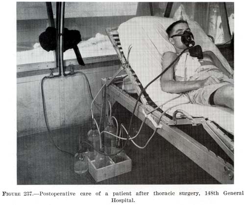
photo courtesy of the US Army

As it turns out – non-intubated thoracic surgery is not a new idea or concept.  It was developed early in the 20th century and was used successfully for many years for even the most complex thoracic cases such as esophagectomies until the development of double lumen intubation in the 1950’s made the use of single lung ventilation possible (Gonzalez-Rivas et. al. 2015, Pompeo 2015, Kiss & Castillo 2015).  Since its rediscovery in the last several years, many of the problems that plagued this technique during its inception over a century ago have been addressed through better understanding of human physiology.  Now, this seemingly fringe technique has been shown to be a feasible approach for treating the very margins of the thoracic surgery population (the extreme elderly, patients with advanced respiratory disease or other serious medical co-morbidities) that are often deemed inoperable using current techniques.

The dreaded complication (spontaneous pneumothorax) of early use of this technique by pioneers in thoracic surgery has now become one of the main advantages.  Surgically created pneumothorax results in almost perfect deflation of the operative lung, achieving better results than even the most experienced of anesthesiologists using traditional single lung ventilation.  Surprisingly to many observers, instead of creating a ventilatory emergency, this process is readily tolerated by most patients, even those with poor baseline pulmonary function (David, Pompeo, Fabbi & Dauri, 2015).

Current research

The majority of the current series of research on this topic are being performed by a small group of surgeons which includes Dr. Diego Gonzalez Rivas (Spain), Dr. Eugenio Pompeo and the Awake Thoracic Surgery Research Group in Italy and Dr. Ming-Hui Hung and colleagues (Taiwan).  Pompeo’s group (Drs Benedetto Cristino, Augusto Orlandi, Umberto Tarantino, Tiziana Frittelli (General Director of the Policlinico Tor Vergata), Leonardo Palombi, Paola Rogliani, Roberto Massa, Mario Dauri) has been especially prolific in 2015 after several of their works were published in a special issue of Annals of Translational Medicine.

In multiple studies, these researchers have reported successful thoracic surgery outcomes in non-intubated patients, thus eliminating the majority of risks related to general anesthesia as well as uni-lung ventilation via mechanical ventilation and intubation.  In several of these studies, the authors were also able to successfully perform these surgeries in fully awake patients (versus consciously sedated), making surgery possible for even the frailest of candidates.  These studies included a small number of comparisons between traditional and non-intubated surgeries.  While the numbers of patients enrolled have been small, and there are few randomized studies, the results have been encouraging.

Chen et. al

Chen et. al’s 2012 study has been one of the largest studies to date, with 285 cases.  In this study, patients underwent lobectomies, wedge resections and segmentectomies with 4.9% requiring conversion with tracheal intubation.  Lung resection was undertaken with traditional (3 port) VATS or a needlescopic approach.

The authors report the biggest problem they encountered was increased bronchial tone and airway hyper-reactivity during manipulation of the pulmonary hilum during lobectomies and segmentectomies.  This was effectively treated without significant alteration in hemodynamics via intrathoracic vagal blockage which eliminated the cough reflex in these patients.

The authors caution judicious patient selection to prevent emergent conversion (intubation) particularly while surgeons are initially attempting this technique.  Chen et al. also believe that non-intubated thoracic surgery is best suited for petite or small-sized female patients because the small tracheal size of these patients predisposes them to a higher rate of complications and potential tracheal injury with traditional surgery and intubation.

Awake anesthesia and lung volume reduction surgery

Pompeo et. al’s review of the literature surrounding of the use of this technique in patients with severe emphysema undergoing nonresectional lung volume reduction surgery (LVRS by awake anesthesia) showed significant treatment advantages for patients undergoing lung volume reduction surgery without intubation or administration of general anesthesia.

With an average mortality of 5% and a morbidity of 59% for traditional lung volume reduction surgery as reported during the National Emphysema Treatment Trial, findings from Tacconi et al.’s 2009 study of 66 LVRS awake patients (matched with 66 patients undergoing traditional surgery) appears promising.  The authors report a reduced incidence of prolonged air leaks (18%) versus 40% in the traditional surgical group as well as a decreased length of stay.  In this study, 3 patients required conversion to general anesthesia – one patient due to an elevated paCo2 of 83% and the remaining two patients for anxiety attacks.

Rate of intubation/ respiratory failure/ mortality in Tacconi et al.

Mortality in both non-intubated and the traditional surgical group was the same, with one patient from each group.  In both cases, the patients had developed massive airleaks following surgery.   In the non-intubated group, the patient developed acute lung injury requiring intubation of POD#12 and died POD#38.

In the traditional surgical group, 4 patients were unable to be extubated at the end of the case, with one patient requiring an additional day of mechanical ventilation.   Another patient was reintubated on POD#3 for respiratory failure and died on POD#67.

Pompeo et. al, over the course of over eleven years, have also investigated the use of non-intubated (and awake) thoracic surgery for a wide variety of cases including urgent /emergent cases, wedge resections, decortications, talc pleurodesis as well as nonintubated anesthesia combined with single (uniportal) thoracoscopic approaches (aka “microinvasive thoracic surgery”).

Anesthesia for non-intubated thoracic surgery

The role of anesthesiologists in caring for patients undergoing non-intubated or awake thoracic surgery is more challenging than general anesthesia.  While thoracic anesthesia already requires specialized skills for initiating, managing and maintaining uni-lung ventilation, the switch to non-intubated patients with either localized anesthesia or conscious sedation adds a new set of complexity to managing these often frail patients.  Kiss & Castillo (2015) in their review of the literature, provide an excellent overview of the pros and cons of non-intubated anesthesia as well as guidelines for patient selection and eligibility criteria for use of this technique.  Special populations who may benefit from this technique include patients with severe respiratory disease (and a high risk of ventilator dependency with intubation), patients with severe but stable dyspnea, or multiple cardiovascular and respiratory co-morbidities.

Kiss et. al also reviews the contraindications to use of this technique including: phrenic nerve paralysis on the non-operative side, patients at risk for difficult intubation, or patients who are unwilling to undergo awake thoracic surgery.  Wang & Ge (2014) expand on these complications to include ASA status 4 or higher, bleeding disorders, decompensated heart failure, extreme obesity, unfavorable airway or spinal anatomy as well as specific respiratory conditions including bronchiestasis, asthma, sleep apnea, clinically significant sputum production and strict contralateral lung isolation.

Wang & Ge also give specific anesthesia dosing guidelines for  conscious sedation, local anesthesia and regional blocks in additional to monitoring parameters.

Alterations in oxygenation and ventilation

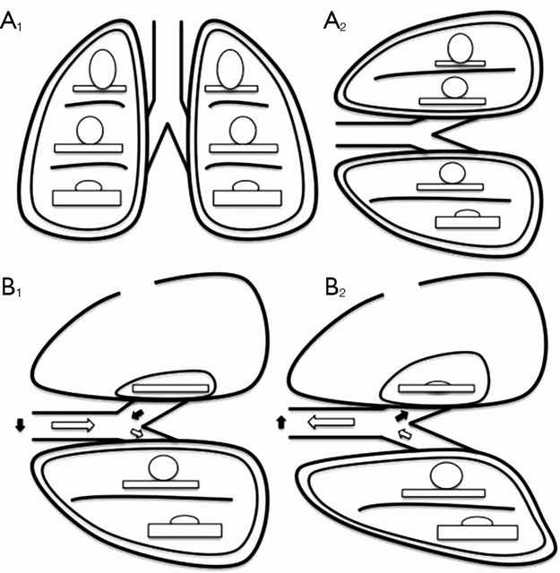
David et. al. (2015) describe the pathophysiology and alterations in oxygenation and ventilation in surgical pneumothorax including hypercapnia, hypoxia and the associated hypoxic pulmonary vasoconstriction that occurs along with the development of  intrapulmonary shunt as the deflated (and unventilated lung) maintains perfusion.  The authors also explain how this effect can be either exacerbated or minimized thru the choice of anesthetic agents,  and the administration of supplemental oxygen, which further demonstrates the importance of involving the thoracic anesthesia team in preparation for non-intubated cases.

The relationship between ventilation and perfusion. (A) Relationship between ventilation (roundes) and perfusion (rectangles) in different lung zones, in upright (A1) and lateral position (A2); (B) relationship between ventilation (roundes) and perfusion (rectangles) in lateral position with surgical pneumothorax, during spontaneous inspiration (B1) and exhalation phase (B2). Black arrows show paradoxical ventilation and mediastinal shift. (Illustration and caption from David et. al, 2015)

This “permissive hypercapnia” has been reported in multiple articles as having minimal to no clinical effects and is easily treated with supplemental oxygen by nasal cannula or facemask.

Editor’s note: In advance of this article, Dr. Pompeo, Dr. Gonzalez Rivas and Dr. Min-Hui Hung were contacted for their additional comments and insights on non-intubated thoracic surgery. This and subsequent articles may be augmented, as applicable with their replies.

Conclusion

Should we really abandon pursuit of better patient outcomes, faster mobility, recovery and reduced length of stay in lieu of the security to tell off-color jokes with our patients safely under general anesthesia?  Should we abandon all hope in treating patients previously deemed inoperable due to our own fears and hesitations to embrace newer techniques and procedures?

Or as Mineo et al, suggests, should we enlist our colleagues to design and devise several large scale studies at multiple institutions so that we can move to the next level of investigation and answer the question: “Should my patient be awake for this?”

References

Ke-Cheng Chen1,3, Ya-Jung Cheng2, Ming-Hui Hung2, Yu-Ding Tseng3, Jin-Shing Chen (2012).  Nonintubated thoracoscopic lung resection: a 3-year experience with 285 cases in a single institution.  Journal of Thoracic Disease, Aug 2012, 4(4).

David P, Pompeo E, Fabbi E, Dauri M. (2015). Surgical pneumothorax under spontaneous ventilation-effect on oxygenation and ventilation. Ann Transl Med. 2015 May;3(8):106. doi: 10.3978/j.issn.2305-5839.2015.03.53. Review.

Gonzalez-Rivas D1, Bonome C2, Fieira E3, Aymerich H4, Fernandez R5, Delgado M3, Mendez L3, de la Torre M5. (2015).  Non-intubated video-assisted thoracoscopic lung resections: the future of thoracic surgery?  Eur J Cardiothorac Surg. 2015 Apr 19. pii: ezv136.  article requires subscription, no free full-text available.  Nice discussion of the risks of traditional anesthesia and mechanical ventilation in thoracic patients.

Gonzalez-Rivas D1, Fernandez R2, de la Torre M2, Rodriguez JL3, Fontan L4, Molina F4. (2014).  Single-port thoracoscopic lobectomy in a nonintubated patient: the least invasive procedure for major lung resection? Interact Cardiovasc Thorac Surg. 2014 Oct;19(4):552-5. doi: 10.1093/icvts/ivu209. Epub 2014 Jul 7.

Kiss G, Castillo M. (2015).  Nonintubated anesthesia in thoracic surgery: general issues.  Ann Transl Med. 2015 May;3(8):110. doi: 10.3978/j.issn.2305-5839.2015.04.21. Review.

Hung MH, Hsu HH, Cheng YJ, Chen JS. (2014).  Nonintubated thoracoscopic surgery: state of the art and future directions. J Thorac Dis. 2014 Jan;6(1):2-9. doi: 10.3978/j.issn.2072-1439.2014.01.16. Review. (Best read in pdf form).

Liu YJ, Hung MH, Hsu HH, Chen JS, Cheng YJ. (2015).  Effects on respiration of nonintubated anesthesia in thoracoscopic surgery under spontaneous ventilation.  Ann Transl Med. 2015 May;3(8):107. doi: 10.3978/j.issn.2305-5839.2015.04.15. Review

Mineo TC, Tacconi F. (2014). Nonintubated thoracic surgery: a lead role or just a walk on part? Chin J Cancer Res. 2014 Oct;26(5):507-10. doi: 10.3978/j.issn.1000-9604.2014.08.11. No abstract available.  Very enjoyable, almost conversational article with the authors sharing their experiences with non-intubated thoracic surgery while calling for larger clinical research studies on the topic.

Noda, M., Okada, Y., Maeda, S., Sado, T., Sakurada, A., Hoshikawa, Y. et al. (2012).   Is there a benefit of awake thoracoscopic surgery in patients with secondary spontaneous pneumothorax?. J Thorac Cardiovasc Surg. 2012; 143: 613–616

Pompeo E, Cristino B, Rogliani P, Dauri M; Awake Thoracic Surgery Research Group (ATSRG). (2015).  Urgent awake thoracoscopic treatment of retained haemothorax associated with respiratory failure.  Ann Transl Med. 2015 May;3(8):112. doi: 10.3978/j.issn.2305-5839.2015.04.13.  Authors review their experiences treating emergent and trauma patients with awake, nonintubated VATS.

Pompeo E, Sorge R, Akopov A, Congregado M, Grodzki T; ESTS Non-intubated Thoracic Surgery Working Group. (2015).  Non-intubated thoracic surgery-A survey from the European Society of Thoracic Surgeons. Ann Transl Med. 2015 Mar;3(3):37. doi: 10.3978/j.issn.2305-5839.2015.01.34.

Pompeo, E. (2015).  Non-intubated thoracic surgery: nostalgic or reasonable?  Annals of Translational Medicine, 2015; 3(8): 99.  Review of the historical development on non-intubated thoracic surgery  and techniques in regional anesthesia for complicated thoracic surgery procedures including esophagectomies in the era predating the development of double lumen intubated and unilung ventilation.  A timely reminder that some of the greatest developments in medicine and surgery are ‘rediscoveries’ of our predecessors.

Pompeo E. (2014).  State of the art and perspectives in non-intubated thoracic surgeryAnn Transl Med. 2014 Nov;2(11):106. doi: 10.3978/j.issn.2305-5839.2014.10.01.  Nicely written article.  Has link to video presentation for purchase.

Pompeo, E. (2014).  Non-intubated vodeo-assisted thoracic surgery under epidural anesthesia – encouraging early results encourage randomized trials.  Chinese Journal of Cancer Research 2014, 26(4); 364-367.

Pompeo E1, Rogliani P1, Palombi L1, Orlandi A1, Cristino B1, Dauri M1; Awake Thoracic Surgery Research Group (ATSRG). (2015).  The complex care of severe emphysema: role of awake lung volume reduction surgery.  Ann Transl Med. 2015 May;3(8):108. doi: 10.3978/j.issn.2305-5839.2015.04.17. Related table:  Lung Volume Reduction Surgery Criteria – Awake Anesthesia

Pompeo E, Dauri M; Awake Thoracic Surgery Research Group (2014).  Is there any benefit in using awake anesthesia with thoracic epidural in thoracoscopic talc pleurodesis? J Thorac Cardiovasc Surg. 2013 Aug;146(2):495-7.e1. doi: 10.1016/j.jtcvs.2013.03.038. Epub 2013 Apr 17. No abstract available.

Pompeo E; Awake Thoracic Surgery Research Group (2012). To be awake, or not to be awake, that is the questionJ Thorac Cardiovasc Surg. 2012 Jul;144(1):281-2; author reply 282. doi: 10.1016/j.jtcvs.2012.01.083. No abstract available.  Comment on article by Noda et. al.

Pompeo E, Mineo D, Rogliani P, Sabato AF, Mineo TC (2004).  Feasibility and results of awake thoracoscopic resection of solitary pulmonary nodules.  Ann Thorac Surg. 2004 Nov;78(5):1761-8.  One of the earlier modern publications on awake thoracic surgery.

Tacconi F, Pompeo E, Mineo TC. (2009). Duration of air leak is reduced after awake nonresectional lung volume reduction surgery. Eur J Cardiothorac Surg 2009;35:822-8; discussion 828.

Wang B, Ge S. (2014).  Nonintubated anesthesia for thoracic surgery. J Thorac Dis. 2014 Dec;6(12):1868-74. doi: 10.3978/j.issn.2072-1439.2014.11.39. Review.   Related table: the advantages and disadvantages of nonintubated anesthesia for thoracic surgery.

Note: This is not an exhaustive list of literature available on this topic but a select listing of the most recent and relevant citations (and are available as free full text).

**Long time readers of thoracics.org may have noticed that we no long cover or report on ‘debates’ or discussions as to whether VATS can be used in oncology cases, or whether an adequate lymph node dissection can be performed using VATS.  The literature clearly demonstrates that it can – and clinical guidelines reflect this, making the discussion one-sided, tedious, out-dated and repetitious.

Interview with the Brazilian Society of Thoracic Surgery

Thoracics.org talks to the Brazilian Society of Thoracic Surgery and result isn’t what you might expect.

A very different article here at Thoracics.org!  For starters, I’m the interviewee – which doesn’t happen very often.  This interview was a joint collaboration after meeting and talking about issues in thoracic surgery with several Brazilian surgeons including Dr. Sergio Tadeu Pereira, at the ALAT conference in Medellin last July.

at ALAT with Bolivian surgeon, Dr. Cristian Anuz

Translation of interview from the December 2014 Journal of the Brazilian Society of Thoracic Surgery*:

The positive outcome of a thoracic surgery depends on several aspects, among them is the teamwork, the harmony between the various professionals involved in making decisions and actions. All experts have an instrumental part in restoring the health and maintenance the patient’s life. Each with its due importance, towards a single goal. The SBCT ratifies such thinking, and this issue of the Journal conducted an interview with K. Eckland, an acute care nurse practitioner in thoracic surgery, writer, and also the founder of Thoracics.org” – a blog about thoracic surgery with an international focus.  She has written several books on surgery in Latin America, including a community sociological examination thoracic surgery in Bogotá, Colombia.

In this conversation, K. Eckland talks about the future of thoracic surgery worldwide and recognizes the contribution of Brazilian surgeons for growth in the art.

Journal SBCT: For us at SBCT is a great pleasure to have their participation in our newspaper. How do you evaluate the specialty in Brazil?
K. Eckland: First, I would like to thank the editors this paper for the invitation to forward my message to Brazilian doctors. More importantly, I would like to serve as call to action to all the experts and future thoracic surgeons. When I look at Brazil, I see the future of thoracic surgery. While, in my own country, our thoracic surgeons are aging with an average age of 60 years, Brazil is full of young, dynamic and innovative surgeons.

Journal SBCT: This predisposition to new techniques of many the Brazilian thoracic surgeons implies an increase in research?

K. Eckland: The high fluency in minimally invasive techniques (in Brazil) combined with some of the largest academic and clinical settings worldwide, outside China, affords unique opportunities in research, development and discovery. Brazil is already home to many of the modern masters of thoracic surgery, names that resonate worldwide.

These surgeons have brought Brazil to the forefront, but it is up to the newest crop of thoracic surgeons to maintain Brazil’s forward momentum for the future.  However, this is hampered by a lack of awareness of the contributions of many Brazilian and other Latin American surgeons.

As a foreigner, writing about developments within the international surgical community, I have noted a large dearth in published research from much of Latin America including Brazil.  What research I do find, is often not widely dispersed or readily available to the rest of the world.  It has taken several years and many thousands of dollars for Cirugia de Torax to acquire and publish information about your many triumphs.  However, this is not the most efficient way for research to be disseminated.

Journal SBCT: In addition to increased investment in research, what more should be done in its assessment to mitigate this  gap in publications and contributions (to the specialty of thoracic surgery)?

K. Eckland: It’s possible to change this story from one of limited international exposure to greater recognition.  But for that to happen, several things need to occur. Firstly, the momentum must be Brazilian thoracic community to participate  and publish research on a large-scale.

Surgeons in São Paulo, for example, have unique opportunities to publish practice-changing work. The Department of Thoracic Surgery, University of São Paulo tracks more thoracic cases in a year than many American institutions have access in a decade. This gives greater impact to studies from this institution than anything that their (North) American colleagues could expect do.

Second, Brazilian surgeons need push for further publication in international journals, and in the international literature language, in English.

Lastly, surgeons need to look outside their corner of the globe and present their findings internationally and outside Latin America on a greater scale.  More groups of Brazilian surgeons should attend international conferences to gain knowledge,and take the opportunity to spread their own knowledge and research findings. Surgeons should not depend on the United States and Europe to take the lead in surgical innovation or research.

There is no reason why these findings will not occur at home, but research needs to be part of your daily practice. It should be more than reading the occasional surgical journal.  It should be a part of active problem solving and solution-seeking.

Journal SBCT: The wide practical experience associated with the host new techniques can be considered as a basis for the growth of the specialty and development more innovative research in Brazil?

K. Eckland:

For this to happen, each surgeon needs ask yourself**:

–  How can I improve my practice? – How can I improve the lives of my patients?

– What can I do to identify and document the phenomena I’m seeing?  – What we are doing now that we need to change? How can we implement these changes? How does this apply to people outside my immediate environment?

Once a potential search area is identified, other questions to ask include:

How I can improve my specialty? How can I represent my country to the world? Sometimes the answers
involve the development of new technologies, sometimes a reframing of the information we already know,
to apply the new clinical scenarios. Other times, we simply need to identify the phenomena and document it to serve as guidance to other professionals. That is what drives the research, and this combined insight with professional curiosity are essential for growth within the specialty. By embracing these concepts, we can begin a new era of thoracic surgery in Brazil and worldwide.

* Corrections to the English translation have been made for ease of reading.  This is an excerpt from a larger interview.

** This is how we identify research to discuss and publish here at Thoracics.org/ Cirugiadetorax.org

For the original article, click here.

XIX Congresso Brasileiro de Cirurgia Torácica

ikone

If you are interested in learning more about the latest research and developments in thoracic surgery in Brazil, the annual conference is this May.

As Florida’s population booms, surgeon shortage becomes acute

As the Florida legislature and medical community considers the impending physician shortage, many of the critical concerns regarding the on-going shortage of surgeons remain unaddressed

Now that Florida is the third most populated state (behind first ranking California and # 2 Texas) in the United States with a census of almost 20 million residents, the ongoing shortage of surgeons is predicted to become more dire over the next ten years.

the thoracic surgeon: a disappearing breed?

The problem is multi-factorial: Training, debt, compensation (financial and otherwise)

In a recent article by Donna Gehrke – White at the Sun Sentinel, the need for over 7,000 additional physicians (in a variety of specialties) highlights some of the difficulties in training and retaining specialty physicians in American medicine.  Lengthy training regimens coupled with high student loan debt as well as feelings of frustration and ‘burnout‘ plague a medical landscape that is already burdened with concerns over the fragile state of American health care, escalating healthcare costs and the impact of Obamacare and other recent federally mandated changes to the health care system.

“More schools” are not the answer

While Florida is responding to the impending crisis by opening new residency programs, this doesn’t address some of the more crucial concerns – high vacancy rates in existing programs, the exorbitant costs of a surgical education, and a growing dissatisfaction with current working conditions.

How about better loan repayment programs/ debt forgiveness?  Or greater access to patients (and less time dealing with paperwork/ EMR and reimbursement issues)?  Instead of lengthening/ shortening training programs and relying on computerized models, maybe consider improving the quality of American surgical training by separating the specialty into two separate tracks (like most countries)?

Florida’s shortage of thoracic surgeons: 14?

While the estimated shortage of thoracic surgeons in Florida is only projected to be 14, existing problems in retaining surgical residents and low specialty board pass rates and echos both nationwide and global shortage concerns.  With an aging population, rising rates of esophageal cancer and earlier detection (of surgically treatable) cancers, these numbers may not tell the whole story.

Additional Recommended Reading:

Gordon, D. (2014) 15 things to know about the physician shortage.  Becker’s Hospital Review (on-line).

Seaman, A. M. (2012). Surgeon’s pressures may worsen shortage.  Reuters.  As we’ve noted previously, this is not a new concern, and the latest studies and reports only confirm this data.  In fact, this report from the Robert Wood Johnson Foundation from 2011, highlights the fact that surgeon shortages are impacting emergency departments ability to provide emergency life-saving treatment.

Aliwadi, G. & Kron, Irving (2008).  The challenges facing thoracic surgeons.  Vascular disease management.  This 2008 article highlights some of the difficulties in attracting and retaining medical students and surgical residents to the cardiothoracic surgery specialty.  While mainly geared at cardiac surgery – and the issues raised by interventionalists and catheter based interventions, it also touches on some of the educational issues that affect both cardiac and general thoracic surgeons.

Single port surgery conference in Berlin

the latest trailer about the documentary film on single port surgery and information about an upcoming training course.

For everyone that’s interested in learning more about the single port surgery technique, as taught by its creator, Dr. Diego Gonzalez Rivas – here’s another opportunity which may be closer to home for some readers.

DSC_0027
Now, which way to Berlin?


The February conference takes place in Berlin, Germany on the 19th thru 21st.  While Dr. Gonzalez Rivas, Dr. Delgado and Dr. Prado are headlining the event, other prominent thoracic surgeons such as Gaetano Rocco (Italy) and Alan Sihoe (Hong Kong) will also be lecturing at this event.

The conference includes live surgery demonstrations as well as a wet-lab for hands-on practice.

Deadline for registration is February 6th.  Interested surgeons should contact:

R. Mette, M. Schmitt
Charité – Universitätsmedizin Berlin
Tel. +49 30 450 622 132 | Fax +49 30 450 522 929
E-mail: thoraxchiurgie@charite.de

To download the event brochure, click:  VATS_Course_2015 brochure


In other news – the newest trailer for the documentary about Dr. Gonzalez Rivas and his work was recently released.  I encourage all thoracic surgery personnel to see (and promote) this movie, which highlights the work of one of our own.


Thoracic surgeon shortage in Australia

When you need surgery, How long will you wait? (and will there be anyone to perform the operation?) The on-going surgeon shortage now affecting South Australia.

There is more evidence of the changing demographics of healthcare and an aging population and its effects world-wide on cardiothoracic surgery as the Australian newspaper, Herald Sun published reports of lengthy patient waiting lists for surgical procedures including procedures categorized as “urgent.”

waiting list

Long lines and waiting lists

More concerning, is the report of the number of patients who have died waiting for surgery*.

The report, which focuses on the Southern region of Australia highlights the shortage of specialty surgeons and the growing numbers of patients affected by these shortages.

Critics of the Australian government have also voiced concerns over the Surgeon Workforce project which aims to partially alleviate these shortages by using foreign-trained surgeons and imported labor.  This comes at a time when Australia actually has an oversupply of general practice or internal medicine physicians.

The shortage of well-trained surgeons is affecting all surgical specialties but the cardiac and thoracic surgery specialties are particularly hard hit due to the lengthy, rigorous (and often costly) training regimen  in many countries.

In the United States, this process is also exacerbated by an antiquated, sometimes arbitrary or impractical practice for residency placement that discourages international medical students.  This, along with other concerns (legitimate as well as political) that govern the regulations that permit overseas graduates to practice in the United States restricts the possibility of reducing the growing shortage in a similar manner.

The lengthy educational process is not the only factor.  Many medical students cite the strenuous work schedules, diminished job satisfaction and physically challenging surgical lifestyles as reasons medical students are choosing other specialties which are seen as being equally or more lucrative but allowing more work-life balance.

Similar shortages have been reported in Canada, the United States and the United Kingdom.

* Many international readers have asked if this is what is affecting the Americans Veteran’s Administration hospital system.  Not really, (or if so, only partially).  Th VA Scandal is a tragic example of the bureaucratic red tape that is becoming far too frequent for Americans seeking medical services.

Dr. Gonzalez Rivas, Johnson & Johnson and Single-port thoracic surgery

the 2013 S.W.A.T conference, presented by Johnson & Johnson. Featured presenters Dr. Diego Gonzalez Rivas and Dr. Paula Ugalde discuss single port thoracoscopy and topics in minimally invasive surgery

Very pleased that despite the initial difficulties, we are able to provide information regarding the recent conference.

Talking about Single-port surgery in Bogotá, Colombia – 2013 S.W.A.T. Summit

Dr. Diego Gonzalez Rivas and Dr. Paula Ugalde were the headliners at the recent Johnson and Johnson thoracic surgery summit on minimally invasive surgery.  Both surgeons gave multiple presentations on several topics.  They were joined at the lectern by several local Colombian surgeons including Dr. Stella Martinez Jaramillo (Bogotá), Dr. Luis Fernando Rueda (Barranquilla), Dr. Jose Maineri (Venezuela) Dr. Mario Lopez (Bogotá) and Dr. Pardo (Cartagena).

Thoracic surgeons at the 2013 S.W.A.T Summit in Bogota, Colombia. Drs. Gonzalez-Rivas and Dr. Paula Ugalde are center, front-row

Target audience missing from conference

The audience was made up of thirty Latin American surgeons from Colombia, Costa Rica and Venezuela.  This surgeons were hand-picked for this invitation-only event.  Unfortunately, while Johnson and Johnson organized and presented a lovely event; their apparent lack of knowledge about the local (Colombian) thoracic surgery community resulted in the exclusion of several key surgeons including Dr. Mauricio Velasquez, one of Colombia’s earliest adopters of single-port thoracoscopy.  Also excluded were the junior members of the community, including Dr. Castano, Dr. Carlos Carvajal, and current thoracic surgery fellows.  It was an otherwise outstandingand informative event.

However, this oversight represents a lost-opportunity for the parent company of Scanlon surgical instruments, the makers of specialized single port thoracoscopic instrumentation endorsed and designed by Dr. Gonzalez-Rivas himself, including the Gonzalez-Rivas dissector.

The Gonzalez - Rivas dissector, photo courtesy of Scanlon International
The Gonzalez – Rivas dissector, photo courtesy of Scanlon International

As discussed in multiple publications, previous posts as well as during the conference itself, it is these younger members who are more likely to adopt newer surgical techniques versus older, more experienced surgeons.  More seasoned surgeons may be hesitant to change their practices since they are more comfortable and accustomed to open surgical procedures.

Despite their absence, it was an engaging and interesting conference which engendered lively discussion among the surgeons present.

Of course, the highlight of the conference actually occurred the day before, when Dr. Gonzalez- Rivas demonstrated his technique during two separate cases at the National Cancer Institute in Bogotá, Colombia. (Case report).

Dr. Gonzalez-Rivas and Dr. Ricardo Buitrago performing single port thoracoscopy at the National Cancer Institute

Featured presenters:

Dr. Diego Gonzalez – Rivas is a world-renown thoracic surgeon jointly credited (along with Dr. Gaetano Rocco) with the development of single-port thoracoscopic (uni-port) surgery.  He and his colleagues at the Minimally Invasive Surgery Unit in La Coruna, Spain give classes and lectures on this technique internationally.  Recent publications include three papers in July alone detailing the application of this surgical approach, as well as several YouTube videos demonstrating use of this technique for a wide variety of cases.

Dr. Gonzalez Rivas
Dr. Gonzalez Rivas

Dr. Paula Ugalde, a Chilean-borne thoracic surgeon (from Brazil) who gave several presentations on minimally-invasive surgery topics. She is currently affiliated with a facility in Quebec, Canada.

Dr. Paula Ugalde
Dr. Paula Ugalde

Refuting the folklore

Part of the conference focused on refuting the ‘folklore’ of minimally-invasive procedures.   Some of these falsehoods have plagued minimally-invasive surgery since the beginning of VATS (in 1991), such as the belief that VATS should not be applied in oncology cases. The presenters also discussed how uniportal VATS actually provides improved visibility and spatial perception over traditional VATS (Bertolaccini et al. 2013).

However, Gonzalez-Rivas, Ugalde and the other surgeons in attendance presented a wealth of data, and publications to demonstrate:

–          VATS is safe and feasible for surgical resection in patients with cancer. (Like all surgeries, oncological principles like obtaining clear margins, and performing a thorough lymph node dissection need to be maintained).

–          Thorough and complete lymph node dissection is possible using minimally invasive techniques like single-port surgery.  Multiple studies have demonstrated that on average, surgeons using this technique obtain more nodes than surgeons using more traditional methods.

–          Large surgeries like pneumonectomies and sleeve resections are reasonable and feasible to perform with single-port thoracoscopy.  Using these techniques may reduce morbidity, pain and length of stay in these patients.

–          Rates of conversion to open surgery are very low (rare occurrence).  In single-port surgery, “conversion” usually means adding another port – not making a larger incision.

–          Learning curve fallacies:  the learning curve varies with each individual surgeon – but in general, surgeons proficient in traditional VATS and younger surgeons (the “X box generation”) will readily adapt to single-port surgery.

–          Bleeding, even significant bleeding can be managed using single-port thoracoscopy.  Dr. Gonzalez Rivas gave a separate presentation using several operative videos to demonstrate methods of controlling bleeding during single-port surgery – since this is a common concern among surgeons hesitant to apply these advanced surgical techniques.

Additional References / Readings about Single-Port Thoracoscopy

 Scanlon single-port thoracoscopy kits  – informational brochure about specially designed instruments endorsed by Dr. Gonzalez Rivas.

Dr. Diego Gonzalez Rivas – YouTube channel : Dr. Gonzalez Rivas maintains an active YouTube channel with multiple videos demonstrating his surgical technique during a variety of cases.  Below is a full-length video demonstrating the uniportal technique.

Additional posts at Cirugia de Torax about Dr. Diego Gonzalez- Rivas

2012 interview in Santiago, Chile

Dr. Gonzalez-Rivas “TedTalk” –

SITS lobectomy – discussion on previous publication/ case report.

Dr. Gonzalez Rivas and the future of thoracic surgery

Upcoming conference in Florida – information about registering for September conference for hands-on course in single-port thoracoscopic surgery with Dr. Gonzalez-Rivas

Youtube video for web conference on Single-port thoracoscopic surgery

Bertolaccini, L., Rocco, G., Viti, A. & Terzi, A. (2013).  Surgical technique: Geometrical characteristics of uniportal VATSJ. Thorac Dis. 2013, Apr 07.    Article from thoracic surgeons at the National Cancer Institute in Naples, Italy explains how the geometric advantages of uniportal VATS improves visibility and spatial perception over traditional VATS, and mimics open surgery.

Calvin, S. H. Ng (2013). Uniportal VATS in Asia.  J Thorac Dis 2013 Jun 20.  Article discussing the spread of uniportal techniques in Taiwan, China and other parts of Asia.

Gonzalez Rivas, D., Fieira, E., Delgado, M., Mendez, L., Fernandez, R. & De la Torre, M. (2013). Surgical technique: Uniportal video-assisted thoracoscopic lobectomyJ. Thorac Dis. 2013 July 4.

Gonzalez Rivas, D., Delgado, M., Fieira, E., Mendez, L. Fernandez, R. & De la Torre, M. (2013).  Surgical technique: Uniportal video-assisted thoracoscopic pneumonectomy.  J. Thorac Dis. 2013 July 4.

Rocco, G. (2013). VATS and uniportal VATS: a glimpse into the future.  J. Thorac Dis. 2013 July 04.  After coming across several articles by Dr. Gaetano Rocco, and actively pursuing several other publications by this Italian thoracic surgeon, I have become increasingly convinced that Gaetano Rocco, along with Dr. Gonzalez Rivas is one of the world’s leading innovators in thoracic surgery.  Hopefully, cirugia de torax will be able to catch up to Dr. Rocco at some point for an in-depth discussion.

While I advance criticism of this event – it was a fantastic conference.  My only reservations were to the exclusivity of the event.  While this was certainly related to the costs of providing facilities and services for this event – hopefully, the next J & J thoracic event will be open to more interested individuals including young surgeons and nurses.

Bilateral surgery using a unilateral single-port approach: the Chen technique

a sneak peak at a game-changer in thoracic surgery – unilateral single-port surgery for bilateral disease

More news out of Taiwan from the innovative and dynamic Dr. Chih – Hao “Roy” Chen, this time in the form of a soon-to-be published case report in the prestigious Asian journal, the Annals of Thoracic & Cardiovascular Surgery.

Dr. Chen, Thoracic Surgeon

The article, entitled, “Treatment of bilateral empyema thoracis using unilateral single port approach,” details one of his recent cases and discusses the use of unilateral single port surgery for the treatment of bilateral conditions.  (For the uninitiated – that’s one small incision to treat an infection on both sides of the chest.)

Case report: bilateral empyema

In this case report, a 28 year old male presented with dyspnea, sore throat, malaise, fever and weakness. Patient was admitted with a diagnosis of sepsis and started on antibiotics.

Labs showed an elevated WBC count (19,300), C-reactive protein and D-dimer.  Subsequent imaging confirmed the presence of pulmonary emboli, and with serial imaging showing worsening bilateral pleural effusions. Thoracic surgery was consulted for definitive treatment.

Dr. Chen discusses this technique, as well as considerations for using this novel approach.

First look at innovative approach

Other that his recent discussions here at Cirugia de Torax, this is the first time that surgery utilizing this technique has been discussed in a medical forum.  This represents a ground-breaking advance in thoracoscopic surgery, single port surgery and thoracic surgery as a whole.

Update:  Article published June 18, 2013 in the Annals of Thoracic and Cardiovascular Surgery.  A pdf of the full article is available.

Chih-Hao Chen), Wei-Sha Lin, Ho Chang, Shih-Yi Lee, Tzu-Ti Hung, Chih-Yin Tai (2013). Treatment of Bilateral Empyema Thoracis Using Unilateral Single-Port Thoracoscopic Approach. http://dx.doi.org/10.5761/atcs.nm.13-00051


The cowboys and rodeo stars of thoracic surgery

Discussing Dr. Joseph Coselli and ‘the cowboys of cardiac surgery’ along with some of our own heros of thoracic surgery here at Cirugia de Torax.

There’s a great article in this month’s Annals of Thoracic Surgery, by Dr. Joseph Coselli, from Texas Heart Institute and the Michael DeBakey Department of Surgery at Baylor.   His article, entitled,” My heros have always been cowboys” is more than just a title torn from the song sheets of Willie Nelson.  It’s a look back at both the pioneers of cardiac surgery and his own experiences as a cardiac surgeon.  He also discusses the role of surgeons, and medical practitioners in American society in general and the promises we make to both society at large and our patients.

Here at Cirugia de Torax, I’d like to take a moment to look back at the surgeons that inspired and encouraged me in this and all of my endeavors.  Some of these surgeons knew me, and some of them didn’t – but their encouragement and kindnesses have spurred a career and life that have brought immense personal and professional satisfaction.

Like Dr. Coselli, I too, took inspiration from the likes of Dr. Denton Cooley.  But our stories diverge greatly from there.  I never met Dr. Cooley and I probably never will.  But it was a related story, from my former boss (and cardiothoracic surgeon), Dr. Richard Embrey that led to an email to Dr. Cooley himself.  My boss had too trained under Dr. Cooley, Dr. Debakey and the Texas Heart Institute, the citadel of American heart surgery.   Then, somehow, along the way – Dr. Embrey stopped to work at our little rural Virginia hospital.  We were the remnants of a larger Duke cardiothoracic program but we were a country hospital all the same.

While I learned the ins and outs of surgery from Dr. Embrey (and Dr. Geoffrey Graeber at West Virginia University) on a day-to-day basis, I was also weaned on the folklore of cardiothoracic surgery – stories of the giants of history, like the ones mentioned in Dr. Coselli’s article, as well as local Duke legends who occasionally roamed the halls of our tiny ICU and our two cardiothoracic OR suites; Dr. Duane Davis, Dr. Shu S. Lin and Dr. Peter Smith.  While never working side-by-side, Dr. D’Amico’s name was almost as familiar as my own.  As the sole nurse practitioner in this facility, without residents or fellows, there was no buffer, and little social divide in our daily practice.  Certainly, this changed me – and my perceptions.  I asked the ‘stupid’ questions but received intelligent and insightful answers.  I asked even more questions, and learned even more..

These opportunities fed my mind, and nurtured my ambitions.  Not to be a physician or a doctor, but to learn as much as possible about my specialty; to be the best nurse possible in my field.  It also nurtured a desire to share these experiences, and this knowledge with my peers, my patients and everyone else who ever had an interest.

It was that tiny little email, a gracious three-line reply from Dr. Cooley himself that made me realize that I didn’t have to rely on folklore and second-hand stories to hear more.  That’s critical; because as we’ve seen (here at Cirugia de Torax) there are a quite of few of “Masters of thoracic surgery” or perhaps future giants that haven’t had their stories told.  Dr. Coselli and his fellow writers haven’t written about them yet.. So I will.

Sometimes I interview famous (or semi-famous) surgeons here, but other times, I interview lesser-known but equally talented/ innovative or promising surgeons.  All of them share similar traits; dedication and love for the profession, immense surgical talent and proficiency and sincere belief in the future of technology of surgery.

So, let’s hope that it won’t take forty more years for these surgeons to be recognized for their contributions to thoracic surgery in the way that Cooley, DeBakey and Crawford are heralded in cardiac surgery.

K. Eckland, ACNP-BC

Founder & Editor -in – chief

Thoracic surgery shortage worsens as graduates fail to pass exams

a record number of surgeons fail to pass the American thoracic surgery certification exam, in the midst of a deepening shortage of surgeons.

A new report from the (American) Board of Thoracic Surgery shows a growing number of eligible surgeons are failing the thoracic surgery certification examination.

Record Failure Rate

As stated in the article published at Family Practice News, the failure rate has doubled to 28% in just a few short years.  This comes at a critical period in American medicine as shortages in specialty surgeons have emerged around the country due to an aging workforce.  This shortage is not confined to the United States – and has been echoed in Canada, the UK and several other industrialized nations.

Decrease in resident hours = decreased surgical knowledge

This record failure rate comes in the wake of recent reforms to resident surgical education  – including several reductions in resident training hours, and the push for a condensed 6 year residency program.

Rapidly evolving surgical technology

At the same time, rapidly evolving surgical technology and research in thoracic surgery may actually require significant curriculum changes and increased length of specialty training, according to this report at Thoracic Surgery News.

But, as previously reported, the extensive training requirements for cardiothoracic surgery have led to fewer residents and widespread vacancies in residency programs as fewer and fewer surgical residents elect to devote themselves to cardiothoracic surgery due to concerns about diminishing financial returns, reduced economic opportunities, excessive student loan burdens and concerns related to the hardships of the ‘cardiothoracic lifestyle’.

Solo Cardiac, General Thoracic tracks may trump combined “Cardiothoracic”

Alternatively, North American surgeons may need to follow the example of many of their international peers and diverge into two separate tracks: cardiac surgery and general thoracic to maintain surgical proficiency without excessive education burden in an era of rapidly evolving surgical knowledge.


Additional Recommended Reading:

Ann Thorac Surg. 2009 Aug;88(2):515-21; discussion 521-2. doi: 10.1016/j.athoracsur.2009.04.010.

Awake Epidural Anesthesia for thoracoscopic pleurodesis

Awake epidural anesthesia for thoracoscopic pleurodesis: A prospective cohort study. a new publication from Dr. Mauricio Velasquez and his surgical team reviewing results from their 36 month study

On the heels of a recent announcement on CTSnet.org soliciting surgeon input on their experiences with non-general anesthesia for thoracic surgery procedures, Cirugia de torax is revisiting one of the surgeons we interviewed last year, Dr. Mauricio Velasquez at Fundacion Valle de Lili in Cali, Colombia.

Dr. Velasquez in the operating room with Lina Caicedo Quintero (nurse) Valle de Lili, Cali, Colombia
Dr. Velasquez in the operating room with Lina Caicedo Quintero (nurse) Valle de Lili, Cali, Colombia

The trip to Cali was primarily to discuss Dr. Velasquez’s Thoracic Surgery Registry, and to observe him performing several single port surgery cases.  However, during the trip, Dr. Velasquez also spoke about several other aspects of his current practice including some of his recent cases, and the thoracic surgery program at Fundacion Valle de Lili.

Dr. Mauricio Velasquez after another successful case

We also talked with his wife, (and lead author), the talented Dra. Cujiño, an anesthesiologist who subspecializes in thoracic anesthesia.   Together, they have successfully performed several thoracic cases using thoracic epidural anesthesia on awake patients.

By chance, they published articles in both  Revista Colombianas de anesthesia and Neumologia y cirugía de torax in the last few weeks.

Revista Colombianas de anesthesia

Patients receiving epidural anesthesia received a small dose of midazolam prior to insertion of epidural needle at the T3 – T4 intervertebral space.  During the case, patients received bolus administration via epidural of 0.5% bupivacaine on a prn basis.

Short surgeries, single port approach

All patients, regardless of anesthesia type underwent single port thoracoscopic surgery for the talc pleurodesis procedure.  Surgery times were brief, averaging 30 to 35 minutes  for all cases (range 25 – 45 minutes) with the epidural patient cases being slightly shorter.

Dr. Mauricio Velasquez performing single port thorascopic surgery

Dramatic reduction in length of stay

In their study, patients receiving awake anesthesia had an average length of stay of four days compared with ten days for the general anesthesia group.

Decreased incidence of post-operative complications

There was a marked reduction in the incidence of post-operative respiratory complications (19 in general anesthesia group) versus 3 patients in the awake anesthesia group.  Post-operative mortality was also decreased (six in general anesthesia) versus two deaths in the awake anesthesia group.  However, the mortality statistics may also be impacted by the overall poor prognosis and median survival time of patients presenting with malignant effusions.

Post-operative pain

Study patients also self-reported less post-operative pain in the awake anesthesia group – with only one patient reporting severe pain versus seven patients in the general anesthesia group.

Conclusions

Cujiño, Velasquez and their team found awake thoracic epidural anesthesia (ATEA) was a safe and effective method for intra-operative anesthesia and was associated with a decreased post-operative pain, decreased length of stay (LOS) and decreased incidence of post-operative complications.

Notes

This study was funded by the authors with no relevant disclosures or outside financial support.

References

Indira F. Cujiño,  Mauricio Velásquez,  Fredy Ariza,  Jhon Harry Loaiza (2013).    Awake epidural anesthesia for thoracoscopic pleurodesis: A prospective cohort studyRev Colomb Anestesiol. 2013;41:10-5.  A 36 month study involving 47 cancer patients comparing (standard) general anesthesia outcomes with awake epidural anesthesia.

en Espanol: Anestesia epidural para pleurodesis por toracoscopia: un estudio prospectivo de cohort.

The second article has not been posted online yet.  Look for updates in the coming weeks.

TedTalks about the New Masters of Thoracic Surgery

TedTalks sits up and takes notice of one of the New Masters and Superstars of modern thoracic surgery, Dr. Diego Gonzalez Rivas.

It looks like even the illustrious and élite Ted Talks have taken notice of the New Masters of Thoracic Surgery – these visionary, game-changing surgeons who are revolutionizing the thoracic surgery specialty.

The Spanish-language lecture entitled, “El viaje de los pioneros: Dr. Diego Gonzalez Rivas” should be just as inspiring to readers/ and viewers as it is to Cirugia de Torax.

If you don’t speak Spanish – don’t despair!  Dr. Gonzalez’ TED talk is now available with captions in multiple languages.  (Click on the closed captioning icon for translation options.)

Sometimes, it’s lonely out front – and being innovative is difficult.  It’s one thing to be Ivor Lewis, Pearson or McKeown but it’s another to be the first or sole surgeon to challenge edicts and procedures laid down by the giants of the specialty.  But without the modern-day Dylewskis, Gonzalez Rivas, Chen, (and others) – technology within the specialty would remain static.

Changing the future of thoracic surgery

These surgeons take big risks with their careers and reputations by attempting to deviate from long-standing surgical traditions.  But sometimes, it pays off – and when it does, it is wonderful to see these daring and forward thinkers receive the admiration and appreciation they deserve for their contributions to the field and to their patients.

Dr. Santolaya, Dr. Sales dos Santos, Dr.Berrios and Dr. Diego Gonzalez Rivas

Congratulations, Dr. Diego Gonzalez Rivas!  Here’s to your continued success..

Creative Commons License
Cirugia de Torax by K. Eckland is licensed under a Creative Commons Attribution-NonCommercial-NoDerivs 3.0 Unported License.
Based on a work at cirugiadetorax.org.
Permissions beyond the scope of this license may be available at cirugiadetorax.org.

This article is one of several articles and content from Cirugia de Torax that has been used commercially without consent, permission or attribution/ proper citation.

Evolving thoracic surgery: from open surgery to single port thoracoscopic surgery and future robotic

the future of thoracic surgery as seen by one of the New Masters, Dr. Diego Gonzalez Rivas.

A new editorial by ‘New Master‘, Dr. Diego Gonzalez Rivas explores the evolution of thoracic surgery from traditional open surgery to minimally invasive technologies such as robotic surgery and single port surgery.  The article is available on-line and as a free pdf download over at the Chinese Journal of Cancer Research.

Dr. Gonzalez at a conference in Chile

We’ve also posted it here for our readers.

Gonzalez Rivas, D. (2013).  Evolving thoracic surgery: from open surgery to single port thoracoscopic surgery and future robotic.  Chinese Journal of Cancer Research, 25 (1) 4-6.  Editorial pdf download.

Surgeon shortage to hit rural areas the hardest

the latest predictions on the impending shortage of surgeons in the United States

Unsurprisingly – rural area hospitals face additional challenges in attracting and retaining specialty surgeons in comparison to big cities/ metropolitan areas.  However, as reported by Patrice Welding at Thoracic Surgery News in a report on the annual meeting of the Central Surgical Association, this may be viewed as a boon for the surgeons themselves as hospitals may devise new and enhanced incentives to attract surgeons to their facilities.  The surgical specialties most likely to benefit from this strategy include (as previously reported), obstetrics and gynecology, or­thopedic surgery, general surgery, otolaryngology, urology, neurosurgery, and thoracic surgery.

The article which quotes Dr. Thomas E. Williams, Jr. predicts that hospitals and institutions may break out into a ‘bidding war’ over surgeons.

While this is dire news for rural hospitals  and the estimated 56 million patients served by these facilities, it comes as a relief for current thoracic surgery fellows and new thoracic surgeons who have faced an increasingly bleak economic landscape over the last few years.

Of course, more sanguine experts note that the impact of the impending shortage has been reported for several years – with little impact on the current job market for new graduates.

The United States isn’t the only nation to be suffering from a shortage of surgeons, particularly in thoracic surgery.  So, maybe this is one of the questions we should be asking.

Dr. Thomas E. Williams Jr. is one of the main researchers on the impending shortage in the United States and published a book based on his findings in 2009, entitled, “The coming shortage of surgeons: why they are disappearing and what that means for our health“. (Praeger, ISBN #978-0313380709).  His work has also be published in multiple journals, and presented in meetings and conferences across the country.

Williams, T. E & Ellison, E. C. (2008). Population analysis predicts a future critical shortage of general surgeons.   Surgery, 144 (4): 548-556, October 2008.

“General Thoracic Surgery” is thoracic surgery

what is the future of thoracic surgery education? A new American study asks the if it is time to separate the specialties of cardiac and thoracic surgery.

A new study by Cooke & Wisner performed at a large medical center in California (UC Davis) and published in the Annals of Thoracic Surgery  provides additional weight to the idea that Thoracic Surgery has increasingly developed into it’s own subspecialty away from the traditional cardiothoracic surgery model (seen in the United States and several other countries.)

In an article published in Medical News Today, the authors of the study explained that the increased complexity of (noncardiac) thoracic surgery procedures for general thoracic conditions has led to increased referrals and utilization of general thoracic surgeons (versus cardiac or general surgeons).  This shows a reversal in a previous trend away from specialists – with more patients now receiving “complex” thoracic surgery procedures from specialty trained, board-certified thoracic surgeons.  Previously up to 75% of all thoracic surgery procedures were performed by general surgeons.

As the authors of the study discussed; this has serious implications for the curriculum of thoracic surgery fellowship programs, particularly as the specialty tries to attract more residents to stem an on-going and critical shortage.

With lung cancer rates expected to climb dramatically in North America and Europe, particularly in women – along with esophageal cancer, and   long waits already common, support and on-going discussion about the evolution of resident and fellow education is desperately needed.

Reference

Cooke, D. T. & Wisner, D. H. (2012).  Who performs complex noncardiac thoracic Surgery in United States Academic Medical Centers? Ann Thorac Surg 2012;94:1060-1064. doi:10.1016/j.athoracsur.2012.04.018

Single port thoracoscopy for wedge resection – does size matter?

Dr. Chen discusses single port thoracoscopy – and specimen size.

Single port thoracoscopy for wedge resection – does size matter?
Dr. Chih-Hao Chen, Thoracic Surgeon, Mackay Memorial Hospital, Taiwan
Correspondence: musclenet2003@yahoo.com.tw

Case presentation and discussion

A 77 y/o woman was found to have a neoplasm in RML during routine health exam. (Full case presentation here.)
The incision was 2.5 cm. The specimen was removed successfully; patient experienced no complications and was discharged from the hospital without incident. However, when discussing this case with surgeon colleagues a specific question arose.

preparing to remove the specimen through the port

Does size matter? How big is too big to remove through a port incision?
An important issue is the theoretically smallest (and effective) incision size.  The issue is very challenging for most surgeons. In the past, other surgeons have questioned me :
“The specimen to be removed is more than 12-15 cm. How can you remove the specimen from a 3.5 cm incision?”

BUT, the fact is “the smallest size depends mostly on the solid part of the neoplasm “.

The lung is soft in nature. In my experience, a 3.5 cm port is usually enough for any lobe.  The only problem is that if the solid part is huge, pull-out of the specimen may be difficult.
Therefore, the solid part size is probably the smallest incision size we have to make, usually in the last step when dissection completed , if needed.

successful removal of lung specimen thru small port
Port with chest tube in place

Using endo-bags to avoid larger incisions
There is an exception. I have never do this but I think it may be possible.  We usually avoid opening the tumor (or cutting tumor tissue into half ) within the pleural space in order to prevent metastasis from spillage of cancerous tissue.

What if we can protect the tumor from metastasis while we cut it into smaller pieces? For example, into much smaller portions that will easily pass through the small port incisions?
My idea is to use double-layer or even triple layers of endo-bag to contain the specimen. If it is huge, with perfect protection, we cut it into smaller parts within the bag. Then the huge specimen  can be removed through a very tiny incision. This is possible.

We still have to avoid “fragmentations” since this might interfere or confuse the pathologist for accurate cancer staging. Therefore, in theory, we can cut it into smaller parts but not into fragments. However this idea is worthy of consideration, and I welcome debate from my fellow surgeons and medical colleagues.

Thank you to Dr. Chen.

Single-port thoracoscopy as a first-line approach & the “Chen esophagectomy”

Dr. Chih-Hao Chen at Mackay Memorial Hospital in Taiwan talks about his experiences with single port/ single incision thoracic surgery (SITS) as well as the “Chen esophagectomy”; a new single port approach to esophagectomies.

Single-port thoracoscopic surgery (SITS) as a first-line approach
With the advent of minimally invasive surgical techniques such as VATS, surgeons now have the ability to perform multiple surgical procedures such as lobectomy, decortication and even esophagectomy through 1 – 2 cm port incisions instead of traditional open surgery. However, as mentioned during an interview with Dr. Mark Dylewski, few American* surgeons have fully embraced this technology. Even fewer surgeons internationally have embraced the emerging single port techniques that have developed from VATS. One of these surgeons is Dr. Chih-Hao Chen at Mackay Memorial Hospital in Taiwan. We previously discussed one of his studies here at Cirugia de Torax, so it was with great delight when we had an opportunity to discuss his continuing research and development in this area in a series of emails.
Dr. Chen is currently in the forefront of the movement to make SITS a first-line approach for majority of thoracic surgery procedures that can currently be approached with traditional VATS. The biggest risk to this “less is more” approach to port placement is needing to add additional ports during the case (thus converting to traditional VATS 3-4 port approach).

As Dr. Chen explains, “In contrast to creating 3 small wounds, I always try single-port first. If it is technically unavoidable, I would make the second port incision. If it is still difficult, a third port incision would be made. The conversion rate (to 2-port or three port methods not open) is very low in most conditions.
“I believe the role of SITS as a first-line endoscopic approach is possible in nearly all patients. So far, I have performed roughly, SITS in more than 150 patients with various thoracic diseases, including esophagectomy in 5 cases using 2.5 cm single incision”.
However, the contraindications for the SITS approach are mainly those with “unstable hemodynamics in trauma”, “highly complicated cancer resection(such as sleeve lobectomy, etc)” and “thick and dense peel in chronic empyema”.

Dr. Chen was kind of the provide this clip of him performing single port thoracoscopy.

Over 150 cases, so far.

“According to my experience, patients with spontaneous pneumothorax and acute stage empyema as well as solitary pulmonary nodules are the best candidates for such procedure. The time required for the same operation is much shorter in single-port approach. For simple spontaneous pneumothorax, the time may be as short as 20-25 minutes. ( from skin incision to suture )”.
As I mentioned in my report (see publications linked below), the conversion rate of such condition is pretty low and worthy to try. In my experience, SITS w/o trocar greatly decrease incisional pain and have pleasant cosmetic results, as the wound can be extremely small”.

A recent case: Wedge resection by SITS

Procedure: single-port approach for a case of lung cancer in a 77 year-old woman.

Multiple wedge resections, pleural biopsy and LN smapling were performed.

single incision (now with chest tube) and specimen removed (photo courtesy of Dr. Chen)

The wound was 1.5 cm in length and the specimen is 7cm X 4cm ( solid part :2.5 cm ).  The specimen was removed within an endo-bag. (From previous experience, I knew that a specimen of this size can be safely removed through a tiny incision w/o destruction of the specimen.

Her chest tube was removed within 24 hrs and patient reports minimal discomfort. ( I injected Marcaine in ICS to prevent neuralgia in all cases.)

Sometimes innovation is hard
As we’ve seen frequently in the history of medicine / surgery, early innovators and adopters of new technology are often face significant resistance from their colleagues despite utilizing ‘best-evidence’ to support their ideas. People, many people, including surgeons – don’t like change and are sometimes hesitant to learn and practice techniques that develop in the years following fellowship.
One of the reasons Dr. Chen contacted Cirugia de Torax is to share his experiences and this technique with other interested thoracic surgeons. ‘Unfortunately, only a small portion of thoracic surgeons would like to try such procedure in Taiwan. Actually, most of them considered the procedure not valuable. Therefore, I would like to publish more experiences in the journals, which is one way to tell them “to try”.

Wait.. Did you say single-port thoracoscopy for esophagectomy?

“Esophagectomy in my team was performed by single-port thoracoscopic approach (in the chest). However, the abdominal portion was performed with four-port or 5-port laparoscopic approach, because the abdominal part was done by another doctor who is not familiar with single-incision laparoscopy (SILS). However, I have to admit that esophagectomy through single-port approach is much more difficult than other procedures. The main reason for this is that the esophagus is located in posterior mediastinum.”

While I usually utilize a more anterior ICS as my port incision for other single-incision procedures because the anterior ICS is very easy, with low conversion ( to 2- or 3-port ) rate. However, the same port is not appropriate for esophagectomy because of poor visualization.

New Approach, the “Chen esophagectomy” but ergonomic considerations
“For the reason, I tried a more lateral port incision (usually 5 ICS along the mid-axillary line. ) This is a BIG problem for me due to ergonomic issues. Manipulation of endoscopic instruments and the endoscope through the port is uncomfortable. At times, I have to rest for a while in order to alleviate soreness in my arm”.
“The time-determining step is to loop the esophagus. Proximal and distal dissection as well as lymph node dissection would be done with a harmonic scalpel. (We resected the esophagus, the anastomosis is in the neck ). For uncomplicated case, the procedure in the chest takes aroud 1-2.5 hours”.

*American research data suggests that VATS is used for less than 30% of all thoracic surgery procedures. However, anecdotal evidence suggests that internationally, VATS is utilized with much higher frequency outside of the United States.

Articles about single-incision thoracoscopic surgery (SITS) by Dr. Chih-Hao Chen

Chih-Hao Chen, Shih-Yi Lee, Ho Chang, Hung-Chang Liu, Chao-Hung Chen (2012). The adequacy of single-incisional thoracoscopic surgery as a first-line endoscopic approach for the management of recurrent primary spontaneous pneumothorax: a retrospective study. Journal of Cardiothoracic Surgery 2012, 7:99  [abstract only, full article pending publication.]

Chih-Hao Chen, Shih-Yi Lee, Ho Chang, Hung-Chang Liu, Chao-Hung Chen (2012). Technical Aspects of Single-Port Thoracoscopic Surgery for Lobectomy. Journal of Cardiothoracic Surgery 2012, 7:50.

Chih-Hao Chen, Ho Chang, Tzu-Ti Hung, Hung-Chang Liu (2012). Single Port Thoracoscopic Surgery can be a First-line Approach for Elective Thoracoscopic Surgery. Revista Portuguesa de Pneumologia, Portuguese Journal of Pulmonology, 2012, Sept 22.

Dual port VATS for recurrent spontaneous pneumothorax: Foroulis et. al

A newly published study comparing dual port thoracoscopy with mini-thoracotomy for the treatment of recurrent spontaneous pneumothorax

Here at cirugia de torax, we take a keen interest in the development of increasingly minimally invasive technologies from dual (and single-port) thoracoscopy for a variety of conditions to RATS (robot- assisted thoracic surgery).  It is our belief that by embracing these emerging technologies, we help to advance the thoracic surgery specialty.

This spring, we have had the pleasure of publishing case reports on dual port thoracoscopy for decortication of parapneumonic effusions and empyema and catching up with one of the leaders in single incision thoracic surgery, Dr. Diego Gonzalez Rivas.

This month, another entry, “A modified two-port thoracoscopic technique versus axillary minithoracotomy for the treatment of recurrent spontaneous pneumothorax: a prospective randomized study,”  by Dr. Christophoros N. Foroulis at the Aristotle Medical School in Thessaloniki, Greece was published in Surgical Endoscopy.  As noted in a previous post, there have been few (if any) published papers on dual port thoracoscopy, and no comparison studies of these two techniques.

This study, which was conducted during 2006 to 2009 followed 66 patients who were randomly assigned to receive either mini-thoracotomy or dual-port VATS for surgical pleurodesis/ bullectomy / blebectomy.

In this study, despite random assignment, each group of 33 patients were well matched in all characteristics such as age, operative side and BMI.   After surgical treatment, patients were followed for a median of 30 months (range 3 – 53 months) for development of late complications or recurrent pneumothorax.

Each treatment group – VATS versus open surgical was overseen by one surgeon with Dr. Foroulis performing all of the dual port surgeries, and Dr. Papakonstantinou performing all of the open procedures.  Outcomes were independently reviewed / evaluated by the remaining authors.

Study Findings

–  No conversions to open thoracotomy from the VATS group.

– Similar rate of recurrence between open (2.7%) and VATS (3%) group (but timing of recurrence differed.)  Both recurrent pneumothoraces in the VATS group occurred early post-operatively (POD#5) compared to the open surgical group – 13 months post-op.

– Rate of complications the same between groups but the type of complications differed. 2 patients in each group required reoperation:

VATS – reoperation for prolonged airleak

Minithoracotomy group – hematomas/ evacuation of clots

Length of stay (LOS) and post-operative pain

Surprisingly, length of stay and post-operative pain – two of the outcomes that are usually cited in favor of minimally invasive procedures – were not significantly different between the groups.

Differences

Patient satisfaction

However, patient satisfaction was significantly higher in the dual port group.  This was related to an earlier return to normal activities, and earlier full mobilization of the affected arm.

Longer procedures

VATS procedures were longer than open procedures – by a mean of 21 minutes (87.1 minutes for VATS versus 66.7 minutes for minithoracotomy) with associated increases in uni-lung ventilation time.

Discussion/ Conclusions

While previous studies had reported a recurrence rate that was significantly higher in the VATS group – that was not seen here.  The ability to detect blebs/ bullae (and thus treat) with VATS remains limited in comparison to a mini-thoracotomy, but does not appear to change outcomes after a successful pleurodesis procedure.  Dual port thoracoscopy does take more time but both procedures appear equally effective.

Reference:

Foroulis, C. N., Anastasiadis, K., Charokopos, N., Antonitisis, P., Halvatzoulis, H. V., Karapanagiotidis, G. T., Grosomanidis, V. & Papakonstantinou, C. (2012).  A modified two-port thoracoscopic technique versus axillary minithoracotomy for the treatment of recurrent spontaneous pneumothorax: a prospective randomized study.  Surg Endosc 2012 May 12.  Includes color photographs of procedures.

Thank you to Dr. Foroulis for your assistance.

For more on related topics:

Case study: spontaneous pneumothorax

About spontaneous pneumothorax

Minimally invasive surgery: SITS

Block, Tuffier, Blalock & Gonzalez? Returning to single incision thoracoscopic surgery with Dr. Diego Gonzalez – Rivas

Checking in with Dr. Diego Gonzalez – Rivas and his team in Coruna, Spain – the innovators in single incision thoracic surgery, as Dr. Gonzalez publishes a new report on single incision pneumonectomy.

After speaking with Dr. Raimundo Santolaya last week – I contacted Dr. Diego Gonzalez over at UTCMI in Coruna, Spain  to see what he’s been doing since our last contact, and discuss a possible interview in the future.

The single-port thoracic surgery superstar and his colleagues are certainly keeping busy – and continue to push the edges of modern thoracic surgery firmly into more and more advanced minimally invasive techniques.

Last fall, he published another case report on single incision VATS – lobectomy, and since then he has continued to operate and publish reports on his successes.  Now he has an upcoming case report on a right-sided single-port pneumonectomy, which was largely held as one of the last frontiers in VATS procedures.   (Pneumonectomy by standard VATS, despite being reported in the literature several years ago, remains a relatively uncommon procedure.)

While a common criticism of his work is related to the fact that removing a portion of the lung as large as a lobe, or an entire lung requires a small additional incision at the conclusion of the case – but these criticisms are weak at best – and fail to see the true clinical importance of his continued innovation and investigation in advancing video-assisted thoracoscopic surgery for the benefits of our patients.  Ten years from now – single incision VATS will be a common procedure, and Drs. Gonzalez, Fernandez and De la Torre will be the ones responsible.


References: Single port pneumonectomy

Gonzalez-Rivas D, de la Torre M, Fernandez R, Garcia J. (2012). Single-incision video-assisted thoracoscopic right pneumonectomy.  Surg Endosc. 2012 Jan 11. [Epub ahead of print – abstract re-posted below]

Abstract

BACKGROUND: The most common approach for Video-assisted thoracoscopic (VATS) lobectomy is undertaken with three or four incisions, including a utility incision of about 3-5 cm. However, major pulmonary resections are amenable by using only a single utility incision. This video shows the technical procedure of a right pneumonectomy by single-incision approach with no rib spreading.

METHODSA 52-year-old woman was proposed for single-incision VATS resection of a 5-cm right lower lobe adenocarcinoma. A 4-cm incision was made in the fifth intercostal space. We placed a 30-degree, high-definition, 10-mm thoracoscope in the posterior anterior part of the incision. Digital palpation confirmed that the tumor involved the fissure and the posterior portion of the upper lobe, which indicated the need for right pneumonectomy. We inserted the instruments through the anterior part of the utility incision to start the detachment of the right upper lobe by using a harmonic scalpel. The first step was dissecting the inferior pulmonary vein. The hilar structures were exposed by using harmonic scalpel and a long dissector (Fig. 1A). The upper and middle-lobe pulmonary veins were dissected and transected, allowing visualization of truncus anterior, which was then stapled. The inferior pulmonary vein and the intermediate truncus artery were divided, allowing optimal exposure to the main bronchus, which was stapled. The lung was removed in a protective bag by adding 1 cm to the incision, and a systematic lymph node dissection was performed. A single chest tube was placed in the posterior part of the utility incision.

RESULTS: Total surgery time was 210 min. The chest tube was removed on postoperative day 2, and the patient was discharged home on day 4 with no complications.

CONCLUSIONS:  Single-port VATS pneumonectomy for selected cases is a feasible procedure, especially when performed from a center with previous experience in double-port VATS approach.

DISCUSSION: Recent advances in surgical and video-assisted techniques have allowed minimally invasive pneumonectomy to be undertaken safely. VATS pneumonectomy is not a new procedure and in fact was initially reported 15 years ago and was felt to result in less postoperative pain and a faster return to normal activities [1]. Despite this, there have been only a few case reports or series published of VATS pneumonectomies [2,3].

Additional References/ Resources

Gonzalez – Rivas, D., Fernandez, R., De la Torre, M., & Martin – Ucar, A. E. (2012).  Thoracoscopic lobectomy through a single incision.  Multimedia manual cardio-thoracic surgery, Volume 2012This is an excellent article which gives a detailed description, and overview of the techniques used in single incision surgery.  Contains illustrations, full color photos and videos of the procedure.

Gonzalez-Rivas D, Paradela M, Fieira E, Velasco C. (2012).  Single-incision video-assisted thoracoscopic lobectomy: initial results.  J Thorac Cardiovasc Surg. 2012 Mar;143(3):745-7.

Gonzalez-Rivas D, de la Torre M, Fernandez R, Mosquera VX. (2011).  Single-port video-assisted thoracoscopic left upper lobectomyInteract Cardiovasc Thorac Surg. 2011 Nov;13(5):539-41.

Thoracic Surgery Training

Reviewing the literature regarding training in thoracic surgery

Thoracic surgery differs across the globe, but is one style of training superior to another? If it is the sheer amount of training, and time devoted to training in thoracic surgery, then by all accounts – Japan (with a twelve-year residency for potential thoracic surgeons*) is way out in front.  Or, as Komatsu suggests, is it more cases in a shorter, more intensive training period?

Of course, part of the confusion regarding the training and practice of thoracic surgery is related to the nomenclature itself.  As many of the surgeons I have interviewed have remarked or advocated – “Thoracic surgery, is in itself a complex surgical specialty required advanced skills and knowledge.”  Few would dispute that.  But then again, how would they define it?

In many countries, including my own, “thoracic surgery” may actually specify ‘cardiothoracic surgery’.  In fact, cardiac surgeons in the USA (and several other countries) are not board certified in cardiac surgery – but within the broad umbrella of ‘thoracic surgery,’ and that’s where the confusion begins.

For dedicated thoracic surgeons (versus the more broad-based ‘cardiothoracic surgeons’) this is just a symptom of the problems within the specialty AND its training programs.  As many of these surgeons have suggested, as general thoracic surgery has expanded and become more complex (with robotic, thoracoscopic and minimally invasive techniques being developed to treat more advanced diseased and conditions), a broad-based training program may no longer suffice.  Indeed, with recent studies showing that over one-fifth of thoracic surgery residents in the USA being devoted to sole thoracic practice only, the time may have come for significant changes in our training programs.  Gasparri, Tisol & Masroor (2012) recently published an article on the six-year integrated thoracic surgery program but these programs seek to integrate cardiothoracic training into what was previous the fifth year of general surgery (to shorten training by one year rather than to segregate cardiac and thoracic surgery into separate kingdoms.

In comparison to the United States – several countries offer specialty surgical training as thoracic surgery only.   Conceptually, this makes more sense, as this training method may give better grounding than mixed programs.  Unfortunately, there isn’t a lot of definitive data to suggest one method is superior to another.

However, in my humble opinion, as thoracic surgery continues to advance into more specialized and technically challenging procedures to treat a widening array of thoracic and mediastinal disease – thoracic surgery will require surgeons to devote themselves to continuing knowledge and skills acquisition to the exclusion of cardiac surgery.  It’s already started, on some level – most of the great thoracic surgeons (and many of the surgeons profiled here) have elected to forgo cardiac surgery.

Komatsu T. (2011).  Reflecting the thoracic fellowship in Canada as a Japanese thoracic surgeon: is there anything we should follow?  Ann Thorac Cardiovasc Surg. 2011;17(4):327-331.  (I must admit a partiality to Dr. Kotmatsu for his work on the role of nurse practitioners in thoracic surgery.)

Reddy VS, Calhoon JH (2010).  Cardiothoracic surgical education: the ideal platform for tomorrow’s surgeon.  Tex Heart Inst J. 2010;37(6):656-7.

Tchantchaleishvi et al (2010). Comparison of cardiothoracic surgery training in usa and germany.  Journal of Cardiothoracic Surgery.
The ’12 year residency’ actually includes a separate five year certification period..

Rising debts and decreased employment opportunities: the American landscape of thoracic surgery

$200,000 & counting: the escalating educational debt of thoracic surgery residents in the United States. Presenting the results of the 2010 Workforce Survey Report (Sarkaria et. al). Despite impending surgeon shortages of catastrophic proportions today’s new surgeons are unable to find full-time employment.

Sarkaria et al. (2011) recently published data from the 2010 Thoracic Surgery Residents Association Workforce Survey Report and the results are alarming.

In 2010 – there were 299 thoracic surgery* residents training in the United States.  Of these, 76% were US residents.   These residents are among the 11% of all graduating general surgery residents who are considering cardiothoracic surgery as a career.  Despite the low numbers of residents in a specialty anticipated to have severe shortages, the realities for many emerging thoracic surgeons remains bleak.

* 22% of thoracic surgery residents are pursuing a career in general thoracic surgery (versus cardiac only, or combined cardiothoracic).

With escalating outstanding educational debt and diminished job opportunities – the future of thoracic surgery in the United States remains uncertain.  But several facts are clear – the enormous costs of specialty surgical training are crushing obstacles for many residents considering a future in thoracic surgery.

In just three short years – the percent of thoracic surgery residents owing in excess of $200,000 more than doubled – from 8% in 2007 to 17 percent in 2010.  (Almost half of all thoracic surgery residents (46%) reported owing more than $100,000.)

At the same time – decreased cardiac surgery volumes, program closures, and delayed retirement among older surgeons (due to the prolonged economic recession in North America) decreased the amount of available positions for new and practicing surgeons.  This means that in spite of projected shortages of cardiothoracic surgeons in 2020, today’s new surgeons face an increasingly competitive and limited employment opportunities.  According to Sarkaria et. al.  while 80% of thoracic surgery residents had gone on job interviews, 47% were still actively looking for a position (at the time of the survey).  In fact, according to the study, 30% of residents were still jobless two weeks before completing their specialty training.

None of these issues are new – high vacancy rates have existed within specialty training programs for years.  In fact, many argue that these statistics argue for an overabundance of specialty training programs, creating an excess of surgeons.  But, we are now faced with a critical cross-roads between medicine and society, as record numbers of baby boomers (surgeons included) and their co-morbid conditions tax our medical system beyond all known resources.

Advances in technology (mainly in interventional cardiology) have falsely decreased surgical volumes to a point where numerous surgical programs have been vastly reduced or shuttered entirely.  But now, these [cardiac] patients are back for ’round two’ as preliminary and stopgap measures fail.  At the same time, skyrocketing rates of diabetes have led to an even younger generation of candidates for revascularization.  All of these cardiac surgery issues play into the development and training of thoracic surgery residents due to the current structure of the majority of American training programs.  (In this study, 30% of residents surveyed planned to practice in cardiac surgery only, with another 20 to 25% planning to perform both cardiac and thoracic procedures.)

As thoracic surgery techniques such as VATS, RATS, HITHOC and uni-port procedures grow increasingly complex, and specialized, only one-fifth of all cardiothoracic surgery residents devote themselves exclusively to thoracic surgery.  Lung cancers, and esophageal cancer cases are at an all time high, yet, without significant changes to our existing medical system and resident education programs, our emerging surgeons will have nowhere to practice.

Talking with William Serpa about the da Vinci robot

a sit down interview with William Serpa of Intuitive Surgical to discuss RATS (robot-assisted thoracic surgery) with the da Vinci robot.

As we look towards the future of thoracic surgery, at emerging technologies and procedures – one technology stands apart from the rest: robot-assisted surgery.  Love it or hate it – all thoracic surgeons have heard of it. So you can imagine my excitement this week when I had the opportunity to sit down and talk to one of the representatives of Intuitive Surgical, the makers of the best-known robotic surgery device, the da Vinci robot.

William ‘Al’ Serpa sat down with me to discuss robotic technology and the growing use of robotic technology in thoracic surgery.  While the da Vinci robot is used fairly frequently in urological and gynecological surgery, it is just now making inroads into other specialties.  The robot, which costs in excess of one million dollars, is more than a financial investment; it is an investment into the future of modern surgery – and Intuitive Surgical understands this.  The company maintains a long term mentoring relationship with surgeons trained on the da Vinci, and they take the training process seriously.

Interested surgeons of facilities with da Vinci equipment participate in multiple in-services, high-level on-site observations, and on-line training modules prior to beginning actual training on the robot in a 1 to 2 day skills lab.  After completing this initial training, surgeons are mentored through several cases, increasing in complexity as they become more familiar with the robot.

Mr. Serpa reports that most surgeons demonstrate surgical proficiency with the da Vinci system after completing about twenty cases.  This is also the minimal volume of annual cases required to be listed on the da Vinci website as a specialty provider.

Mr. Serpa and I discussed the perceptions that many physicians have of the difficulty of the learning curve for robotic surgery.  We discussed multiple published reports that robotic surgery lengthens case times, and the realities behind robotic surgery.  “Actually, after surgeons become familiar with using the robot, it doesn’t take more than a moment to re-position it.”  That’s sounds similar to what several previous surgeons [using the robot] have reported – so I guess the only way to find it is to see for myself.

Hopefully, my next post about the da Vinci robot will come to you from the OR.

Additional References

Giulianotti PC, Buchs NC, Caravaglios G, Bianco FM.  Robot-assisted lung resection: outcomes and technical details.  Interact Cardiovasc Thorac Surg. 2010 Oct;11(4):388-92.

Ninan M, Dylewski MR.  Total port-access robot-assisted pulmonary lobectomy without utility thoracotomy.  Eur J Cardiothorac Surg. 2010 Aug;38(2):231-2.

Kajiwara N, Kakihana M, Usuda J, Uchida O, Ohira T, Kawate N, Ikeda N. Training in robotic surgery using the da Vinci® surgical system for left pneumonectomy and lymph node dissection in an animal modelAnn Thorac Cardiovasc Surg. 2011 Oct 25;17(5):446-53.

Palep JH.  Robotic assisted minimally invasive surgeryJ Minim Access Surg. 2009 Jan;5(1):1-7.  Indian article – gives nice overview of robotic surgery.

Bodner J, Augustin F, Wykypiel H, Fish J, Muehlmann G, Wetscher G, Schmid T.  The da Vinci robotic system for general surgical applications: a critical interim appraisal.  Swiss Med Wkly. 2005 Nov 19;135(45-46):674-8.

Obasi PC, Hebra A, Varela JC.  Excision of esophageal duplication cysts with robotic-assisted thoracoscopic surgery.  JSLS. 2011 Apr-Jun;15(2):244-7.

Schmid T, Augustin F, Kainz G, Pratschke J, Bodner J.  Hybrid video-assisted thoracic surgery-robotic minimally invasive right upper lobe sleeve lobectomy.  Ann Thorac Surg. 2011 Jun;91(6):1961-5.

Melfi FM, Viti A, Davini F, Mussi A.  Robot-assisted resection of pulmonary sequestrations.  Eur J Cardiothorac Surg. 2011 Oct;40(4):1025-6.

Spaggiari L, Galetta D.  Pneumonectomy for lung cancer: a further step in minimally invasive surgery.  Ann Thorac Surg. 2011 Mar;91(3):e45-7.  Case reports of two pneumonectomies with the daVinci robot.

Kajiwara N, Kakihana M, Kawate N, Ikeda N.  Appropriate set-up of the da Vinci Surgical System in relation to the location of anterior and middle mediastinal tumors Interact Cardiovasc Thorac Surg. 2011 Feb;12(2):112-6. [this article has been cited in previous articles on the da Vinci robot.

Augustin F, Bodner J, Wykypiel H, Schwinghammer C, Schmid T.  Initial experience with robotic lung lobectomy: report of two different approaches.  Surg Endosc. 2011 Jan;25(1):108-13.

Al-Mufarrej F, Margolis M, Tempesta B, Strother E, Najam F, Gharagozloo F.  From Jacobeaus to the da Vinci: thoracoscopic applications of the robot.  Surg Laparosc Endosc Percutan Tech. 2010 Feb;20(1):1-9. Review.

Campos JH.  An update on robotic thoracic surgery and anesthesia.  Curr Opin Anaesthesiol. 2010 Feb;23(1):1-6. Review.

In-press:

Kajiwara N, Taira M, Yoshida K, Hagiwara M, Kakihana M, Usuda J, Uchida O, Ohira T, Kawate N, Ikeda N.  Early experience using the da Vinci Surgical System for the treatment of mediastinal tumors.  Gen Thorac Cardiovasc Surg. 2011 Oct;59(10):693-8. doi: 10.1007/s11748-010-0790-9.

History of Robotic Surgery – link to website

More about robotic surgery and the da Vinci surgical system

History of Intuitive Surgical and the da Vinci robot

Not enough surgeons = not enough surgery for lung cancer patients in the UK

a new article in The Guardian – discusses the impact of the shortage of thoracic surgeons in the United Kingdom – and estimates that 1500 people die annually because of the lack of available surgeons.

A new article in the British newspaper, the Guardian highlights the growing shortage of thoracic surgeons in the United Kingdom.  Unfortunately, these shortages mean that patients with potentially resectable cancers are going without surgery.

The UK boasts a total of 70 thoracic surgeons for their population of almost 62 million.  Sources cited in the article estimate a shortage of approximately 30 surgeons..

This is a drop in the bucket of the estimated shortage of over 2,000 cardiothoracic surgeons that is anticipated to affect the United States over the next ten to fifteen years as surgeons retire in large numbers.   This underlines the mission of Cirugia de Torax, to form alliances with thoracic surgeons internationally, cement the important of specialty practice and build bridges with the general public.

Additional articles:

Cancer Research UK statement

Related articles in Welsh paper – rate of lung cancer in the UK

Lung cancer kills more poor people – due to late diagnosis

Robotic Surgery training centers

Where do thoracic surgeons go to learn robotic surgery techniques?

Along with minimally invasive surgery (such as single port surgery), robotic surgery is one of the evolving therapies in thoracic surgery.  While the first-generation robots were unweldly, and awkward to position and use in the operating room, newer models and increased collective and individual experience has addressed some of the initial problems as well as the steep learning curve.

The most well known of these devices is the da Vinci robot.  While the robot has been used for several years, there still aren’t many thoracic surgeons using this technique.  (I have emailed a couple thoracic surgeons who practicing robotic techniques – so hopefully I can bring more information about their programs soon.)

But where do interested, practicing thoracic surgeons go for training?  What kind of training is available?  There are several programs nationwide, and more coming.

The Nicholson Center has recently expanded their robotic surgery training center.

Across the country, at UC San Diego, another new training site (which includes robotic surgery) has opened their doors.

This February (2012), Baptist Health South Florida is hosting the second annual Miami Robotics Symposium.

Surgery for pleural infection: Empyema

second in a series of articles questioning whether thoracic surgery remains a relevant treatment for pleural diseases – as discussed in an article by Davies et. al.

In a previous post, we presented an article by Australian pulmonologists that challenged several of the current surgical treatments utilized in thoracic surgery for different pleural conditions.  In today’s post we will discuss Davies, Rosenstengel & Lee’s contention that fibrinolytics and thoracostomy drainage are superior versus surgical decortication and evacuation for treatment of empyema.  (An empyema is a collection of purulent material or pus from a lung infection that collects in the pleural space.  Additional references and information on this condition are listed below.)

Unfortunately, Davies et.al are operating on a faulty premise – that all empyemas are currently managed with surgery or that current treatment theories support the use of surgery for uncomplicated empyemas.  For the most part, in early, and uncomplicated empyemas (stage I) – thoracostomy (chest tube placement) and antibiotics are the most common first line treatment. In fact, Na, Dikensoy & Light at Vanderbilt (2008) attributed the high mortality rates in this condition to the failure to pursue more aggressive( surgical) treatment after early evidence of treatment failure (with antibiotics, and thoracentesis.)  They, along with most of the thoracic surgery community, advocate surgery as treatment once initial conservative measures fail (as discussed in this article by Barbetakis  et. al(2011).

Davies also contends that thoracentesis is an effective measure noting that thoracostomy catheter size is not an issue, stating “Empyema fails to drain most commonly because of multiple septations, a hurdle which large drains will not overcome; increasing numbers of studies now show that larger drain size does not increase efficacy, even in empyema” as an argument against surgery – however – it is this very condition (septations) that is best served by surgery, where surgeons can physically break up and remove these pocketed areas of infectious material.

.

Photo courtesy of CTSnet – CT scan showing loculatations

While the Austrailian authors argue that the use of antibiotics has changed the treatment regimen of empyema in recent times, a look back at a previous article by our guest commentator shows this too, to be a dated approach.

Surgery is usually reserved for advanced empyemas, with patients presenting in septic conditions, or failure of conservative measures (antibiotics & chest tube treatment) or residual trapped lung following attempted drainage. (However, multiple authors content that the problem with the treatment of empyema is that surgery is not utilized early enough).

As commonly reported in the literature, advanced empyema (stage II or III) or empyema with septic presentation is a serious condition with patient mortality approaching or exceeding THIRTY percent.  In these cases, it can easily be argued that more aggressive (and rapid) treatment of these critically ill patients is warranted.  Many of these patients have already failed multiple rounds of antibiotics. Evacuation of the infected fluid is key to survival in these patients – and VATS decortication is the most effective way to remove the fibrinous material (that causes loculations and trapped lung.)  In these patients – treatment failures lead to rapid reaccumulation of purulent material (pus), and worsening of patient condition.

Another factor to be considered – is that many of these patients initially present to hospitals with later stage empyemas due to delayed diagnosis in outpatient settings.  These patients have loculations and evidence of trapped lung on initial CT evaluation.  Given the gravity of this condition, and the relative ease (and safety) of modern-day surgery by VATS – surgical intervention at this time is not unreasonable.   If we take practical issues into consideration – the risk of hemorrhage and bleeding with fibrinolytics not withstanding – VATS allows for direct visualization and manipulation within the pleural space.

Surgeons can physically and manually remove purulent material and necrotic tissue, and free compressed lung.  (in comparison – fibrinolytics such as t-Pa are injected blindly into the pleural space in an attempt to chemically dissolve fibrous tissue.)  These medications (which are also known as ‘clot busters’) can cause severe bleeding – particularly in these patients which often have very friable (or delicate) pleural tissue due to the extensive infection.

Conclusion: A review of existing literature and available studies shows mixed results – making Davies et.al.’s conclusions premature at best – and potentially harmful to this fragile subset of patients. For patients with advanced empyema, or empyema complicated by sepsis – surgical intervention remains the best course of treatment to reduce morbidity and mortality.

Additional references: (limited selection of more recent literature)

Overview and explanation of pleural abscess and empyema on Medscape.

Guidelines for surgical treatment of Empyema and Related Pleural Diseases (note these are pediatric guidelines but the article is clear, concise and well-written.)

Tuberk Toraks. 2008;56(1):113-20.  New trends in the diagnosis and treatment in parapneumonic effusion and empyema.  Na MJ, Dikensoy O, Light RW

Rahman et. al (2011) reported modest results in their double-blind randominzed study using fibrolytics versus placebo in “Intrapleural Use of Tissue Plasminogen Activator and DNase in Pleural Infection,” with use of a combination of agents showing modest decrease in hospital stays and surgical referrals.  No benefit was seen with a single agent alone versus placebo.  There was no difference in the incidence of adverse outcomes in the treatment group versus placebo.  

Curr Opin Pulm Med. 2011 Jul;17(4):255-9.  Comparison of video-assisted thoracoscopic surgery and open surgery in the management of primary empyema.   Zahid I, Nagendran M, Routledge T, Scarci M.  (no free full text available.)   In comparison to Davies et. al,  Zahid et. al, contend that current evidence supports the use of early VATS decortication rather than conservative measures in this article, published in the same issue of Current Opinions in Pulmonary Medicine.

Clin Med Insights Circ Respir Pulm Med. 2010 Jun 17;4:1-8.  Empyema thoracis.  Ahmed AE, Yacoub TE.  While the authors cite fibrinolytics and thoracostomy for first line treatment in children (who rarely have underlying co-morbidities) – the authors readily concede that VATS decortication is the treatment of choice in adults.

Monaldi Arch Chest Dis. 2010 Sep;73(3):124-9.  Practical management of pleural empyema.  Tassi GF, Marchetti GP, Pinelli V, Chiari S.  (No free full text available).  The authors in this review of the literature acknowledge the effectiveness of VATS decortication for the treatment of empyema but recommend additional consideration of medical manangement particularly in more fragile patients.

Prilozi. 2010 Dec;31(2):61-70.  Indications for VATS or open decortication in the surgical treatment of fibrino-purulent stage of parapneumonic pleural empyema.  Colanceski R, Spirovski Z, Kondov G, Jovev S, Antevski B, Cvetanovski M V.  Article linked in text above, recommending early surgical treatment for better patient outcomes.  However, this study did not compare surgical treatments to medical therapies.

Asian Cardiovasc Thorac Ann 2010;18:337–43. Thoracic empyema in high-risk patients: conservative management or surgery?   Bar I, Stav D, Fink G, Peer A, Lazarovitch T, Papiashvilli M.  Limited study of 119 patients showing benefit in both groups of patients with surgery used as primary management strategy in clinically unstable patients.  (Increased mortality in this limited study of surgical interventions versus medical management can be easily attributed to the fact that surgery was used as a last resort in the sicker, more debilitated patients by the authors descriptions).

older references on VATS decortication:

J Thorac Cardiovasc Surg 1999;117:234-8. Video-assisted thoracoscopy in the treatment of pleural empyema: stage-based management and outcome.  Cassina PC et al.  Authors discuss the results of VATS decortication in 45 patients after failed medical treatment and attempted thoracostomy drainage.  Several patients required open thoracotomy due to late organized infectious process.

Ann Thorac Surg 2006;81:309-313.  Video-Assisted Thoracic Surgery for Pleural Empyema.  Wurnig, S. S.,Wittmer, V., Pridun, N., & Hollaus, P. H. (2006).  Linked in text above. Austrian study of 130 patients.

Pulmonology throws down the gauntlet..

Evaluation and discussion of a new article by Davies et. al. (2011) which calls many of the current practices in thoracic surgery into question. Is this a legitimate assessment of evolving treatment strategies or another attempt for pulmonology to encroach on the thoracic surgery specialty? In this multi-part series, we will address the major points debated in this article.

In a recent article by several pulmonologists in Australia (Helen E. Davies, Andrew Rosenstengel and Y.C. Gary Lee) the authors contend the recent developments in pulmonology have largely made the thoracic surgery specialty obsolete – particularly in the treatment of pleural disease. Are there merits to their claims?  or is this just another example of an expanding turf war, reminiscent of recent battles between cardiology and cardiac surgery?

We will re-post the article here, and discuss their findings at length in a multi-part series.

From Current Opinion in Pulmonary Medicine, “The Diminishing Role of Surgery in Pleural Disease”

Helen E. Davies; Andrew Rosenstengel; Y.C. Gary Lee

 Curr Opin Pulm Med. 2011;17(4):247-254.

Abstract and Introduction

Abstract

Purpose of review Pleural disease is common. Traditionally, many patients were subjected to surgery for diagnosis and treatment. Most pleural surgical procedures have not been subjected to high-quality clinical appraisal and their use is based on anecdotal series with selection bias. The evidence (or the lack) of benefits of surgery in common pleural conditions is reviewed.
Recent findings Recent studies do not support the routine therapeutic use of surgery in patients with malignant pleural effusions, empyema or mesothelioma. Four randomized studies have failed to show significant benefits of thoracoscopic poudrage over bedside pleurodesis. Surgery as first-line therapy for empyema was studied in four randomized studies with mixed results and no consistent benefits. Cumulative evidence suggests that radical surgery in mesothelioma, especially extrapleural pneumonectomy, is not justified. Advances in imaging modalities and histopathological tools have minimized the need for surgery in the workup of pleural effusions. Complications associated with surgery are increasingly recognized.
Summary Surgery has associated perioperative risks and costs, and residual pain is not uncommon. Many conventional pleural surgeries have not been assessed in randomized studies. Pulmonologists should be aware of the evidence that supports surgical interventions, or the lack of it, in order to make informed clinical decisions and optimize patient care.

Introduction

Pleural diseases are common; over 1 500 000 patients develop a new pleural effusion annually in the USA alone.[1] Pleural effusion can arise from more than 60 causes, and establishing the cause and effective treatment can be challenging.

Thoracic surgery traditionally plays a major role in the workup and management of pleural effusions, from pleural biopsies to pleurodesis and from empyema to pneumothorax. Various aggressive pleural surgeries have been developed over the years: from the description of Clagett procedure in 1963[2] – a three-stage radical procedure with chest wall resection to create a permanent open window for pleural empyema – to modern day extrapleural pneumonectomy (EPP) for mesothelioma, which involves resection of lung, chest wall, hemidiaphragm, pericardium and regional lymph nodes. Most pleural surgical procedures have not been subjected to high-quality clinical appraisal (let alone randomized studies) and their use is based largely on anecdotal series often flawed with selection bias.

The aim of management of pleural diseases is to deliver the diagnosis and best management with the least invasive procedure(s), shortest hospitalization period and lowest procedural morbidity and cost. Realization of the lack of evidence for many pleural surgeries, and the growing documentation of their procedure-related complications, has prompted the pleural community to examine ‘conventionally accepted’ pleural surgical approaches using randomized trials. Not surprisingly, many (e.g. thoracoscopic poudrage) have failed to demonstrate any significant benefits. Advances in imaging techniques, histopathology methods and therapeutic protocols further contribute to a reduction in need for invasive surgeries. Worldwide, in recent decades, the role of surgical intervention for the diagnosis and management of pleural disease has diminished significantly.

Clinicians must be critically aware of the evidence (or lack of evidence) supporting a specific surgical intervention before subjecting their patient to an operation. Progress can only be made if clinicians continue to challenge the truthfulness of ‘conventional wisdom’ and work toward less invasive means to achieve better patient care.

In this review, we discuss the role of surgery in commonly encountered pleural diseases and highlight the deficit in evidence that supports many ‘accepted’ surgical interventions, and the advances in pleural research which suggest parity or superiority of noninvasive approaches.

Surgery for Diagnosis of Pleural Effusions

A significant shift in the choice of diagnostic procedure for undiagnosed pleural effusion has been seen in recent years. Open thoracotomy, once the gold standard, has given way to less invasive video-assisted thoracoscopic surgery (VATS). In turn, VATS is giving way to the less invasive pleuroscopy (or medical thoracoscopy). VATS requires general anesthesia and is performed usually through two to four portals of entry. Pleuroscopy is performed usually by pulmonologists under conscious sedation with a single or double port of entry, often as a day case.

In the UK, the number of centers offering pleuroscopy has jumped from 11 to 37 in the past decade, significantly reducing the need for VATS or open pleural biopsies.[3] Flexi-rigid pleuroscopy further increases the ease of the procedure over traditional rigid thoracoscopy and is gaining popularity.

This march toward less invasive procedures is in part driven by the realization that surgery carries a risk of chronic complications. Furrer et al.[4] reported that chronic intercostal neuralgia (persistent pain) occurred in up to 44% of patients at 6 months postthoracotomy. In another series (n = 56), 9% of patients suffered from chronic postthoracotomy pain severe enough to require daily analgesia, nerve blocks, acupuncture or specialist pain clinic visits.[5] It is not surprising that a systematic review favored VATS over thoracotomy, reporting lower analgesia requirements and a shorter length of hospital stay. However, VATS is still associated with persistent pain or discomfort at the operation site in over a third of patients after 3–18 months.[4]

No studies directly compare VATS with pleuroscopy, but several large case series have suggested similar diagnostic efficacy for malignancy. Pooled results from case series evaluating pleuroscopy show a diagnostic sensitivity for malignant pleural disease of 92.6% (95% confidence interval 91.0%–93.9%),[6–25] comparable to those achieved following VATS pleural biopsy.[26,27] Pleuroscopy is a well-tolerated, cost-effective procedure. Mortality rates are low (<0.01%) and, in a series of over 6000 cases, surgical intervention was never required for hemostasis.[28] Pleuroscopy is preferable over VATS if initial fluid analyses were uninformative, especially in suspected cases of malignant or tuberculous pleural effusions.

Furthermore, technological improvements in diagnostic imaging modalities have reduced the need for thoracoscopic biopsies. Computed tomography with pleural phase enhancement provides closer definition of the pleural surfaces and circumferential, nodular or mediastinal thickening, and a parietal pleural thickness of more than 1 cm provides diagnostic specificities of 100%, 94%, 88% and 94%, respectively, for malignant disease.[29] Similar results were recently demonstrated by Qureshi et al.[30] using pleural ultrasound.

In patients with radiological evidence of pleural thickening, the diagnostic sensitivity of imaging-guided and thoracoscopically obtained pleural biopsy samples is comparable (approaching 90%).[3]

Advances in laboratory tests and biomarkers for pleural diseases also significantly reduce the need for pleural tissue biopsies. In many endemic countries, adenosine deaminase is used in the diagnosis of tuberculous effusion especially in patients with a compatible clinical picture and a lymphocyte-predominant effusion, negating thoracoscopic biopsies.[27,31] Other examples include flow cytometry for diagnosing lymphoma from pleural fluids, amylase for pancreatic effusions or ruptured esophagus and beta-2 transferrin for duropleural fistulae.

In patients with suspected mesothelioma, the use of a rapidly growing number of biomarkers has been proposed to aid the diagnosis through serum or pleural fluid analyses (reviewed elsewhere[32,33]). Although none can substitute a histocytological diagnosis, a high mesothelin level in cases with suspicious cytology of mesothelioma can add confidence to diagnostic certainty and may obviate the need for surgery.[34] In a study of 167 prospective patients presenting with undiagnosed pleural effusion, a negative mesothelin level together with negative pleural fluid cytology for malignancy yield a negative predictive value of 94%[34] – highly comparable to the false negative rate for pleuroscopy in three large series.[13,35,36] It is anticipated that within the next decade, these biomarkers will have an established place in the diagnostic algorithms for common pleural conditions, further minimizing the need for thoracoscopy.

Surgery for Pleural Infection

Pleural infection is a centuries’ old problem, but its incidence continues to rise despite better medical care and antimicrobials. The principle of therapy is control of sepsis (antibiotics) and drainage of the infected pleural fluid collection by thoracentesis, and if this fails, surgical evacuation.

Empyema is still considered in many centers as a ‘surgical’ disease, where surgeons will insert large bore chest tubes and have a low threshold of performing thoracoscopy for fluid evacuation if there are residual radiographic opacities. The conventional belief of the benefits of surgery stemmed from many anecdotal series, flawed by selection bias. The magnitude of that bias has recently been quantified in a retrospective series of 4424 empyema patients in the USA over 20 years.[37] Empyema patients selected for surgery were significantly younger by almost 10 years (52.9 vs. 61.5 years, P < 0.001) and had a significantly lower comorbidity index (0.8 vs. 1.4, P < 0.001).[37] VATS procedures often (up to 17%[38]) require conversion to open thoracotomy, thus increasing postoperative morbidity (see above). Many aspects of these ‘conventional practices’ are now shown to be overaggressive and unnecessary. There are several factors to consider.

First, the majority of patients with pleural infection can be adequately treated with antibiotics and chest tube drainage, without needing surgery. In the Multicentre Intrapleural Sepsis Trial (MIST) (n = 454), only 18% (n = 74) failed the above approach and required surgical treatment.[39•] [This is akin to pneumothorax management, where a 20% recurrence risk after the first episode does not warrant automatic surgery.[40] Routinely, subjecting all empyema patients to surgery is, therefore, unnecessary.

Four randomized clinical trials (RCTs) have now compared first-line VATS with conservative treatment (antibiotics and chest tube drainage with/without fibrinolytics). No major advantage (e.g. on mortality) has been documented with early surgical approach in all the trials. Two RCTs of pediatric empyema, comparing primary VATS intervention with chest drain and intrapleural fibrinolytic, both showed no advantages of early surgery. On the contrary, Sonnappa et al.[41] showed that surgery was more expensive ($11 379 vs. $9127) but did not alter outcome over conservative management in 60 children with pleural infection. Higher hospital charges were observed in the study by St Peter et al.[42] and similarly no significant differences in length of stay, oxygen requirement, days until afebrile or analgesia needed (n = 36). The two trials on adult empyema were small (n = 19 and 70, respectively) and difficult to interpret. Clear criteria to guide the need for surgical decortication, following the initial treatment administered postrandomization, are lacking in both studies.[43,44] Bilgin et al.[43] and Wait et al.[44] both randomized patients for immediate VATS or chest drain and antibiotics +/− intrapleural fibrinolytic. Neither study showed a major benefit other than shorter hospital stays (8.7 vs. 12.8 and 8.3 vs. 12.8 days, respectively). Hence, a recent Cochrane review concluded that further studies are needed to determine best practice.[45]

Supplementing improvements in antimicrobial therapies, imaging guidance of chest tube drainage is now increasingly used in place of surgical evacuation of pleural collections. This practice has reduced the amount of patients subjected to surgery, though the exact magnitude of the reduction is difficult to quantify.

Intrapleural therapy to aid the drainage also can negate the need for surgical evacuation. A large randomized study (n = 454) and subsequent meta-analysis have shown no benefit from intrapleural streptokinase therapy alone.[39•,46] However, the combined intrapleural use of tissue plasminogen activator (tPA) and deoxyribonuclease (DNase) to breakdown adhesions and thin pus has synergistic benefits in preclinical models.[47,48] This has led to a factorial trial of intrapleural tPA and DNase in patients with pleural infection. Preliminary results from the MIST-2 study (presented at the British Thoracic Society 2009 scientific meeting[49]) appear promising: tPA and DNase improved radiological clearance of pleural abnormalities and reduced hospital stay. Only 5% of patients treated with this combination required surgical debridement.

Second, surgical decortication postempyema is grossly overemployed. Many centers submit patients to surgical decortication because of residual radiologic changes, even when sepsis had subsided. This practice is not supported by current clinical practice guidelines, which recommend surgery only in patients with persistent sepsis and a residual pleural collection despite appropriate drainage and antimicrobial therapy.[50] Longitudinal follow-up data from large clinical studies showed that residual pleural opacities will resolve with time, as the inflammatory changes settle.[39•,51] This is akin to radiographic parenchymal changes in patients with pneumonia.

Third, conventional teaching advocates large bore chest tube drainage for empyema and, in many centers, large drains are inserted only by thoracic surgeons. Traditionally, the main arguments for large catheters have been a better drainage rate, especially in draining pus, and a lower blockage rate. However, no evidence-based data concur with this supposition.[52•] The difference in drainage rate for pus is not significant once the size of internal diameter of the catheter reaches at least 8F or above (~12–14F external diameter). Rates of drain blockage in empyema, another conventional concern, are similar in published literature between large and small bore drains; and regular flushing of small bore catheters often overcomes the problem of blockage.[53]

Empyema fails to drain most commonly because of multiple septations, a hurdle which large drains will not overcome; increasing numbers of studies now show that larger drain size does not increase efficacy, even in empyema. In their study, analysing data on 405 patients with empyema, Rahman et al.[52•] showed no significant difference in mortality, need for subsequent thoracic surgery, length of hospital stay, lung function or radiographic resolution in patients with chest tubes of varying sizes (<10F, 10–14F, 15–20F or >20F).

The main drawback of the large bore catheters is pain secondary to the larger incision and subcutaneous/transpleural tract required, as reported in several series.[52•,54] Others have shown higher rates of infection with large tubes.[55,56]

Surgery for Malignant Pleural Effusions

As many as 100 000 patients in Europe develop a malignant pleural effusion from lung cancer alone[57] and 150 000 cancer patients in USA have a malignant pleural effusion each year.[58] Little evidence suggests thoracic surgery has a salient therapeutic role in malignant effusion management, even though it is often employed worldwide.

Pleurodesis is considered the best therapy wherever suitable and, in head-on comparisons, talc has been shown to be superior to bleomycin, tetracycline or doxycycline.[59–65] The optimal route for delivery of talc is controversial.

Talc poudrage administered by VATS is traditionally thought to be more effective than bedside slurry instilled via a chest tube. However, talc induces pleural mesothelial damage with subsequent pleural inflammation and symphysis, rather than acting as a glue;[66–69] therefore, the supposed even distribution which results from insufflation is not essential for successful pleurodesis. Radioactive isotope studies have shown that talc can distribute around the pleural cavity by respiratory motions even if administered as slurry.[70]

All randomized trials to date have failed to show a benefit of thoracoscopic talc poudrage over bedside chemical pleurodesis; three recent studies have compared talc poudrage with talc slurry,[71•,73,74] and one, by Mohsen et al.,[72] with povidone iodine. These are outlined in Table 1.[71•,72–74]

Table 1. Recent trials comparing talc slurry and bedside chemical pleurodesis

Reference; study design Patient number Primary cancer (TP/TS) Length of follow up Outcome measures Result
Dresler et al. [71•]; TP=251 Lung: 89/93 Until death Recurrence rate at 30 days: No significant difference
RCT TS=250 Breast: 59/56 TP: 145/251 Similar success rates at 30 days (75%) and efficacy at 6 months (50%)
TS: 126/250 (P=NS)
Yim et al. [74]; TP=28 Lung: 18/15 Until death Recurrence rate: No significant difference
RCT TS=29 Breast: 6/9 TP: 1/28
GIT/other: 4/5 TS:3/29
(P=NS)
Complications:
TP:3/28
TS:2/29
Terra et al. [73]; TP=30 Breast: 15/19 6 months Postpleurodesis lung expansion No significant difference in Clinical outcome complications or quality of life
RCT TC=30 Lung: 11/6 Radiological recurrence
Lymphoma: 2/1 Clinical recurrence (requiring intervention):
Unknown: 1/1 TP: 5/30
Other: 1/3 TS: 4/30
(P=NS)
Mohsen et al. [72]; TP=22 All breast 4 years Recurrence requiring intervention: No significant difference
RCT PI=20 TP: 2/22
PI: 3/20

GIT, gastrointestinal; NS, not significant; PI, povidone iodine; RCT, randomized controlled trial; TP, talc poudrage; TS, talc slurry

The largest trial by Dresler et al.[71•] showed that talc poudrage at thoracoscopy induced significantly more complications than talc slurry pleurodesis. Rates of pneumonia requiring antibiotics, respiratory failure, bronchopleural fistulae, requirement for blood transfusion, atelectasis requiring more than two bronchoscopies, dysrhythmia, deep vein thrombosis, pulmonary embolism and postoperative death rates were all increased in the talc poudrage compared with the bedside talc slurry group.[71•] Success rates of both techniques were similar (~75%) at 30 days after procedure. Efficacy reduced with time to approximately 50% at 6 months and a suggestion of a trend toward talc slurry being more effective.[71•]

Indwelling Pleural Catheters

One major recent advance has been the increased utility of indwelling pleural catheters (IPC). These may be inserted as a day-case procedure, with local anesthesia and conscious sedation, thus reducing hospital time and avoiding the risks inherent to a general anaesthetic. It is now the preferred treatment method for patients with an underlying trapped lung and those who fail initial pleurodesis.[75] Extending the use of IPC as a first-line treatment for patients with malignant pleural effusion is the subject of randomized trials in Europe. Recent series suggest that bedside insertion of IPC by pulmonologists or interventional radiologists is as well tolerated as surgical placement in the operating rooms.[76]

Surgery for Malignant Pleural Mesothelioma

Perhaps the most aggressive pleural surgery performed nowadays is EPP. EPP is usually part of trimodality treatment in combination with chemotherapy and hemithoracic radiotherapy. Little high-quality evidence supports its use.

Even in the most experienced centers and despite surgical advances, the perioperative mortality rate remains approximately 4%.[77] Other centers have observed similar findings; e.g. Rice et al.[78] had 8% mortality in 100 cases of EPP; Stewart et al.[79] had 7% mortality in 74 patients and Hasani et al.[80] had 11% mortality in a series of 18 patients. There is also significant associated morbidity: Sugarbaker et al.[77] report a complication rate in excess of 60%, a finding echoed by Schipper et al.[81] (who also report a 3-year survival rate of only 14%). Life-threatening complications affect 25% of patients, including surgical complications requiring re-exploration (7%), cardiac arrest/myocardial infarction (5%), prolonged intubation (8%), deep vein thrombosis and renal failure.[77] Late mortality (days 30–180) is significant, killing as many patients as in the first 30 days in one report. Additional morbidity arises from the chemotherapy and radiotherapy arms of the trimodality regime.

Despite this unacceptable safety profile, the trimodality approach does not cure mesothelioma. Alarmingly, though not surprisingly, Weder et al.[82] reported worsening of quality of life in patients who underwent EPP, especially in physical, psychological and activity scores for at least up to 6 months after surgery. Although improved long-term survival has been claimed, the data are almost certainly a result of selection bias.

The Mesothelioma and Radical Surgery (MARS) trial was designed to address the role of EPP as a component of trimodality treatment in malignant mesothelioma.[83] Even in the 300 patients believed to be potentially suitable and referred, only 50 were ultimately eligible after screening and were randomized; further confirming that EPP, even if useful, is applicable only to a minority of patients and will not make an impact on the global burden of mesothelioma.[84]

Increasing data confirmed that EPP has a worse outcome than less radical surgery, for example pleurectomy/decortication. Flores et al.[85] showed in a large but nonrandomized series that patients who underwent pleurectomy had improved survival compared with those who underwent EPP. Nakas et al.[86] reported significant improvements in pain and dyspnea with VATS pleurectomy/decortication (n = 67) compared with EPP (n = 112), with improved 30 day mortality (VATS group 7.1% vs. EPP 23%), reduced hospital stay (14.3 days vs. 36.6 days) and overall mean survival (14.0 months vs. 11.5 months) in patients aged more than 65 years.

The most striking data to show the lack of surgical benefits came from Flores et al.,[87] who in a large retrospective series showed a median survival of 14.3 months in patients undergoing EPP (n = 208) compared with 15.8 months (n = 176) following pleurectomy/decortication. Both were only marginally better than the median survival for patients (n = 174) who underwent explorative thoracotomy and were found to have extensive and inoperable disease (12.7 months).

Mesothelioma is not a solitary tumor but spreads along serosal surfaces. Surgery is not likely therefore to provide cure, as has been the observation to date. Because of the lag time between exposure and disease onset, the patients are often elderly with significant comorbidity, and current data do not support aggressive operations.

Surgery for Chylothorax

Although dietary manipulation may reduce chyle flow, patients with refractory chylothoraces often require surgical ligation of the thoracic duct if this fails, necessitating either VATS or thoracotomy. Increasing reports suggest that percutaneous thoracic duct embolization using fluoroscopic guidance may be effective and can obviate the need for invasive surgery.[88,89]

Surgery for Pneumothorax

The majority of pneumothoraces can be managed without surgery. Patients with small primary spontaneous pneumothoraces (PSP) or with no symptoms, regardless of the size of the pneumothorax, may be safely treated with observation alone. Guidelines recommend initial pleural aspiration for patients with PSP and significant symptoms, and that any patient with a secondary spontaneous pneumothorax (SSP) has an intercostal chest drain inserted.[41]

No evidence exists on which to base timing of referral for surgical intervention in patients with an ongoing air leak. International guidelines recommend that an opinion is sought within 2–5 days; however, this timeline is largely arbitrary.[41,90]

Several retrospective studies argue against early surgical treatment. One retrospective review (n = 115) reported spontaneous resolution rates of 74% and 100% for those with PSP at 7 and 15 days, respectively; and 61% and 79% (at 7 and 14 days, respectively) for patients with SSP. Only five patients required surgical intervention.[91] Two further studies of PSP showed that only 37% had an air leak at presentation, resolving in two thirds of cases within 1 week without intervention.

Ferraro et al.[92] compared conservative (including tube thoracostomy) to surgical intervention (apical resection with either pleurectomy of pleural abrasion) for 366 patients with 508 episodes of spontaneous pneumothorax (239 patients with PSP, 127 with SSP). No significant difference was noted between the two groups in terms of recurrence rates.

Other nonsurgical approaches under exploration include ambulatory management with chest tube and one-way valve, and pleuroscopy. For patients with SSP, who are more likely to have a prolonged air leak and less likely to tolerate surgical intervention, prolonged observation, intercostal catheter drainage and use of flutter valves may preclude the need for surgery. Medical thoracoscopy as an alternative to VATS has increasingly been used for the management of spontaneous pneumothorax. Tschopp et al.[93] in a RCT compared the efficacy of VATS pleurodesis (via abrasion or talc poudrage) to poudrage via medical thoracoscopy, showing no difference in long-term recurrence rate (approximately 5%).

Conclusion

For centuries, different surgical procedures have been used for various pleural diseases, without any quality data to prove their benefits over conservative alternatives. Surgery has associated perioperative risks and costs; and residual pain is not uncommon. To date, the randomized studies on surgical approaches have not shown a significant advantage in the settings of pleural infection, malignant effusions and mesothelioma. Pulmonologists should be aware of the evidence that supports surgical interventions, or the lack of it, in order to make well-informed clinical decisions and optimize patient care.

Sidebar

Key Points

  • The overall aim of medical practice is to diagnose and treat with the least invasive methods.
  • There is a paucity of randomized evidence to support surgical intervention for many pleural diseases and physicians need to be aware of this in order to make well-informed clinical decisions to optimize patient care.
  • Radical surgery, especially extrapleural pneumonectomy, is not justified for patients with mesothelioma.
  • Advances in pleural research suggest parity or improved outcomes with less interventional approaches for patients with empyema.

References

  1. Light RW. Pleural diseases. 5th ed. Philadelphia: Lippincott Williams & Wilkins; 2007.
  2. Clagett OT, Geraci JE. A procedure for the management of postpneumonectomy empyema. J Thorac Cardiovasc Surg 1963; 45:141–145.
  3. Rahman NM, Ali NJ, Brown G, et al. Local anaesthetic thoracoscopy: British Thoracic Society Pleural Disease Guideline 2010. Thorax 2010; 65(Suppl 2):ii54–ii60.
  4. Furrer M, Rechsteiner R, Eigenmann V, et al. Thoracotomy and thoracoscopy: postoperative pulmonary function, pain and chest wall complaints. Eur J Cardiothorac Surg 1997; 12:82–87.
  5. Dajczman E, Gordon A, Kreisman H, Wolkove N. Long-term postthoracotomy pain. Chest 1991; 99:270–274.
  6. Blanc FX, Atassi K, Bignon J, Housset B. Diagnostic value of medical thoracoscopy in pleural disease: a 6-year retrospective study. Chest 2002; 121:1677–1683.
  7. Boutin C, Rey F. Thoracoscopy in pleural malignant mesothelioma: a prospective study of 188 consecutive patients. Part 1: Diagnosis. Cancer 1993; 72:389–393.
  8. Davidson AC, George RJ, Sheldon CD, et al. Thoracoscopy: assessment of a physician service and comparison of a flexible bronchoscope used as a thoracoscope with a rigid thoracoscope. Thorax 1988; 43:327–332.
  9. Debeljak A, Kecelj P. Medical thoracoscopy: experience with 212 patients. J Buon 2000; 5:169–172.
  10. Fielding D, Hopkins P, Serisier D. Frozen section of pleural biopsies at medical thoracoscopy assists in correctly identifying benign disease. Respirology 2005; 10:636–642.
  11. Fletcher SV, Clark RJ. The Portsmouth thoracoscopy experience: an evaluation of service by retrospective case note analysis. Respir Med 2007; 101:1021–1025.
  12. Hansen M, Faurschou P, Clementsen P. Medical thoracoscopy, results and complications in 146 patients: a retrospective study. Respir Med 1998; 92:228–232.
  13. Janssen JP, Ramlal S, Mravunac M. The long-term follow up of exudative pleural effusion after nondiagnostic thoracoscopy. J Bronchol 2004; 11:169–174.
  14. Lee P, Hsu A, Lo C, Colt HG. Prospective evaluation of flex-rigid pleuroscopy for indeterminate pleural effusion: accuracy, safety and outcome. Respirology 2007; 12:881–886.
  15. Macha HN, Reichle G, von Zwehl D, et al. The role of ultrasound assisted thoracoscopy in the diagnosis of pleural disease. Clinical experience in 687 cases. Eur J Cardiothorac Surg 1993; 7:19–22.
  16. McLean AN, Bicknell SR, McAlpine LG, Peacock AJ. Investigation of pleural effusion: an evaluation of the new Olympus LTF semiflexible thoracofiberscope and comparison with Abram’s needle biopsy. Chest 1998; 114:150–153.
  17. Menzies R, Charbonneau M. Thoracoscopy for the diagnosis of pleural disease. Ann Intern Med 1991; 114:271–276.
  18. Munavvar M, Khan MA, Edwards J, et al. The autoclavable semirigid thoracoscope: the way forward in pleural disease? Eur Respir J 2007; 29:571–574.
  19. Oldenburg FA Jr, Newhouse MT. Thoracoscopy. A safe, accurate diagnostic procedure using the rigid thoracoscope and local anesthesia. Chest 1979; 75:45–50.
  20. Sakuraba M, Masuda K, Hebisawa A, et al. Diagnostic value of thoracoscopic pleural biopsy for pleurisy under local anaesthesia. ANZ J Surg 2006; 76:722–724.
  21. Schwarz C, Lubbert H, Rahn W, et al. Medical thoracoscopy: hormone receptor content in pleural metastases due to breast cancer. Eur Respir J 2004; 24:728–730.
  22. Simpson G. Medical thoracoscopy in an Australian regional hospital. Intern Med J 2007; 37:267–269.
  23. Smit HJ, Schramel FM, Sutedja TG, et al. Video-assisted thoracoscopy is feasible under local anesthesia. Diagn Ther Endosc 1998; 4:177–182.
  24. Tassi G, Marchetti G. Minithoracoscopy: a less invasive approach to thoracoscopy. Chest 2003; 124:1975–1977.
  25. Wilsher ML, Veale AG. Medical thoracoscopy in the diagnosis of unexplained pleural effusion. Respirology 1998; 3:77–80.
  26. Waller DA, Hasan A, Forty J, Morritt GN. Videothoracoscopy in the diagnosis of intrathoracic pathology: early experience. Ann R Coll Surg Engl 1994; 76:123–126.
  27. Hooper C, Lee YC, Maskell N. Investigation of a unilateral pleural effusion in adults: British Thoracic Society Pleural Disease Guideline 2010. Thorax 2010; 65(Suppl 2):ii4–ii17.
  28. Loddenkemper R. Thoracoscopy: state of the art. Eur Respir J 1998; 11:213–221.
  29. Leung AN, Muller NL, Miller RR. CT in differential diagnosis of diffuse pleural disease. AJR Am J Roentgenol 1990; 154:487–492.
  30. Qureshi NR, Rahman NM, Gleeson FV. Thoracic ultrasound in the diagnosis of malignant pleural effusion. Thorax 2009; 64:139–143.
  31. Liang QL, Shi HZ, Wang K, et al. Diagnostic accuracy of adenosine deaminase in tuberculous pleurisy: a meta-analysis. Respir Med 2008; 102:744–754.
  32. Scherpereel A, Lee YC. Biomarkers for mesothelioma. Curr Opin Pulm Med 2007; 13:339–443.
  33. Tung A, Porcel JM, Bilaceroglu S, et al. Biomarkers in pleural disease. US Respiratory Diseases 2011 (in press).
  34. Davies HE, Sadler RS, Bielsa S, et al. Clinical impact and reliability of pleural fluid mesothelin in undiagnosed pleural effusions. Am J Respir Crit Care Med 2009; 180:437–444.
  35. Davies HE, Nicholson JE, Rahman NM, et al. Outcome of patients with nonspecific pleuritis/fibrosis on thoracoscopic pleural biopsies. Eur J Cardiothorac Surg 2010; 38:472–477.
  36. Venekamp LN, Velkeniers B, Noppen M. Does ‘idiopathic pleuritis’ exist? Natural history of nonspecific pleuritis diagnosed after thoracoscopy. Respiration 2005; 72:74–78.
  37. Farjah F, Symons RG, Krishnadasan B, et al. Management of pleural space infections: a population-based analysis. J Thorac Cardiovasc Surg 2007; 133:346–351.
  38. Landreneau RJ, Keenan RJ, Hazelrigg SR, et al. Thoracoscopy for empyema and hemothorax. Chest 1996; 109:18–24.
  39. Maskell NA, Davies CW, Nunn AJ, et al. U.K. Controlled trial of intrapleural streptokinase for pleural infection. N Engl J Med 2005; 352:865–874.
    • In this double-blind randomized placebo-controlled trial of 454 patients, no improvement in mortality, rate of surgery or length of hospital stay was shown with the intrapleural administration of streptokinase among patients with pleural infection.
  40. MacDuff A, Arnold A, Harvey J. Management of spontaneous pneumothorax: British Thoracic Society Pleural Disease Guideline 2010. Thorax 2010; 65(Suppl 2):ii18–ii31.
  41. Sonnappa S, Cohen G, Owens CM, et al. Comparison of urokinase and video-assisted thoracoscopic surgery for treatment of childhood empyema. Am J Respir Crit Care Med 2006; 174:221–227.
  42. St Peter SD, Tsao K, Spilde TL, et al. Thoracoscopic decortication vs tube thoracostomy with fibrinolysis for empyema in children: a prospective, randomized trial. J Pediatr Surg 2009; 44:106–111.
  43. Bilgin M, Akcali Y, Oguzkaya F. Benefits of early aggressive management of empyema thoracis. ANZ J Surg 2006; 76:120–122.
  44. Wait MA, Sharma S, Hohn J, Dal Nogare A. A randomized trial of empyema therapy. Chest 1997; 111:1548–1551.
  45. Cameron R, Davies HR. Intra-pleural fibrinolytic therapy versus conservative management in the treatment of parapneumonic effusions and empyema. Cochrane Database Syst Rev 2004:CD002312.
  46. Tokuda Y, Matsushima D, Stein GH, Miyagi S. Intrapleural fibrinolytic agents for empyema and complicated parapneumonic effusions: a meta-analysis. Chest 2006; 129:783–790.
  47. Light RW, Nguyen T, Mulligan ME, Sasse SA. The in vitro efficacy of varidase versus streptokinase or urokinase for liquefying thick purulent exudative material from loculated empyema. Lung 2000; 178:13–18.
  48. Simpson G, Roomes D, Heron M. Effects of streptokinase and deoxyribonuclease on viscosity of human surgical and empyema pus. Chest 2000; 117:1728–1733.
  49. Rahman NM, Maskell NA, Davies CWH, et al. Primary result of the second multicentre intrapleural sepsis (MIST2) trial; randomised trial of intrapleural tPA and DNase in pleural infection [abstract]. Thorax 2009; 64: A1.
  50. Davies HE, Davies RJ, Davies CW. Management of pleural infection in adults: British Thoracic Society Pleural Disease Guideline 2010. Thorax 2010; 65(Suppl 2):ii41–ii53.
  51. Diacon AH, Theron J, Schuurmans MM, et al. Intrapleural streptokinase for empyema and complicated parapneumonic effusions. Am J Respir Crit Care Med 2004; 170:49–53.
  52. Rahman NM, Maskell NA, Davies CW, et al. The relationship between chest tube size and clinical outcome in pleural infection. Chest 2010; 137:536–543.
    • This analysis of patients with pleural infection shows that smaller bore chest tubes cause less pain than larger tubes, inserted using blunt dissection, with no impairment of clinical outcome.
  53. Davies HE, Merchant S, McGown A. A study of the complications of small bore ‘Seldinger’ intercostal chest drains. Respirology 2008; 13:603–607.
  54. Clementsen P, Evald T, Grode G, et al. Treatment of malignant pleural effusion: pleurodesis using a small percutaneous catheter. A prospective randomized study. Respir Med 1998; 92:593–596.
  55. Benton IJ, Benfield GF. Comparison of a large and small-calibre tube drain for managing spontaneous pneumothoraces. Respir Med 2009; 103:1436–1440.
  56. Parulekar W, Di Primio G, Matzinger F, et al. Use of small-bore vs large-bore chest tubes for treatment of malignant pleural effusions. Chest 2001; 120:19–25.
  57. Ferlay J, Autier P, Boniol M, et al. Estimates of the cancer incidence and mortality in Europe in 2006. Ann Oncol 2007; 18:581–592.
  58. Marel M, Zrustova M, Stasny B, Light RW. The incidence of pleural effusion in a well defined region. Epidemiologic study in central Bohemia. Chest 1993; 104:1486–1489.
  59. Diacon AH, Wyser C, Bolliger CT, et al. Prospective randomized comparison of thoracoscopic talc poudrage under local anesthesia versus bleomycin instillation for pleurodesis in malignant pleural effusions. Am J Respir Crit Care Med 2000; 162(4 Pt 1):1445–1449.
  60. Fentiman IS, Rubens RD, Hayward JL. A comparison of intracavitary talc and tetracycline for the control of pleural effusions secondary to breast cancer. Eur J Cancer Clin Oncol 1986; 22:1079–1081.
  61. Haddad FJ, Younes RN, Gross JL, Deheinzelin D. Pleurodesis in patients with malignant pleural effusions: talc slurry or bleomycin? Results of a prospective randomized trial. World J Surg 2004; 28:749–753.
  62. Hamed H, Fentiman IS, Chaudary MA, Rubens RD. Comparison of intracavitary bleomycin and talc for control of pleural effusions secondary to carcinoma of the breast. Br J Surg 1989; 76:1266–1267.
  63. Noppen M, Degreve J, Mignolet M, Vincken W. A prospective, randomised study comparing the efficacy of talc slurry and bleomycin in the treatment of malignant pleural effusions. Acta Clin Belg 1997; 52:258–262.
  64. Ong KC, Indumathi V, Raghuram J, Ong YY. A comparative study of pleurodesis using talc slurry and bleomycin in the management of malignant pleural effusions. Respirology 2000; 5:99–103.
  65. Zimmer PW, Hill M, Casey K, et al. Prospective randomized trial of talc slurry vs bleomycin in pleurodesis for symptomatic malignant pleural effusions. Chest 1997; 112:430–434.
  66. Genofre EH, Vargas FS, Antonangelo L, et al. Ultrastructural acute features of active remodeling after chemical pleurodesis induced by silver nitrate or talc. Lung 2005; 183:197–207.
  67. Idell S, Pendurthi U, Pueblitz S, et al. Tissue factor pathway inhibitor in tetracycline-induced pleuritis in rabbits. Thromb Haemost 1998; 79:649–655.
  68. Kennedy L, Harley RA, Sahn SA, Strange C. Talc slurry pleurodesis. Pleural fluid and histologic analysis. Chest 1995; 107:1707–1712.
  69. Marchi E, Vargas FS, Acencio MM, et al. Evidence that mesothelial cells regulate the acute inflammatory response in talc pleurodesis. Eur Respir J 2006; 28:929–932.
  70. Mager HJ, Maesen B, Verzijlbergen F, Schramel F. Distribution of talc suspension during treatment of malignant pleural effusion with talc pleurodesis. Lung Cancer 2002; 36:77–81.
  71. Dresler CM, Olak J, Herndon JE, et al. Phase III intergroup study of talc poudrage vs talc slurry sclerosis for malignant pleural effusion. Chest 2005; 127:909–915.
    • In this phase III study of 501 patients, no difference was seen in pleurodesis efficacy between thoracoscopic talc poudrage and talc slurry tube pleurodesis.
  72. Mohsen TA, Zeid AA, Meshref M, et al. Local iodine pleurodesis versus thoracoscopic talc insufflation in recurrent malignant pleural effusion: a prospective randomized control trial. Eur J Cardiothorac Surg 2010 [Epub ahead of print].
  73. Terra RM, Junqueira JJ, Teixeira LR, et al. Is full postpleurodesis lung expansion a determinant of a successful outcome after talc pleurodesis? Chest 2009; 136:361–368.
  74. Yim AP, Chan AT, Lee TW, et al. Thoracoscopic talc insufflation versus talc slurry for symptomatic malignant pleural effusion. Ann Thorac Surg 1996; 62:1655–1658.
  75. Roberts ME, Neville E, Berrisford RG, et al. Management of a malignant pleural effusion: British Thoracic Society Pleural Disease Guideline 2010. Thorax 2010; 65(Suppl 2):ii32–ii40.
  76. Suzuki K, Servais EL, Rizk NP, et al. Palliation and pleurodesis in malignant pleural effusion: the role for tunnelled pleural catheters. J Thorac Oncol 2011 (in press).
  77. Sugarbaker DJ, Jaklitsch MT, Bueno R, et al. Prevention, early detection, and management of complications after 328 consecutive extrapleural pneumonectomies. J Thorac Cardiovasc Surg 2004; 128:138–146.
  78. Rice DC, Stevens CW, Correa AM, et al. Outcomes after extrapleural pneumonectomy and intensity-modulated radiation therapy for malignant pleural mesothelioma. Ann Thorac Surg 2007; 84:1685–1692.
  79. Stewart DJ, Martin-Ucar AE, Edwards JG, et al. Extra-pleural pneumonectomy for malignant pleural mesothelioma: the risks of induction chemotherapy, right-sided procedures and prolonged operations. Eur J Cardiothorac Surg 2005; 27:373–378.
  80. Hasani A, Alvarez JM, Wyatt JM, et al. Outcome for patients with malignant pleural mesothelioma referred for trimodality therapy in Western Australia. J Thorac Oncol 2009; 4:1010–1016.
  81. Schipper PH, Nichols FC, Thomse KM, et al. Malignant pleural mesothelioma: surgical management in 285 patients. Ann Thorac Surg 2008; 85:257–264.
  82. Weder W, Stahel RA, Bernhard J, et al. Multicenter trial of neo-adjuvant chemotherapy followed by extrapleural pneumonectomy in malignant pleural mesothelioma. Ann Oncol 2007; 18:1196–1202.
  83. Treasure T, Tan C, Lang-Lazdunski L, Waller D. The MARS trial: mesothelioma and radical surgery. Interact Cardiovasc Thorac Surg 2006; 5:58–59.
  84. Treasure T, Waller D, Tan C, et al. The mesothelioma and radical surgery randomized controlled trial: the Mars feasibility study. J Thorac Oncol 2009; 4:1254–1258.
  85. Flores RM, Pass HI, Seshan VE, et al. Extrapleural pneumonectomy versus pleurectomy/decortication in the surgical management of malignant pleural mesothelioma: results in 663 patients. J Thorac Cardiovasc Surg 2008; 135:620–626.
  86. Nakas A, Martin Ucar AE, Edwards JG, Waller DA. The role of video assisted thoracoscopic pleurectomy/decortication in the therapeutic management of malignant pleural mesothelioma. Eur J Cardiothorac Surg 2008; 33:83–88.
  87. Flores RM, Zakowski M, Venkatraman E, et al. Prognostic factors in the treatment of malignant pleural mesothelioma at a large tertiary referral center. J Thorac Oncol 2007; 2:957–965.
  88. Cope C, Kaiser LR. Management of unremitting chylothorax by percutaneous embolization and blockage of retroperitoneal lymphatic vessels in 42 patients. J Vasc Interv Radiol 2002; 13:1139–1148.
  89. Cope C. Management of chylothorax via percutaneous embolization. Curr Opin Pulm Med 2004; 10:311–314.
  90. Baumann MH, Strange C, Heffner JE, et al. Management of spontaneous pneumothorax: an American College of Chest Physicians Delphi consensus statement. Chest 2001; 119:590–602.
  91. Chee CB, Abisheganaden J, Yeo JK, et al. Persistent air-leak in spontaneous pneumothorax: clinical course and outcome. Respir Med 1998; 92:757–761.
  92. Ferraro P, Beauchamp G, Lord F, et al. Spontaneous primary and secondary pneumothorax: a 10-year study of management alternatives. Can J Surg 1994; 37:197–202.
  93. Tschopp JM, Brutsche M, Frey JG. Treatment of complicated spontaneous pneumothorax by simple talc pleurodesis under thoracoscopy and local anaesthesia. Thorax 1997; 52:329–332.Papers of particular interest, published within the annual period of review, have been highlighted as:
    • of special interest
    •• of outstanding interest
    Additional references related to this topic can also be found in the Current World Literature section in this issue (p. 293).

Acknowledgements
Professor Y.C.G.L. receives research grants from the Western Australian Health Department (State Health Research Advisory Council), Sir Charles Gairdner Research Funds, Raine Medical Research Foundation and the Cancer Council of Western Australia.

Curr Opin Pulm Med. 2011;17(4):247-254. © 2011 Lippincott Williams & Wilkins

We’ll be talking about each of these concepts/ treatments in turn in future posts with related research, and published literature.  However, it is immediately apparent in reviewing this work that the authors selectively chose their references to reflect their pre-existing viewpoints – and that much of data on which their conclusions are based is considerable outdated.  While we respectfully appreciate the historical perspectives inherent in thoracic surgery – this type of ‘data culling’ is a deceptive practice.

Dual port thoracoscopy for diaphragmatic plication with Dr. Edgard Gutierrez Puente

Talking with the energetic and innovative Colombian surgeon, Dr. Edgard Gutierrez Puentes.

Dr. Edgard Gutierrez Puente is a Colombian thoracic surgeon that I had the pleasure of interviewing in February of 2010.  He is a professor of Thoracic Surgery at the University of Cartagena.  As the only board certified thoracic surgeon in that city (of over 1 million people) – he currently operates in several facilities including: Hospital Naval de Cartagena, Clinica Universitaria San Juan de Dios, Hospital universitario del Caribe, Clinica Medihelp.

As part of a previous project on surgeons in Cartagena, I spent a considerable amount of time with Dr. Gutierrez, seeing patients in all of these facilities.  As a result, I have a deep and profound respect for his dedication to his patients and his work.  (As a matter of fact – I saw my first true* uni-port thoracoscopic surgery in Dr. Gutierrez’s operating room at Medi-help.)

I recently contacted Dr. Gutierrez on a return visit to Cartagena, and he was happy to tell me about some of his recent cases including a Diaphragmatic plication utilizing dual port thoracoscopy.  He is currently writing up the case for publication in surgery journals.  (This is more impressive than it may sound to many of us – traditionally Diaphragmatic plication requires open surgery or traditional VATS (with five ports). This is a big development in thoracic surgery, and I will be bringing you more information as soon as possible. (I don’t want to jeopardize his upcoming article – but still wanted to bring it to you first, here at Cirugia de Torax.)

* Often surgeons call a procedure with a small but 3 -5cm surgery a uni-port surgery, but this is actually more akin to a mini-thoracotomy.  A true uniport VATS procedure, is as the name implies – using an incision that is only large enough to accommodate a single port – and is then used with thoracoscopy equipment (not open surgery instrumentation).  This distinction is important because the amount of post-operative pain depends on the size of the incision and trauma to surrounding tissues and nerves.  (A small incision that is heavily stretched from the use of open surgery instrumentation may actually be more painful post-operatively that a sightly larger incision that is under less stress.)

More about Dr. Edgard Gutierrez Puente

Contact details:

Centro Medico Bocagrande
Consultorio 606
Bocagrande Calle 5  #6 -19
Telefonos: 6658300
Celular: 3114115130

Dr. Gutierrez is a specialty trained thoracic surgeon.  After completing medical school at the University of Cartagena, he completed his general surgery residency at the University of Costa Rica.  He returned to Colombia for his thoracic surgery fellowship at Universidad El Bosque.  He has been operating as a thoracic surgeon for over twenty years.

While his English is limited, his surgical skills aren’t.  In reviewing cases and spending time in the operating room with Dr. Gutierrez, I was very impressed by his extensive use of thoracoscopy for many of the cases that often remain in the realm of open surgery.  Having said that – I would like to clarify that Dr. Gutierrez is no ‘showboat’ – the decision to perform VATS in each of these cases was based on his skills, the patient’s anatomy and the ability to complete the surgery under safe and appropriate conditions via thoracoscopy  Had Dr. Gutierrez been unable to visualize the anatomy easily, or access structures during surgery (or encountered any other problems during the cases) he would have immediately converted to open thoracotomy (as is appropriate.)

SITS lobectomy with Dr. Diego Gonzalez

Discussion of a case report by Gonzalez, Paradela, Garcia & Dela Torre (2011) of a lobectomy by single incision thoracoscopic surgery.

Since there’s been quite a bit of interest in single-port thoracoscopic surgery (SITS) here at Cirugia de Torax.org  – I’ve added information about SITS lobectomy.  British surgeons, Rocco et. al  had previously reported the outcomes of several wedge resections by uni-port (SITS) back in 2004 but this is the first case report that I’ve seen for lobectomies via this technique*.

Gonzalez et al. in Coruna, Spain published a case report of a lobectomy by SITS.  The authors note that they have performed three cases by this technique at the time of article submission (November 2010).

As expected, the authors reported decreased post-operative pain and parathesias when using this technique.   They also reported that while visibility is more limited with this approach, they feel that it is less problematic for surgeons already accustomed to, and familiar with double port lobectomies.  This approach, in their experience, is best used for lower lobe lesions due to difficulties accessing and maneuvering for bronchial resection for upper lobectomies.

* If you’ve seen other published reports – please send the citations to the site.

Update:  25 July 2011

I contacted Dr. Gonzalez to inquire about his surgical experiences since the publication of the article this past March.  Dr. Gonzalez reports that he and his colleagues (Dr. Mercedes De la Torre and Dr. Fernandez) have continued to practice SITS for lobectomies and other thoracic procedures, and that he is now using it for the majority of his cases.

Dr. Gonzalez states that many of his patients are discharged earlier (POD 2 or 3) and are experiencing less post-operative pain.  He is planning future studies to demonstrate this.

Dr. Gonzalez website

I expect we’ll be hearing more about Dr. Gonzalez and his partners in the future.

Note: Dr. Chu in Beijing, China has also published cases in the literature with single port lobectomies.

Reference

Gonzalez D., Paradela M., Garcia J. & De la Torre M. (2011). Single-port video-assisted thoracoscopic lobectomy. Interact Cardiovasc Thorac Surg. 2011 Mar;12(3):514-5. Epub  2010 Dec 5. (free full-text article with photographs).

Rocco,  G.,  Martin-Ucar, A. & Passera, E. (2004).  Uniportal VATS wedge pulmonary resections. Ann Thorac Surg 2004;77:726-728. (free full text aricle with color photographs).

Cytoreductive Surgery with Intraoperative Hyperthermic Intrathoracic Chemotherapy

An introduction to cytoreductive surgery with hyperthermic intrathoracic chemotherapy administrative for the treatment of malignant pleural mesothelioma.

Cytoreductive surgery with Intraoperative Hyperthermic Chemotherapy (HIPEC) has been used for over a decade now for abdominal cancers including metastatic colon cancer (peritoneal carcinomatosis) and malignant peritoneal mesothelioma.  During this lengthy procedure, surgeons remove as much gross disease as possible, and then infuse heated chemotherapy agents directly into the abdominal cavity to kill any residual cancer cells.  One of the benefits of this treatment is that by directly administering chemotherapy to the site of disease – the patient experiences less toxic side effects (versus intravenous or oral ingestion) and higher concentrations can be used, which are more effective at killing the malignant cells.  Research findings have been encouraging, and have shown significant improvement in median survival in comparison to standard treatment.

During my research in Bogotá, Colombia – I interviewed a general surgeon who was responsible for establishing a HIPEC treatment program in a local hospital there.  (There are less than 25 HIPEC treatment centers in the world.)  This spurred my interest in thoracic applications of this procedure (called the Sugarbaker procedure after the inventor, Dr. Paul Sugarbaker, an oncologist.)

In recent years, thoracic surgery has investigated and adopted some of this research for use and treatment of thoracic cancers, in a procedure known as HITHOC.  In thoracic surgery, intrathoracic (inside the chest) administration of heated chemotherapy in the operating room has been used primarily to treat malignant thymomas and malignant pleural mesothelioma.  Results of recent studies have been mixed – with the best results occurring in patients with thymomas.  In patients with mesothelioma, prognosis is dependent on stage.

Rutgers and Bree et. al at the Netherlands Cancer Institute published several additional studies on the subject,  looking at the effectiveness of different chemotherapeutic agents for HITHOC.  Given their extensive experience and knowledge on the subject, I have contacted the researchers at the Netherlands Cancer Institute to invite the authors to submit a guest post.  (I’d rather all of you hear from the experts!)

Additional References: (links when possible)

1. Dutch study using the Sugarbaker procedure for intrathoracic infusion for pleural thymomas and malignant pleural mesothelioma.  Bree et. al (2000) from Chest. Small study with only 14 patients but a nice discussion of the procedure with graphics. Multiple other studies from these authors, as mentioned above.

2. A nice blog that explains the Sugarbaker procedure.

3. Very small Japanese study from 2003 – five patients.  Notably, these patients had a different disease process – lung cancer with pleuritic carcinomatosis. 4 out of five patients demonstrated significant longevity after the procedure with no recurrence.

The Future of Thoracic Surgery

What is the future of thoracic surgery? Who are our brightest and best young surgeons? Who are the upcoming surgeons of tomorrow?

The future of Thoracic Surgery and the impending shortages of thoracic surgeons is something I’ve talked about before on my sister sites, but since it’s integral to any discussion on thoracic surgery – I’ve re-posted some of my thoughts here.

In discussions on the growing medical tourism phenomenon, we talked about the fact that these shortages, not cost, will soon be the driving force behind the outsourcing of American health care.

We also talked about the need to interview thoracic surgeons in other locations, tour their facilities, observe surgeries and evaluate the care – to establish our international networks now, in:
Chasing Thoracics

But, as this site grows and matures, I would also like to start profiling some of the wonderful and talented surgeons I have been interviewing and meeting during my travels. I also [and this is a big leap] would like to do MORE travelling, as part of an effort to meet more of our thoracic surgery counterparts all over the globe – and bring them here, to you, my readers.

K. Eckland ACNP

For a snapshot of Thoracic Surgeons (dated to 2002), the profession, and projections – this article gives an excellent overview.

Impending Thoracic Surgery Shortage – unable to fill residencies (2008)

Thoracic surgery education; Past, Present and Future (2005) – shortage projections, educational requirements and implications for the future