HITHOC goes head to head with VATS talc pleurodesis for treatment of Malignant Pleural Mesothelioma

Here’s an update on our 2016 article: Q & A with Migliore et al. about HITHOC and mesothelioma in Catania, Italy.

Migliore et. al at the University of Catania, Italy have just published the first randomized pilot study that directly compares hyperthermic intrathoracic chemotherapy with VATS pleurectomy / decortication with VATS talc pleurodesis. This is important because it represents a shift in the thinking surrounding treatment of Malignant Pleural Mesothelioma (MPM). For too long, too many surgeons have automatically shunted these patients into the palliative care treatment algorithm, which includes talc pleurodesis.

As we have discussed on multiple previous posts on this topic; treatments like talc pleurodesis are mainly performed for symptom relief. (The instillation of talc into the pleural space does nothing to treat the underlying cancer, but the talc pleurodesis slows the re-accumulation of pleural effusions which are a common cause of shortness of breath in these patients). HITHOC is different; it’s an active treatment aimed at treating the mesothelioma. A related treatment, called HIPEC (which is the same treatment aimed at cancers in the abdominal cavity) has rapidly become the standard of care for carcinomatosis, malignant peritoneal mesothelioma and other abdominally-based cancers.

Another important difference between this study and prior work in this area is the use of minimally invasive surgery for both groups. In several prior research studies, the use of large open operations in combination with HITHOC is believed to have contributed to an increased morbidity and mortality.

Multiple small studies (featured on this site) have shown increased survival and longevity for patients receiving HITHOC but these studies were not randomized. Randomization (while sometimes seeming to be cruel to enrolled patients) is important to eliminate conscious or unconscious treatment bias, and randomized control trials (RCT) are considered the highest level of evidence.

Why randomize?

Treatment bias is when researchers consciously or unconsciously select patients that they think will do better to place into one treatment group versus another. Sometimes this treatment bias is built into the study (ie. sicker patients enrolled into a palliative care arm of a study).

As you can imagine, if all of the high functioning, ambulatory, well-nourished patient with earlier stage cancers go into the treatment arm, and all the cachectic, bedridden patients with advanced cancer go into the other arm of the study, the results are more likely to favor the first group. Surprisingly, this sort of sorting strategy is not uncommon, and is sometimes used along with ‘non-inferiority’ trials to push expensive treatments and technologies. Migliore et al. lessen this by using patients at 3 separate study sites and randomizing them into two groups.

However, some selection bias will usually still exist, particularly when involved in a study in a specialized area like this – meaning that patients have to be referred to the study center in the first place. Hopefully, if the program is large enough and well-publicized in the local medical communities, referring physicians will send any and all of their patients with malignant pleural mesothelioma to be evaluated for enrollment. Once the researchers start receiving the referrals, then they use standardized inclusion criteria to enroll patients. This way, the patients selected are similar to each other, in cancer staging, functional status, age etc. Apples to Apples, so to speak.

How is a pilot study different from a ‘regular’ study?

As a pilot study, the main aim of the study was to recruit patients (to see if a larger future trial is practical or feasible). If you can’t get eligible patients into your studies, it doesn’t matter what medical breakthrough you might be working on.

This pilot study also have secondary goals; determining statistical significance (how many patients do we need to treat to show a statistically significant difference aka Number Needed to Treat (NNT), Survival rates at specific fixed intervals, length of stay, rate of peri and postoperative complications.

Who could participate (aka inclusion criteria)

In this particular study, all of the participants had to have a pleural effusion along performance status equal or below 2. This means that the patients had to be fairly functional and independent.

ECOG/WHO Performance Status (borrowed from verywellhealth.com)

0: Fully active, no restrictions on activities. A performance status of 0 means no restrictions in the sense that someone is able to do everything they were able to do prior to their diagnosis.

1: Unable to do strenuous activities, but able to carry out light housework and sedentary activities. This status basically means you can’t do heavy work but can do anything else.

2: Able to walk and manage self-care, but unable to work. Out of bed more than 50% of waking hours. In this category, people are usually unable to carry on any work activities, including light office work.

3: Confined to bed or a chair more than 50 percent of waking hours.Capable of limited self-care.

4: Completely disabled. Totally confined to a bed or chair. Unable to do any self-care.

5: Death

In addition to this, and patient participant consent, the participants had to agree to undergo VATS pleurodesis. (This last inclusion criteria may sound obvious, but if all your enrollees only agree to take the ‘experimental’ treatment, then the study isn’t random).

Patients with advanced disease, and patients who were too sick/ debilitated to undergo surgery/ anesthesia were excluded.

Potential limitations to randomization with this study design

In this pilot study, the randomization strategy is one of limited utility. In this study, recruited patients were ‘randomized’ based on which medical center they presented to. Now, that probably worked just fine when they were only recruiting 3 to 5 patients per year but this presents a potential problem for future, larger studies. Imagine, dear reader, after reading numerous articles here at thoracics.org, your loved one, family member, or even a neighbor is diagnosed with malignant pleural mesothelioma. Well, as an educated reader, and patient advocate, you are going to send your loved one to the treatment center that you know does the procedure you want. Depending on your oncologist, they might do the same. (We do it all the time in medicine when we refer patients to specific hospitals for “a higher level of care”/ surgical evaluation etc.). It wouldn’t take very long or very many patients for much of the medical community and the educated public to know patients enrolled in the trial at the University of Catania are in the treatment arm of the study, and getting HITHOC (Group B) and that the patients at Morgagni Hospital and University Hospital of Palermo (Group A) receive palliative treatment with talc pleurodesis. But given the relative scarcity of published information on HITHOC for the general public and in Italian, we can argue that for this small pilot study, this strategy worked. As long as the patients in the treatment groups look about the same, it shouldn’t affect the outcomes (that’s where performance status, and degree of disease comes in.)

Also, I would like to point out – that in this study, all of the patients continued to receive adjuvant therapy, which I think is really the only ethical option available. (If you know that talc pleurodesis is only of palliative value, it’s very questionable to require study participants to discontinue adjuvant chemotherapy, which may help slow the spread of their disease. We already know adjuvant chemotherapy doesn’t work that well, (hence the need for discovery of new treatments) but it seems almost punitive to make participants discontinue chemotherapy. So, while some many argue that this adjuvant treatment may impact results, the authors opted to take the more ethical route. Since everyone in the study was getting the adjuvant treatment, it can be factored into the study results.

As a pilot study, comparison groups are small. As we discussed before, one of the primary aims of this study was the recruitment of eligible patients – and it took several years (almost six) for the authors to recruit enough patients to be able to extrapolate data and publish this study. In this study, Group A had 14 people, group B had 13. As a pilot study, that is a respectable size (many pilot studies have groups in the single digits). However, this study size highlights one of the biggest limitations of pilot studies – and it’s also the reason that these authors don’t suggest changes to the treatment algorithm based on their results. Pilot studies are not designed to change treatment regimens – they are designed to see if there is enough of a reason to investigate further. (aka Is there something there? )

It’s just not enough people to make broad statements or changes to current treatment. The authors of this study acknowledge this.

A word about study size

Readers need to be careful to make sure they don’t fall into the trap of forgetting the importance of study and treatment group size. (This commonly occurs when the general media reports on medical findings. One of the best examples is the widespread reporting in the early 2000’s on the use of cinnamon as a treatment for diabetes. Millions of people at home adopted this as a more ‘holistic’ alternative, despite the fact that the preliminary studies had very few patients in the treatment (cinnamon arm). It wasn’t until 2013, that the first meta-analysis was published showing many of these claims to be misleading and exaggerated, and this meta-analysis was still based on multiple small size studies (see figure below)

Looking at these numbers, no one should abandon their medications in favor of cinnamon

So now that we’ve discussed study size for this pilot study, let’s look at their findings and determine, Is there something there – an apparent difference in outcomes between the small groups important enough that a larger study should be conducted.

What were the actual treatments performed?

The patients in Group A had a talc pleurodesis via the Uniportal VATS approach that included a surgical biopsy for final diagnosis and tissue type.

Patients in group B underwent tissue biopsy prior to the procedure to confirm the diagnosis of Malignant Pleural Mesothelioma and tissue type. These patients then underwent pleurectomy / decortication via the VATS approach with mini-thoracotomy followed by the instillation of chemotherapy. The surgeons removed all of the parietal and visceral pleural as well as any visible tumor tissue (debulking). Then cisplatin, diluted with 2-3 liters of saline was heated to 41 degrees in temperature, and then circulated through the chest cavity for 60 minutes.

Results

Since I’ve included the link to the reference article, I am going to skip a lot of the discussion of group comparisons, (they were very similar), hospital stay (very similar) and the rates of post-operative complications were very similar (group A 8 patients, group B 7 patients).

Let’s look at the big question for the participants in the study and their families – and the real reason Dr. Migliore and all of his colleagues are investigating HITHOC as treatment.

Patient survival

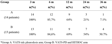
Look at the last column – at 36 months (3 year survival):

4 patients in the HITHOC treatment group were still alive versus just one in the talc pleurodesis cohort. The authors note that this survival for the HITHOC group might even be skewed a little, in that some of the patients in the HITHOC group didn’t receive treatment until SIX months after diagnosis (and all survival rates are calculated as length of survival after diagnosis).

So, yes, even with these small, small numbers, these findings are important enough for researchers to continue investigate in this area. It certainly warrants a larger study, research grants/ and other financial support.

However, it also needs to be noted, that researchers in this study found that the tumor tissue type had a major impact on outcomes. Patients with epithelioid MPM lived on average of 15 months after talc pleurodesis ( 9 patients) versus 45 months after HITHOC ( 9 patients). Patients with biphasic tumor type, or sarcomatoid type were less frequent in this study, but it appears to carry a poorer prognosis.

Reference article:

Migliore, M. et. al. (2021). Comparison of VATS Pleurectomy/Decortication Surgery plus Hyperthermic Intrathoracic Chemotherapy with VATS talc pleurodesis for the treatment of Malignant Pleural Mesothelioma: a randomized pilot study. MedRxIV, 28 Nov 2021.

For more about HITHOC, please see our archives.

Dr. Ahmet F. Işık talks about pleural mesothelioma, HITHOC, and thoracic surgery in Gaziantep, Turkey

updates on the on-going HITHOC project, war surgery, foreign body obstructions and bronchoscopy for infants

DSC_0033

Gaziantep, Southeastern Anatolia 

antep

It’s been over a year since I first read Dr. Isik’s work on treating pleural mesothelioma.  Since that time, Dr. Işik has continued his research into HITHOC and has now enrolled over 79 patients into the hyperthermic treatment group including one of the patients I met during my visit.  (There are 29 surviving patients in the study, 13 in the mesothelioma group, the remainder are secondary pleural cancers.).

(If you are a patient seeking treatment, or would like more information about Dr. Isik (or Dr. Gonzalez Rivas, Dr. Sihoe or any of the other modern Masters of thoracic surgery), we  are happy to assist you.  Contact me at kristin@americanphysiciansnetwork.org

First impressions are deceiving

I don’t know what I expected Gaziantep to look like as one of the world’s oldest cities, but from the moment the airplane begins its descent into a beige dust cloud, to the desolate brush and dirt of the airport outside the city, it isn’t what I expected.  Much of the antiquity of the biblical city of Antiochia has been replaced by a bustling modern city.  Historic ruins and ancient Roman roads marking this as part of the original Silk Road are conspicuous, only by their scarcity.

modern Gaziatep is featureless at first glance
modern Gaziantep is featureless at first glance

There are a handful of museums and monuments to the area’s rich history, but like the new name of Gaziantep (replacing Antep after the first world war), Turkey’s sixth largest city is modern; a collection of traffic and squat square buildings of post-modern architecture.

Kale
Kale

The city is also a mosaic of people.  There are groups of foreign journalists in the lobby of our hotel, and convoys of United Nations vehicles cruising the streets.  Crowds of Syrian children play in the park, calling out in Arabic to their parents resting on the benches nearby.  There is a smattering of Americans and English speakers interspersed, many are college students and other foreign aid workers on humanitarian missions to help alleviate the strain caused by large numbers of people displaced by the Syrian civil war.

Gaziantep is famed for their copper work

But like a mosaic, there is always more to see, the closer you look.  For me, as I look closer, I just want to see more.  I feel the same about Dr. Elbeyli’s thoracic surgery department.

The closer you look, the more you see. photo courtesy of wiki-commons
The closer you look, the more you see.
photo courtesy of wiki-commons

The border (and the largest Syrian city of Aleppo) lies just to the south – and the impact of the Islāmic militants is felt throughout the region.  No where is this more evident than at the local university hospital, where I meet Dr. Ahmet Işık and the Chief of Thoracic Surgery, Dr. Levent Elbeyli.

with Dr. Elbeyli (left) and Dr. Isik

Dr. Ahmet Feridun Işık

I like Dr. Işık immediately.  He is friendly and appears genuinely interested by my visit.  He’s from Giresun in the Black Sea region of northern  Anatolia of Turkey.  He attended medical school at Ankara University and completed his thoracic surgery training in Ankara before going to Adiyaman State Hospital in the bordering Turkish province of Adiyaman in southeastern Turkey.

He was an associate professor of thoracic surgery at Yuzuncu Yil University in the far eastern province of Van, Turkey before coming to Gaziantep in 2005.  He became a full professor at the University of Gaziantep in 2013.  In additional to authoring and contributing to his own publications, he also served as a reviewer for the Edorium series of open access journals.

It helps that his English is miles better than my non-existent Turkish.  (Reading about the Turkish language in phrase books is one thing, pronouncing words correctly is another.)

He doesn’t seem to mind my questions tumbling out one after another.  I’d like to be the cool, sophisticated visitor, but I’ve been waiting so long to ask some of these questions – and frankly, I am just excited to be there.

Dead-ends in medicine

There are a lot of “dead ends” in medicine – treatments that at first appear promising, but then end up being either impractical or ineffective.  In fact, for the first ten years of HIPEC, most surgeons dismissed it as a ‘dead-end’ treatment; the surgery was too radical and mortality too high.  But researchers kept trying experimental protocols; tweaking medications (less toxic) and procedures – and finding the right patients (not too frail prior to surgery) – and the literature shifted; from a largely useless ‘last ditch’ salvage procedure to a large, but potentially life-saving treatment. HITHOC is HIPEC in another color…

So I fire away –

Since our last post about Dr. Işık – he has performed several more cases of HITHOC on patients with pleural mesothelioma, pleural based cancers and advanced lung cancers.  He now has 79 patients in the HITHOC treatment group.  He has been receiving patients from all over Turkey, including Istanbul to be evaluated for eligibility for this procedure.  While the majority of patients are referred by their oncologists, others come to Gaziantep after reading about Dr. Işık on the internet.

None of the original patients (from 2009) are still alive, but their survival still exceeded all expectations, with 13 patients (of 14 HITHOC patients) living 24 to 36 months after the procedure.  (I don’t mean to be vague – but I was asking some of these questions in the operating room and I forgot to stuff my little notebook in my scrub pocket.)

While much of the literature surrounding the procedure cites renal failure as one of the major complications of the procedure, Dr. Işık has had one case of renal failure requiring dialysis.  Any other instances of elevated creatinine were mild and transient.  He doesn’t use any chemical renal prophylaxis but he does use fluid rehydration to limit nephrotoxicity.

He reports that while many surgeons consider sarcomas to be a contraindication to this procedure, he has had good outcomes with these patients.

He does state that diaphragmatic involvement in mesothelioma is an absolute contraindication because while the diaphragm can be resected / patched etc, it is almost impossible to guarantee or absolutely prevent the seeding of microscopic cancer cells from the diaphragm to the abdominal cavity – which increases the risk of disseminated disease.

He still uses Cisplatin – since that is what the original HITHOC researchers were using, but he uses a slightly higher dose of 300mg.  He’d like to do some prospective studies utilizing HITHOC (these have all been retrospective in nature – comparing today’s patients with past patients that received PDD and pleurodesis for similar conditions).  Prospective studies would allow him to better match his patients and to compare treatments head to head.  It would also allow him to compare different techniques or chemotherapeutic agents.

Unfortunately, as he explained, many of these types of studies of ineligible for government funding in Turkey because the government doesn’t want to pay for experimental / unproven treatments for patients even if there are few or no alternatives for treatment.  He is hoping to appeal this regulation so that he can continue his research since there is such a high rate of mesothelioma, that disproportionately affects rural Turkish patients.

 The University of Gaziantep Hospital

The University of Gaziantep Hospital

The University Hospital is one of several hospitals in Gaziantep.  The academic institution has over 900 beds and 20 operating rooms spread out over three floors.  There is a large 24 bed surgical ICU which includes 4 dedicated thoracic surgery beds.

Thoracic surgery may not be the advertised superstar of the hospital but it is the backbone of patient care.  There are three full-time professors of surgery; Dr. Ahmet Isik, Dr. Levent Elbeyli and Dr. Bulent Tunçözgür, along with an associate professor, Dr. Maruf Sanli, several thoracic surgery fellows and research assistants.  Together the thoracic surgery team performs over 1000 cases a year.

Dr. Levent Elbeyli is the driving force for thoracic surgery.  A Gaziantep native, he founded the department in 1992, and has seen it grow from a few scattered beds to a full-fledged program with a full-time clinic, 2 dedicated operating rooms, 4 ICU beds and 15 to 20 cases a week.

Dr. Levent Elbeyli (in loupes) in the operating room

For the thoracic nurse, the department of Thoracic Surgery is a dream come true; tracheal cases, surgical resections, esophagectomies, thoracic trauma – all of the bread and butter that makes our hearts go pitter-pat.  But then there is also plenty of pediatric cases, pectus repair, foreign body removal (oro-esophageal) and on-going surgical research.  They do a large amount of pediatric and infant bronchoscopies (for foreign body obstructions, tracheal malformations etc).

There is the slightly exotic hydatid cysts and the more mundane (but my personal favorite) empyema thoracis to be treated.  Cancers to be staged, and chest wall resections to undertake.  I feel almost overwhelmed in my own petite version of a candy store; everywhere I turn I see opportunities to learn, case reports to write and new things to see.

Dr. Levent Elbeyli operates as Dr. Isik observes.

My non-medical readers might be slightly repulsed by my glee – but it is this intellectual interest that keeps me captivated, engaged and enamored with thoracic surgery and caring for thoracic surgery patients.   And then there is the HITHOC program.  With a large volume of mesothelioma and pleural based cancers due to endemic environmental asbestos in rural regions of Turkey, there is an opportunity to bring hope and alleviate suffering on a larger level.  (Dr. Isik sees more cases here in his clinic in one year than I have seen in my entire career).

What’s not to love about that?

Article updates:

Since our original visit to Dr. Isik, he has continued his work on HITHOC for malignant pleural mesothelioma and other cancers.  You can read his latest paper, “Can hyperthermic intrathoracic perfusion chemotherapy added to lung sparing surgery be the solution for malignant pleural mesothelioma?

In this study, Dr. Isik and hs team looked at 73 patients with malignant pleural mesothelioma (MPM) who were in three different treatment groups.  Group 1 received surgery only (extrapleural pneumonectomy).  Group 2 received palliative treatment only.  Group 3 received lung sparing surgery with hyperthermic chemotherapy (HITHOC).  Lung sparing surgery included pleural decortication.

While the treatment groups are small, the results show a clear survival benefit to the patients receiving HITHOC.   Surprisingly, the palliative group lived longer than the surgery alone group.

Survival based on treatment modality:

Surgery only:  5 months average surgery.  15% survival at 2 years

Palliative treatment only: 6 months average survival   17.6% at 2 years

HITHOC group:  27 months average survival    56.5% at 2 years

Selected Bibliography for Dr. Işık  

Işık AF, Sanlı M, Yılmaz M, Meteroğlu F, Dikensoy O, Sevinç A, Camcı C, Tunçözgür B, Elbeyli L (2013). Intrapleural hyperthermic perfusion chemotherapy in subjects with metastatic pleural malignancies. Respir Med. 2013 May;107(5):762-7. doi: 10.1016/j.rmed.2013.01.010. Epub 2013 Feb 23. The article that brought me to Turkey, and part of our series of articles on the evolving research behind HITHOC.

Isik AF, Tuncozgur B, Elbeyli L, Akar E. (2007).  Congenital chest wall deformities: a modified surgical technique.  Acta Chir Belg. 2007 Jun;107(3):313-6.

Isik AF, Ozturk G, Ugras S, Karaayvaz M. (2005).  Enzymatic dissection for palliative treatment of esophageal carcinoma: an experimental study.  Interact Cardiovasc Thorac Surg. 2005 Apr;4(2):140-2. Epub 2005 Feb 16.

Er M, Işik AF, Kurnaz M, Cobanoğlu U, Sağay S, Yalçinkaya I. (2003).  Clinical results of four hundred and twenty-four cases with chest trauma. Ulus Travma Acil Cerrahi Derg. 2003 Oct;9(4):267-74. Turkish.

Sanli M, Arslan E, Isik AF, Tuncozgur B, Elbeyli L. (2013). Carinal sleeve pneumonectomy for lung cancer. Acta Chir Belg. 2013 Jul-Aug;113(4):258-62.

Maruf Şanlı, MD, Ahmet Feridun Isik, MD, Sabri Zincirkeser, MD, Osman Elbek, MD, Ahmet Mete, MD, Bulent Tuncozgur, MD and Levent Elbeyli, MD (2008). Reliability of positron emission tomography–computed tomography in identification of mediastinal lymph node status in patients with non–small cell lung cancer. The Journal of Thoracic and Cardiovascular Surgery, Volume 138, Issue 5, Pages 1200–1205, November 2009.

Sanlı M, Isik AF, Tuncozgur B, Elbeyli L. (2009).  A new method in thoracoscopic inferior mediastinal lymph node biopsy: a case report.  J Med Case Rep. 2009 Nov 3;3:96. doi: 10.1186/1752-1947-3-96.

Sanli M, Isik AF, Zincirkeser S, Elbek O, Mete A, Tuncozgur B, Elbeyli L. (2009).  The reliability of mediastinoscopic frozen sections in deciding on oncological surgery in bronchogenic carcinoma. J Thorac Cardiovasc Surg. 2009 Nov;138(5):1200-5. doi: 10.1016/j.jtcvs.2009.03.035. Epub 2009 Jun 18.

Sanli M, Işik AF, Tunçözgür B, Arslan E, Elbeyli L. (2009).  Resection via median sternotomy in patients with lung cancer invading the main pulmonary artery.  Acta Chir Belg. 2009 Jul-Aug;109(4):484-8.

Sanli M, Isik AF, Tuncozgur B, Elbeyli L.  (2010).  Successful repair in a child with traumatic complex bronchial rupture.  Pediatr Int. 2010 Feb;52(1):e26-8. doi: 10.1111/j.1442-200X.2009.03000.x

Sanli M, Işik AF, Tunçözgür B, Meteroğlu F, Elbeyli L. (2009).  Diagnosis that should be remembered during evaluation of trauma patients: diaphragmatic rupture].  Ulus Travma Acil Cerrahi Derg. 2009 Jan;15(1):71-6. Turkish.

Pleural fluid cytopathology

How to prepare a proper specimen for pleural fluid cytology & cytopathological analysis.

Pleural Fluid Cytopathology

Pleural fluid analysis is more than a typical ‘rounds’ question for interns and students.  This fluid contains important indicators of disease status.  Who among us hasn’t memorized pH levels, glucose and protein values?  (For a discussion on transudate versus exudate effusions, see the Medscape article by Jeffrey Rubins below.)

While pleural fluid analysis can be used to assist in the differential diagnosis of multiple conditions; pleural fluid cytopathology is often ordered when a more sinister condition like metastatic cancer is suspected.  Therefore, it is especially important for clinicians to ensure that pleural fluid cytopathology samples are collected, and sent in the most efficient and effective manner possible.  While there are few written guidelines regarding this process, here are some helpful tips based on interviews with several pathologists and the available literature.

Biopsy is best but fluid analysis is still helpful

While the gold standard for diagnosis is always tissue biopsy (in this case pleural tissue biopsy), this does not mean that cytopathological analysis is completely unnecessary.  In many cases, this fluid analysis gives a first look that aids in the diagnosis and staging of disease.  It is particularly useful for patients undergoing thoracentesis procedures, particularly when thoracentesis is performed in lieu of a more invasive procedure such as VATS (which allows for direct tissue biopsy.)

But do I still need to do a biopsy if the fluid analysis is negative?

However, there is often a catch-22 in the use of pleural fluid pathology which can lead to some confusion among patients and providers.  This catch-22 is related to the sometimes variable reliability of pleural fluid cytopathology for diagnosis of malignancy.  This means that the results aren’t always accurate.  As anyone in thoracic surgery can tell you, there have been numerous times when the fluid analysis results are reported as negative (for malignancy) even when the surgeon is (literally) staring at a pleural tumor in the operating room.  This means that a negative pleural fluid cytopathology result can not be used to rule out malignancy.

However, when the fluid is positive, it may save the patient from an additional procedure*.

Cytology versus cytopathology

“Cytology” is the generic term for the study of cells.  Cytopathology is the actual pathological investigation of free cells and tissue fragments, often for the diagnosis or treatment of cancer.

When used clinically, cytopathology is often used to distinguish between other more basic studies of bodily fluids or tissues.  This in-depth cellular examination is more critical in many cases than basic pleural fluid analysis.  This examination may include identification of immunological factors and tumor markers.  This is one of the tests that clinicians use to try and answer the question,“Is it cancer?”.  However, the answer is not always as straight forward.

Reliability and Predictive Value

This question is difficult to answer due to sometimes variable prognostic value of the fluid itself.  Even under the best of circumstances, reliability of this test (like most diagnostics) is less than 100%.  Different studies calculate the accuracy of pleural fluid analysis at detecting cancer vary wildly;anywhere from 10 to 80% has been reported in the literature with false negatives as the most frequent error (when discussing sensitivity and specificity). However, poorly prepared specimens may contribute to false results as well.

Ensuring optimal results by obtaining proper specimens

 Over the years, during different discussions with multiple pathologists as well as laboratory technicians, a common theme has emerged regarding the use of pleural fluid for pathology analysis.  Several of these individuals remarked that obtaining an accurate diagnosis was often difficult due to improper or suboptimal preparation of the pleural fluid, in addition to characteristics of the fluid itself.  What constitutes a ‘proper’ or ‘optimal’ pleural fluid is still (among lab technicians and pathologists) up to debate, but here are some general guidelines:

1. Send it all.

Due to the nature of pathology analysis which replies of the presence and identification of malignant cells within the fluid itself, a larger fluid specimen provides for a better sample.  When thoracentesis/ VATS or other drainage is being performed, and this yields 2 liters of fluid – send all two liters.  Don’t select out the first 25ml in a urine specimen cup, send it all.

There are no set guidelines for the amount of fluid necessary for cytopathology analysis. While malignancies have been successfully detected in amounts as small as 4ml, the rationale behind providing larger samples has been explained as ‘increasingly the likelihood of detecting the presence of cells indicative of malignancy’.

While the amount of fluid needed is currently up for debate among pathologists, sending too little fluid may result in a missed diagnosis, whereas an overabundance of fluid is more of an inconvenience to lab technicians.

Be sure to include the last frothy bits, which often contain more sediment/ cellular material than fluid recovered at the beginning of the sample.  (The content of this fluid may even vary due to the patient’s position – which is another reason to take a larger sample.) In a conscious patient, this may mean several minutes of discomfort, but encourage patients to take deep breaths, and cough so that as much fluid as possible can be removed.  (In patients with very large effusions, this may be a lengthy process as ‘short breaks’ are taken during the procedure to accommodate for fluid shifts.  This brings us to # 2.

2.  Keep it fresh: Talk to the lab about whether you should consider adding an additive like heparin or EDTA to your sample at the time of collection to prevent the degradation of cells.  Depending on when / where your sample is collected and sent – there may be significant delays in the processing of the collected sample.  Many pathologists report that after 4 hours there are significant changes in untreated pleural fluid kept at room temperature.

Consider this as you gather your sample;

–          Did you leave it in the patient’s room for the nursing staff to deliver?

–          Is it possible it may sit for several hours before arriving to the lab?

–          Is the lab well-staffed or will the fluid sit waiting for analysis by overworked, and stressed employees at a lab that may be inundated with many more urgent requests?

Guzman et al. (1992) and other researchers found that with the addition of EDTA to pleural fluid specimens, tumor cells were easily identified even after four days of storage.

Even if your facility doesn’t provide EDTA for your specimens, it’s a good bet that sending a syringe full of fluid from the bottom of a week-old pleurovac is probably not your best bet.

3.  Eliminate errors: Don’t make them guess!

Always personally label fluid and tissue samples completely with the patient’s name, reference number (as used by your institution), body site (ie. Right pleural space) as well as the ordering clinician’s name.  Include your phone number if you want to be called with the results or questions.

On the actual order, or lab requisition, provide additional information including patient symptoms, and pertinent history (ie. 63 year old with 40+ pk years of smoking, and history of asbestos exposure in Navy shipyards, now presents with pleural effusion, chest pain and 25 pound weight loss.)  Provide any special instructions as needed.   This allows the pathologist examining the patient’s specimens to correlate clinical history, symptoms and other available diagnostics with cellular findings and stains.

4.  Now do it again.  If the patient develops a second pleural effusion, go ahead and send that fluid too – particularly if the first sample was non-diagnostic.

*Depending on the patient’s clinical status/ symptomology.  As mentioned in a previous post, many patients with malignant pleural effusions may undergo additional procedures at some point in time for palliation of symptoms.

References

 American Society of Cytopathology – a great resource for interested readers.  The website also contains a ‘virtual slide atlas’ which includes case studies and several slides showing pleural fluid cytopathology. Click here for the case study of a 60 year old with pleural effusion.

Antonangelo L, Capelozzi VL. (2006). Collection and preservation of the pleural fluid and pleural biopsy. J Bras Pneumol. 2006;32 Suppl 4:S163-9. Portuguese.  These Brazilian authors from the University of Sao Paulo discuss the proper collection of pleural fluid specimens.  In this article, the authors make recommendations for the collection, storage and examination of pleural fluid for a variety of laboratory and microscopic tests.

Brandstetter RD, Velazquez V, Viejo C, Karetzky M. (1994). Postural changes in pleural fluid constituents. Chest. 1994 May;105(5):1458-61.

Guzman J, Arbogast S, Bross KJ, Finke R, Costabel U (1992).  Effect of storage time of pleural effusions on immunocytochemical cell surface analysis of tumor cells. Anal Quant Cytol Histol. 1992 Jun;14(3):203-9. No free full text available.

Porcel JM.  (2011).  Pearls and myths in pleural fluid analysis. Respirology. 2011 Jan;16(1):44-52.  Porcel advocates for smaller volumes, but an ‘inadequate sample’ should never be a reason for a missed diagnosis.  He also advises the addition of an additive if there are any anticipated delays (4+hours) in specimen processing.

Salyer WR, Eggleston JC, Erozan YS. (1975).  Efficacy of pleural needle biopsy and pleural fluid cytopathology in the diagnosis of malignant neoplasm involving the pleura. Chest 1975 May, (5) 536-9.  Classic article on the predictive value of pleural fluid cytopathology. A  pdf of Salyer et al is available here.

Rubins, J. (2013).  Pleural effusion workup.  From Medscape/ Emedicine.com.  Pleural Effusion Workup pdf version.

Additional Resources

Shidham, V. B. & Falzon, M. (2010). Serous cavities.  Chapter 3 in  Diagnostic Cytopathology: Expert Consult: Online and Print (2010).  Grey & Kocjan (Eds).   Elsevier Health Sciences.

Awake Epidural Anesthesia for thoracoscopic pleurodesis

Awake epidural anesthesia for thoracoscopic pleurodesis: A prospective cohort study. a new publication from Dr. Mauricio Velasquez and his surgical team reviewing results from their 36 month study

On the heels of a recent announcement on CTSnet.org soliciting surgeon input on their experiences with non-general anesthesia for thoracic surgery procedures, Cirugia de torax is revisiting one of the surgeons we interviewed last year, Dr. Mauricio Velasquez at Fundacion Valle de Lili in Cali, Colombia.

Dr. Velasquez in the operating room with Lina Caicedo Quintero (nurse) Valle de Lili, Cali, Colombia
Dr. Velasquez in the operating room with Lina Caicedo Quintero (nurse) Valle de Lili, Cali, Colombia

The trip to Cali was primarily to discuss Dr. Velasquez’s Thoracic Surgery Registry, and to observe him performing several single port surgery cases.  However, during the trip, Dr. Velasquez also spoke about several other aspects of his current practice including some of his recent cases, and the thoracic surgery program at Fundacion Valle de Lili.

Dr. Mauricio Velasquez after another successful case

We also talked with his wife, (and lead author), the talented Dra. Cujiño, an anesthesiologist who subspecializes in thoracic anesthesia.   Together, they have successfully performed several thoracic cases using thoracic epidural anesthesia on awake patients.

By chance, they published articles in both  Revista Colombianas de anesthesia and Neumologia y cirugía de torax in the last few weeks.

Revista Colombianas de anesthesia

Patients receiving epidural anesthesia received a small dose of midazolam prior to insertion of epidural needle at the T3 – T4 intervertebral space.  During the case, patients received bolus administration via epidural of 0.5% bupivacaine on a prn basis.

Short surgeries, single port approach

All patients, regardless of anesthesia type underwent single port thoracoscopic surgery for the talc pleurodesis procedure.  Surgery times were brief, averaging 30 to 35 minutes  for all cases (range 25 – 45 minutes) with the epidural patient cases being slightly shorter.

Dr. Mauricio Velasquez performing single port thorascopic surgery

Dramatic reduction in length of stay

In their study, patients receiving awake anesthesia had an average length of stay of four days compared with ten days for the general anesthesia group.

Decreased incidence of post-operative complications

There was a marked reduction in the incidence of post-operative respiratory complications (19 in general anesthesia group) versus 3 patients in the awake anesthesia group.  Post-operative mortality was also decreased (six in general anesthesia) versus two deaths in the awake anesthesia group.  However, the mortality statistics may also be impacted by the overall poor prognosis and median survival time of patients presenting with malignant effusions.

Post-operative pain

Study patients also self-reported less post-operative pain in the awake anesthesia group – with only one patient reporting severe pain versus seven patients in the general anesthesia group.

Conclusions

Cujiño, Velasquez and their team found awake thoracic epidural anesthesia (ATEA) was a safe and effective method for intra-operative anesthesia and was associated with a decreased post-operative pain, decreased length of stay (LOS) and decreased incidence of post-operative complications.

Notes

This study was funded by the authors with no relevant disclosures or outside financial support.

References

Indira F. Cujiño,  Mauricio Velásquez,  Fredy Ariza,  Jhon Harry Loaiza (2013).    Awake epidural anesthesia for thoracoscopic pleurodesis: A prospective cohort studyRev Colomb Anestesiol. 2013;41:10-5.  A 36 month study involving 47 cancer patients comparing (standard) general anesthesia outcomes with awake epidural anesthesia.

en Espanol: Anestesia epidural para pleurodesis por toracoscopia: un estudio prospectivo de cohort.

The second article has not been posted online yet.  Look for updates in the coming weeks.

Intrathoracic Hyperthermic Chemotherapy (Hithoc) in advanced non-small lung cancer: the Nara Experience

As part of a continuing discussion of HITHOC (Hyperthermic IntraThoracic intraOperative Chemotherapy), today we are talking about the results of a small study conducted at the Nara Medical University, School of Medicine in Nara, Japan.

As part of a continuing discussion of HITHOC (Hyperthermic IntraThoracic intraOperative Chemotherapy), today we are talking about the results of a small study conducted at the Nara Medical University, School of Medicine in Nara, Japan.

While the study is small (just 19 patients in three groups), it’s important because the patients involved all had advanced lung cancer, with malignant pleural effusions or disseminated disease discovered at the time of surgery. This is important, as readers know, because lung cancers are often diagnosed late, (after patients develop malignant effusions), and that the prognosis for patients with malignant effusions is grim.

Population: 19 patients.

Notably, the treatment group C consisting of seven patients (which received no intrathoracic thermic treatment) who were treated during an earlier period (2001 – 2003). Group C had an average age of 64. Essentially a control group.

The remaining patients were treated during 2006 – 2008 and are divided into two groups;

Group A which received hyperthermic (hot) saline infusion with a 30 minute dwell time – consisted of seven patients.  This group was also older (average age 72).

Group B, consisting of five patients who received hyperthermic chemotherapy (cisplatin) infusion into the chest cavity with a 30 minute dwell time.

Note: Infusion in this post refers to instillation of fluid into the chest cavity, not an intravenous treatment.    All patients received post-operative adjuvant chemotherapy.

The grouping of A and B serves to distinguish whether the mechanism of treatment is related to the application of heat alone, or the application of heated chemotherapeutic agents.  Current theories about the effectiveness of HITHOC suggest that the heat of the chemotherapy allows the drugs to penetrate more deeply into the tissues compared to application of chemotherapy alone, but requires studies such as this to support this theory.

Interestingly, the pre-operative staging of these patients differed significantly from intra-operative findings with 8 patients diagnosed with early disease (stage I), five patients with stage II and only six patients as stage IIIA pre-operatively.  (Presence of a pleural effusion denotes stage IV).  Malignant effusions were not seen during pre-operative workup. (It is not uncommon to find more advanced cancer at the time of surgery.)

Surgery: All of the patients underwent a VATS procedure (video-assisted thoracoscopy).  The majority of patients of patients (16) underwent surgery to remove the primary lesion (cytoreductive surgery) with ten patients undergoing lobectomy and six patients undergoing wedge resection.

Intra-operative findings:  16 patients found to have malignant effusions, 10 patients with disseminated disease.

Results: No intra-operative/ post-operative deaths.

Group A (hot saline group): no deaths during follow-up period, with a median follow-up period of almost 20 months.  No recurrence of pleural effusions.

Group B (heated chemotherapy group): 4 deaths in follow-up period; median survival time was 41 months, one patient with recurrent pleural effusion 26 months after treatment.

Group C: (VATs alone): 5 deaths (during follow-up period) median survival 25 months, 4 patients with recurrent pleural effusions (average time to recurrent effusion: 3 months).

While this study is too small (with only five patients receiving intrathoracic chemotherapy) to generalize the results – it should prompt researchers into conducting more studies and trials into the use of hyperthermic intrathoracic chemotherapy in patients with late stage lung cancers.

The decreased incidence of pleural effusion in the treatment groups (A and B) is important also for quality of life issues.  However, these findings are also limited by the small study size.

I have written to Dr. Naito (corresponding author on this article) for further comment and information.

Reference:

1. Kimura, M., Tojo, T., Naito, H., Nagata, Y., Kawai, N., & Taniquichi, S. (2010). Effects of a simple intraoperative intrathoracic hyperthermotherapy for lung cancer with malignant pleural effusion or dissemination. Interactive Cardiovascular & Thoracic Surgery 2010, April, 10 (4); 568 -71.  (linked to pdf).

Outpatient Treatment for Malignant Effusions

Discussion of treatment goals, and patient centered care for Malignant pleural effusions. This is the first in a series of articles on lung cancer, and lung surgery topics. Originally posted at our sister site.

Not all conditions are curable, and not all treatments are curative. Some treatments are based on improving quality of life, and alleviating symptoms. This is a hallmark of patient centered care – doing what we can to make the patient feel better even when we can’t ‘fix’ or cure the underlying disease. No where is this more evident than in the treatment of malignant effusions.

By definition, a Malignant Effusion is the development of fluid in the fluids related to an underlying (and sometimes previously undiagnosed) malignancy. Malignant effusions can be seen with several different kinds of cancers, most commonly lung and breast cancers. The development of a malignant effusion is a poor prognostic sign as it is an indicator of metastasis to the pleural tissue/ space.

The development of a malignant effusion usually presents with symptoms of shortness of breath, and difficulty breathing. While the treatment of the underlying cancer may vary, the primary goal of treatment of an effusion is palliative (or symptom relief). The best way to relieve symptoms is by removing the fluid.

This can be done several ways – but each has its own drawbacks.

Thoracentesis:
The fluid can be drawn out with a needle (thoracentesis) either bedside or under fluroscopy. This procedure is quick, and can be performed on an out-patient basis, in a doctor’s office, or in radiology.

The potential drawbacks with this treatment strategy are two-fold:

1. There is a chance that during the procedure, the needle will ‘poke’ or ‘pop’ the lung, causing a pneumothorax (or collapse of the lung). This then requires a chest tube to be placed so the lung can re-expand while it heals. However, if the procedure is performed uneventfully, (like it usually does) the patient can go home the same day.

2. The other complication – is rapid re – accumulation – since you haven’t treated the underlying cause, but have only removed the fluid. This also happens when the cause of the effusion (nonmalignant) is from congestive heart failure. This means the fluid (and symptoms of shortness of breath) may return quickly, requiring the patient to return to the hospital – which is hard of the patient and their family.

Video- Assisted Thoracoscopy: (VATs)
Malignant effusions can also be treated by VATS – this is a good option if we are uncertain of the etiology (or the reason) for the effusion. While all fluid removed is routinely sent for cytopathology (when removed during surgery, thoracentesis or chest tube placement) – but cytopathology can be notoriously inaccurate with false negative reports, because the diagnosis is dependent on the pathologist actually seeing cancer cells in the fluid.  However, during the VATs procedure – the surgeon can take tissue samples, and photos along with fluid for diagnostic testing.   This is important because I have had cases in the operating room (VATS) where the surgeon actually sees the tumor(s)** with the camera but the fluid comes back as negative.

** in these cases, we send biopsies of the tumor tissue – which is much more accurate and definitive.

But a VATS procedure requires an operation, chest tube placement and several days in the hospital.

Chest tube placement:
Another option is chest tube placement – which also requires several days in the hospital..

During both chest tube placement and VATS, a procedure called pleurodesis can be performed to try to prevent the fluid from re-accumulating.

But what if we know it’s a malignant effusion? What are the other options for treatment?

Catheter based treatments: (aka PleurX style catheter, or Heimlich valve)
(note: catheter means a small tube – a foley catheter is the type used to drain urine, but other types are used for many things – even an IV is a catheter.)
One of the options used in our practice was pleur X (brand) catheter placement. This catheter was a small flexible tube that could be placed under local anesthesia – either in the office or the operating room – as an ambulatory procedure. After some patient teaching, including a short video, most family members felt comfortable emptying the catheter every two or three days at home, to prevent fluid  re -accumulation (and allowing the patient to continue normal activities, at home.)

PleurX catheter placement is preferred in many cases due to ease of use, and patient convenience. The Heimlich valve is messier – as it tends to leak, and harder for patients to hide under clothing.

Sometimes a visiting nurse would go out and empty the catheter, and in several cases, patients would come to the office, where I would do the same thing – it was a nice way to relieve the patient’s symptoms without requiring hospitalization, and several studies have shown that repeated drainage often caused spontaneous pleurodesis (fluid no longer accumulated.) We would then take the catheter out in the office.. Now, like any procedure, there is a chance for problems with this therapy as well, infection, catheter can clog, etc..

But here’s another study, showing that even frail patients benefit from home-based therapy – which is important when we go back and consider our original treatment goals:
-Improving quality of life
-Relieving symptoms

In the article, the authors used talc with the catheters and then applied a Heimlich valve, which is another technique very similar to pleurX catheter placement.  (Sterile talc is used for the pleurodesis procedure – which we will talk about in more detail in the future.)

Another article, this one by Heffner & Klein (2009) published in the Mayo Clinic Proceedings discusses the diagnosis and treatment of malignant effusions.