We are here with the latest HITHOC pilot study updates from Dr. Migliore et al. If you remember, Dr. Migliore and his colleagues in Sicily have been investigating and researching the effectiveness of HITHOC for well over a decade. In fact, we’ve talked about this study before, when we presented preliminary findings. But now the authors are presenting the results of a six-year pilot study with long-term follow-up in a paper that was published in the scientific journal, Cell. (Original paper here).
Why this is important
In the general population, patients diagnosed with malignant pleural mesothelioma (MPM) have a mean survival of 9 to 12 months.
The mean age at time of death was 70, meaning this disease affects relatively young patients after a long incubation period. It affects men at a ratio of almost 4 men (3.6) to one woman. (Presumably, this is related to occupational/ industrial exposures and trends – few women work/ worked in shipyards, construction).
While many people think that asbestos related mesothelioma is a thing of the past (or time limited due to the fact it has known carcinogenic effects), it is still being used in many products in several countries including the United States.
How do we treat Pleural Mesothelioma now?
Despite this, the standard of treatment is generally palliative in nature. Talc Pleurodesis is used to drain existing fluid around the lung, and then the talc is used as a sclerosing (or scarring) agent to prevent the pleural surfaces from being able to secrete more fluid. This makes the patient feel better because they breathe better – which is a very important consideration for patient care – but does nothing to treat the underlying cancer or prevent its spread. Patients generally live around 14 months after this procedure, when it is performed for mesothelioma.
Other surgical treatments have been tried in the past including Extrapleural pneumonectomy (EPP), which has a high mortality rate, and Pleurectomy/ Decortication.
Pleurectomy / Decortication, which has replaced EPP in many cases, has a low surgical mortality (1.8%), but carries a high risk of recurrence, meaning many patients’ cancer will return. The average survival for patients having this procedure is only 17 months.
In contrast, there have been several small studies that suggest that HITHOC offers greater survival for patients with mesotheliomas – with the average survival of 20 to 35 months. But these studies have been small, and many thoracic surgeons and oncologists remain unaware of HITHOC as a potential treatment option. Others remain skeptical of its potential benefits (which is not necessarily a bad thing!) So, Dr. Migliore and his colleagues designed this study to see if a larger scale trial with more participants would be feasible or worthwhile.
Pilot studies like this one are used to determine how many patients need to be enrolled to see a statistically meaningful result, and if there is a “meaningful” result at all.
This is important because many of the studies mentioned in general media do not meet this criteria but are widely reported as successful nonetheless just because of so-called ‘newsworthiness’. Every time readers see a story on a seemingly miraculous cure based on a garden variety supplement for a wide host of medical problems (depression, arthritis, heart disease, Alzheimer’s or you name it), this is an example why pilot studies are needed to both protect the public and to advance medicine using scientific evidence. (We’ve talked about this before.)
So what does this pilot study show?
As you may recall, the preliminary results from this study, (as we reported in 2021) showed favorable results in the HITHOC treatment group, with improved survival rates, despite the fact that many of the patients that were randomized into the HITHOC treatment group actually had more advanced cancers.
The final data confirms this with 30% of the HITHOC patients (4 out of 13) alive at median follow up of 28 months. (Compared to the pleurodesis group which only had one patient (out of 14) still alive at 19 months. For the full results of this trial – please see the original article.
The biggest limitation for the researchers in this trial was recruiting patients for enrollment. They reached out to multiple medical centers and oncology practices – and received referrals for only five patients a year, despite the fact that Biancavilla, in Sicily is one of the mesothelioma hotspots. This small number met the threshold for statistical significance for a pilot study – but falls far short of what would be needed for a larger, more powerful study.
but for large scale, multi-site international trials in the future, we need to do a better job at connecting eligible patients with research studies..
Now what?What’s next ?
Now as we wait to see if other surgeons and oncologists will answer the challenge – and participate in a larger, multi-center (international) randomized control trial with hundreds, if not thousands of patients, to compare HITHOC to other treatments. When, and if, that happens, I will report enrollment information for potential referring physicians and patients.
Other news in the treatment of MPM
In the meantime, we await further results from the MARS2 trial, headed by the dynamic Dr. Eric Lim, in the UK. These results will be presented in Singapore, this fall.
The MARS2 trial, unfortunately, doesn’t use any HITHOC protocols but looks at whether pleurectomy/ decortication in addition to chemotherapy alone enhances survival and quality of life. Now, if only Dr. Lim would introduce some of that chemotherapy (preferably Cisplatin) at 42.5 degrees into the chest cavity. Then we’d really have something to talk about!
As we’ve talked about in a previous interview with Dr. Isik, (Turkiye) there are regional areas where natural conditions (such as asbestos in the soil) lead to mesothelioma clusters. There are also areas, like the naval shipyards in the United States, where occupational/ industrial exposure leads to disease clusters. In this investigation of mesothelioma clusters in Western Europe, the majority were industrial/ occupational in nature.
Is asbestos illegal?, The Mesothelioma Center, consumer information by lawyers for mesothelioma patients.
Banning Asbestos, Mesothelioma.com, consumer information by lawyers for mesothelioma patients.
While working on a recent interview with one of the New Masters of Thoracic Surgery, I talked about one of his biggest contributions to his local community, which was establishing the first dedicated thoracic surgery program in that city. Then I realized that maybe readers wouldn’t know what that was important.. This article came from that interview
Memphis, Tennessee at night
Big hospitals, little hospitals. Major health systems and community facilities battle it out of our insurance dollars. Private wings, VIP suites, catered meals and fancy robots all try and lure patients in the doors. As a writer of several books based on the business of medical tourism – I’ve seen that the appeal of glistening marble floors, free fancy coffees and an aura of exclusivity can trump the principles of safe and effective patient care when it comes to attracting paying patients. This is acutely evident in the surgery wars; the wars to attract referrals between private practice and academic medicine (which usually, but not always – has less glamorous facilities**). But for a person facing a large, and possibly life-saving thoracic surgery, we need to explore the differences that are more than just skin-deep.
Subspecialty interest and skill
The difference between a true thoracic surgery program and a cardiothoracic surgery private practice group is often marked by the degree of continuing competence, subspecialty interest and skill in minimally invasive techniques. (For more about the overall differences between general thoracic and cardiothoracic surgery, read here.) This post is discussing the pitfalls of the private practice medical group and surgical referral patterns. Surgical partners in a lucrative practice don’t have continuing education requirements, but residencies do. In order to teach surgical residents, the attendings themselves need to be well-versed in the latest operating techniques and surgical outcomes research.
Where the patients come from
Private practice groups get their patients thru an ‘old boy network’ particularly in cities with few strong ties to university medical centers. Patients don’t just walk thru the door to see a thoracic surgeon – they are referred to one. Most people have never even heard of a thoracic surgeon before they or a loved one needs one.
As we talked about in one of our very first posts, “Who is performing your thoracic surgery?” – just because you need thoracic surgery, that doesn’t guarantee that a patient will see an actual board certified thoracic surgeon.
In a referral based system, patients are often not referred based on the skills or merits of the surgeon in the operating room, his rates of post-operative infection or even the health system affiliations – but by his charm, wit or connections on the social scene. In a city like Memphis, which is awash in old money, southern tradition and the Junior League, this means that patients are referred to the surgeon based on the friendships amongst wives, college fraternity friendships or 6 am tee-off times.
Cardiothoracic versusgeneralthoracic
Often times, the surgeon is not particularly gifted or even interested in modern lung or esophageal surgery techniques, meaning that the surgeon is most likely to revert to large thoracotomies or median sternotomies because that’s where his comfort lies. There is no standard or requirement to master minimally invasive techniques, so often these surgeons don’t. It’s not a criticism of cardiothoracic surgery, but a basic reality. A heart surgeon wants to be a heart surgeon. He doesn’t necessarily want to do lung or esophageal surgery, but he might not turn away these cases either, because everyone likes to make a living.
In comparison, a dedicated thoracic surgery program, particularly in an academic setting; is made up exclusively of thoracic surgeons who live and breathe general (noncardiac) thoracic surgery. This is what they do, this what they want to do, this is what they have always wanted to do. Academic settings also have more stringent requirements (in general) regarding maintaining clinical and educational competencies. These surgeons are learning or teaching the newer techniques, reading and writing the literature and actively pursuing advances in the field. This dedication is important for more than the most obvious reason – sure, you want your surgeon to be competent in the operating room – but you also want him to be knowledgeable and skilled outside of it.
Academic centers with general thoracic surgery programs are more likely to have a protocol based, formalized multidisciplinary approach to thoracic disease. This means that patients are treated by a team of specialists in a cooperative fashion. There are no conflicts between what the oncologist wants to do and what the surgeon wants. If the patient needs pre-operative radiation or chemotherapy, it’s coordinated in conjunction with surgery, so that the patient receives care in a timely and organized fashion based on the current treatment recommendations and clinical research**.
But American medical care is the best in the world, right?
Multidisciplinary approach, evidence-based practice, ongoing academic research and continuing surgical education: All of these themes don’t sound extraordinarily unusual to readers because I have been discussing and presenting surgeons who work within these types of programs for years here at Thoracics.org.
Not the norm
But it’s actually not the norm in the United States, which means that many American patients get woefully inadequate, outdated or just plain uncoordinated care. These patients have more pain, more suffering, longer lengths of stay, more complications and less quality of life than any of the patients who have been cared for by just about any surgeon ever mentioned on this site. Patients at the University of Pittsburgh, Duke, University of Virginia or John Hopkins were getting great care, but patients here in Memphis, Las Vegas or any of the other cities or regions without these types of specialized programs, weren’t and often still aren’t.
When added to the growing shortages in this specialty area, an appointment with a trained thoracic surgeon may become an elusive endeavor. Especially if patients don’t know to ask.
* A thoracic surgery program that focuses on diseases and conditions of the lungs, esophagus and mediastinum.
** There are several academic medicine facilities that have managed to boast their own celebrity style perks, like the VIP wings at John Hopkins.
An invited report from Dr. Marcello Migliore on the recent Italian conference on VATS and lung cancer
Report from the 3rd Mediterranean Symposium in Thoracic Surgical Oncology on VATS RESECTIONS FOR LUNG CANCER: moving toward standard of care.
Speakers and moderators at the 3rd Mediterranean Symposium on Thoracic Surgical Oncology
The third mediterranean symposium on thoracic surgical oncology was successful. The symposium was held the 21st – 22nd april 2016 at the Aula Magna of the Faculty of Medicine at the University of Catania. More than 150 people attended, and among them there were thoracic surgeons, general surgeons, oncologists, chest physicians, residents and medical students. This year, we had speakers from Europe and the USA. The main topic was VATS resections for lung cancer (Photo 1). During the opening ceremony, the Rector Giacomo Pignataro awarded a medal to Professor Tom Treasure for enhancing our outstanding education and research experience (Photo 2).
Photo 2: The Rector of the University of Catania is giving the medal to Professor Tom Treasure
Although the concept of operating thru a small port was born and developed in Europe (1- 7) it has been noted that 90% of papers on uniportal VATS lobectomy come from East Asian countries (8-11). Throughout the symposium different speakers agreed that a proper definition of uniportal VATS is mandatory to speak the same language worldwide.
Awake thoracic surgery was discussed together with the need of accurate preoperative staging procedures such as endobronchial ultrasound, videomediastinoscopy or Video-assisted mediastinal lympadenectomy. It was concluded that a wide spectrum of factors must be considered when determining the appropriate tests to assess the lymph nodes in NSCLC, which includes not only the sensitivity and specificity of the test, but also the ability to perform the procedure on an individual patient.
Data from New York showed very clearly that there have been no large-scale randomized control trials to compare open and VATS lobectomy. Although most may agree with the short-term superiority of VATs lobectomy over its open counterpart, many argue that is an in adequate oncologic procedure. Hence whether the approach is equivalent in overall and cancer specific survival to its open counterpart is not known. He also reported an important recent analysis of SEER-Medicare which confirmed that VATS lobectomy appears to have similar survival to its open counterparts (12).
A magnificent video was presented to explain every step of the lobectomies performed through a small skin incision. A long discussion followed and all auditorium proposed that ‘single incision’ VATS probably define better than uniportal VATS what surgeons are doing worldwide. Certainly the length of skin incision is important and should be taken in serious consideration. We felt that a consensus conference is probably necessary consensus conference is probably necessary. The indication for a Wedge resection rather than lobectomy in initial stage lung cancer is still weak.
The Italian VATS group was formed in 2013 , and nowadays there are 65 participating centres and that 2800 VATS lobectomy have already been included. In Catania we joined the group few months ago (13)
A very interesting session for juniors and medical students from UK and Italy was carried out, and 12 abstracts have been presented as interactive posters. Two of them have been chosen for possible publication in Future Oncology.
Finally, the first data survival seems to benefit little from the various even growing “personal” modifications of the standard VATS technique. Since there is a limited variation between VATS and uniportal VATS, the likelihood is that either VATS and uniportal VATS will be operative in the near future. Its success will depend on survival advantages and decrease chest pain and not just on new technical instrumentation. To protect patient’s safety, the length of the skin incision should be chosen on the basis of several clinical factors and not in relation of modern “demand”. Although the trial VIOLET is ongoing in UK to demonstrate if VATS resection for lung cancer is better than open thoracotomy, doubts arises as standard postero-lateral thoracotomy for lung cancer seems to be an incision which is performed rarely today. A skin incision of 6-8 cm (mini-thoracotomy) with video assistance is enough for most of lung resections. The question which arises is if a mini-thoracotomy of 6 cm should be called “uniportal” or not.
Marcello Migliore, MD
Thoracic surgeon and invited commentator
Dr. Marcello Migliore
Migliore M Initial History of Uniportal Video-Assisted Thoracoscopic Surgery. Ann Thorac Surg 2016;101 (1), 412-3.
Migliore M, Calvo D, Criscione A, Borrata F. Uniportal video assisted thoracic surgery: summary of experience, mini-review and perspectives. Journal of Thoracic Disease 2015; 7 (9), E378-E380
Migliore, M., Giuliano, R., & Deodato, G. (2000). Video assisted thoracic surgery through a single port. In Thoracic Surgery and Interdisciplinary Symposium on the threshold of the Third Millennium. An International Continuing Medical Education Programme. Naples, Italy (pp. 29-30).
Migliore, M., Deodato, G. (2001). A single-trocar technique for minimally invasive surgery of the chest. Surgical Endoscopy, 8(15), 899-901.
Migliore M. Efficacy and safety of single-trocar technique for minimally invasive surgery of the chest in the treatment of noncomplex pleural disease. J Thorac Cardiovasc Surg 2003;126:1618-23.
Rocco, G., Martin-Ucar, A., & Passera, E. (2004). Uniportal VATS wedge pulmonary resections. The Annals of Thoracic Surgery, 77(2), 726-728.
Gonzalez D, Paradela M, Garcia J, et al. Single-port video-assisted thoracoscopic lobectomy. Interact Cardiovasc Thorac Surg 2011;12:514-5.
Yang HC, Noh D. Single incision thoracoscopic lobectomy through a 2.5 cm skin incision. J Thorac Dis 2015;7:E122-5.
Ocakcioglu I, Sayir F, Dinc M. A 3-cm Single-port Video-assisted Thoracoscopic Lobectomy for Lung Cancer. Surg Laparosc Endosc Percutan Tech 2015;25:351-3.
Kamiyoshihara M, Igai H, Ibe T, et al. A 3.5-cm Single-Incision VATS Anatomical Segmentectomy for Lung Ann Thorac Cardiovasc Surg 2015;21:178-82.
Zhu Y, Xu G, Zheng B, et al. Single-port video-assisted thoracoscopic surgery lung resection: experiences in Fujian Medical University Union Hospital. J Thorac Dis 2015;7:1241-51.
Paul S, Isaacs AJ, Treasure T, Altorki NK, Sedrakyan A. Long term survival with thoracoscopic versus open lobectomy: propensity matched comparative analysis using SEER-Medicare database. BMJ 2014;349:g5575
Migliore, M., Criscione, A., Calvo, D., Borrata, F., Gangemi, M., & Attinà, G. (2015). Preliminary experience with video-assisted thoracic surgery lobectomy for lung malignancies: general considerations moving toward standard practice. Future Oncology, 11(24s), 43-46.
Migliore M. Will the widespread use of uniportal surgery influence the need of surgeons ? Postgrad Med J 2016 (in press).
Details about the upcoming Robotic thoracic surgery course at NYU this June.
New York University School of Medicine has an upcoming CME course on Robotic Thoracic Surgery this June (10th & 11th). The day and a half course will be held at NYU Langone Medical Center in New York City.
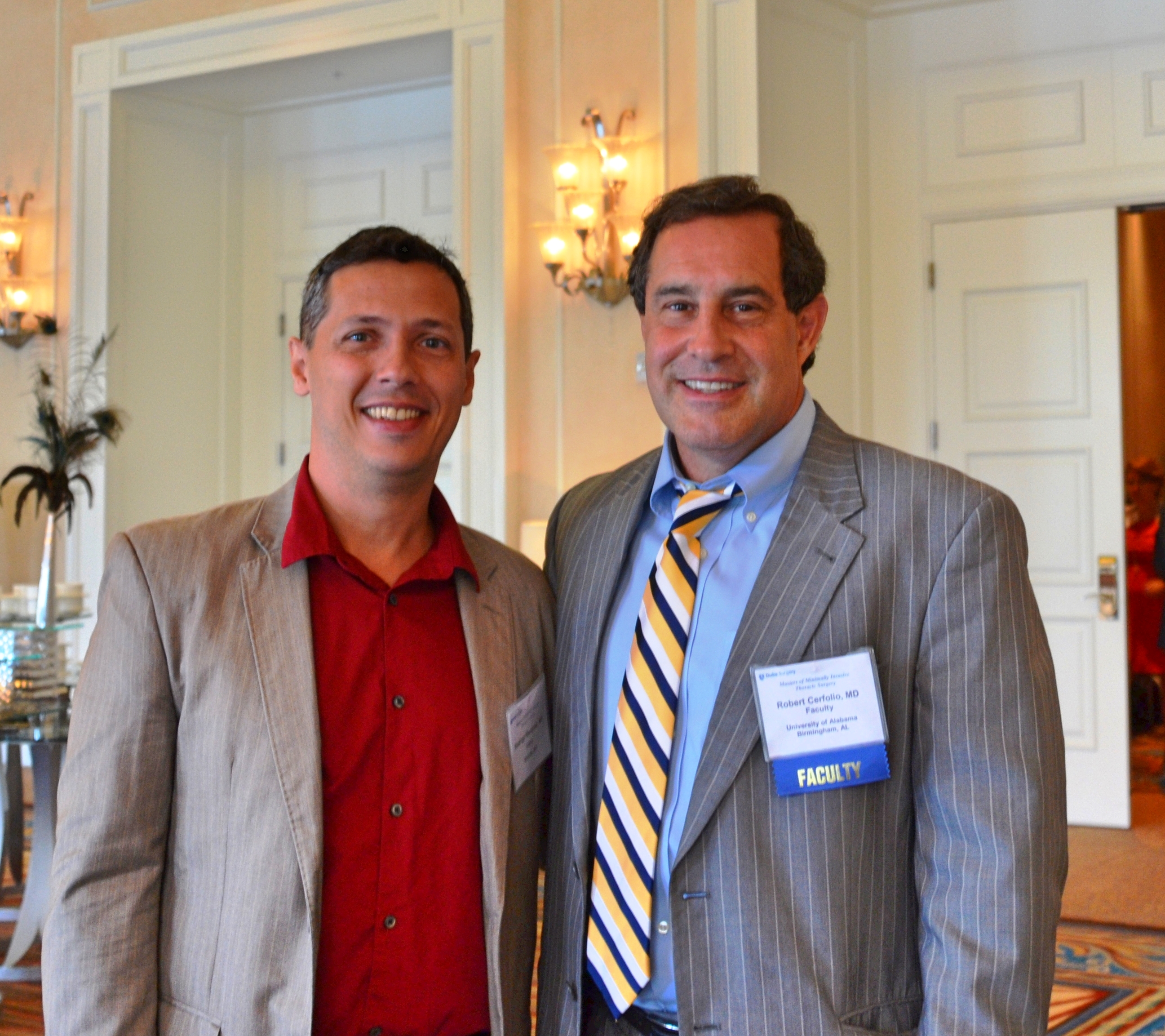
The conference covers robotic surgery basics as well as lectures on robotic esophagectomies and mediastinal surgery. Robotic master surgeon, Dr. Robert Cerfolio will be giving two presentations.
Dr. Robert Cerfolio with a Latin American thoracic surgeon at a conference in Orlando, Florida 2015
Dr. Inderpal Sarkaria from the renown University of Pittsburgh Esophageal & Lung Surgery Institute will be giving a presentation on esophagectomies by the robotic approach. Dr. Sarkaria is the newest thoracic surgeon at the UPMC program run by Dr. James Luketich.
While it is a short conference, it’s a chance for interested thoracic surgery professionals to learn more about establishing a robotic surgery program. It is also part of a larger robotic surgery conference, the Second Annual NYU Langore Multi-Specialty Robotic Surgery Course.
All robotic surgery enthusiasts, fellows and interested surgeons – can register for the course here. Allied health professionals are encouraged to attend.
Interested surgeons, don’t worry – there’s still plenty of time of register for the upcoming Minimally Invasive Thoracic Surgery course offered by the Duke Center for Surgery Innovation. The course will be held September 24th – 26th, 2015 at the Waldorf Astoria in Orlando, Florida.
Dr. D’Amico is organizing the event – which will surely be one of the highlights of the 2015 conference circuit. (Alas! No live surgery).
Several of his Duke colleagues will be presenting including Dr. Matthew Hartwig, Dr. David Harpole and Scott Balderson PA-C.
Surgery with Dr. Shu S. Lin and Dr. Matthew Hartwig at Duke University Medical Center
Dr. Diego Gonzalez Rivas will be talking about uniport lobectomies and segmentectomies in two separate segments, as well as participating in a case presentation.
Dr. Diego Gonzalez Rivas
Lovers of esophageal surgery take note: there will be an entire session devoted to minimally invasive esophageal surgery.
Dr. Todd Demmy will be talking about the use of 3D optics as part of a segment on recent advances in thoracic surgery.
Dr. Jiang Gening (Shanghai Pulmonary Hospital) performs dual port thoracoscopy using a 3D monitor
For more of the course schedule – please see the course agenda.
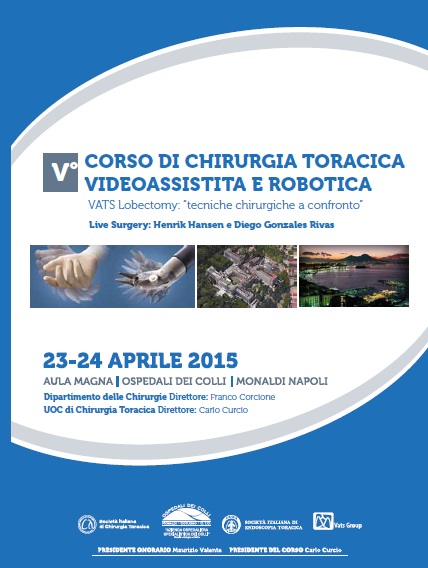
Minimally invasive surgery course in Naples at Hospital Monaldi (April 23 – 24th, 2015)
Munich airport, Germany
I am on the last leg of a long journey to the beautiful southern Italian coastal city of Naples. Best known for its claim as the home of pizza and the nearby ruins of Pompeii, for the next few days, the department of thoracic surgery at Hospital Monaldi will be hosting surgeons (and one wee writer) from around the world for a two day course on minimally invasive and robotic surgery.
The event features live surgery demonstrations by Dr. Henrik Hansen and Dr. Diego Gonzalez Rivas, with a host of other speakers. For more details on the V corso VATS Monaldi Napoli dr Curcio dr Amore dr Casazza click the highlighted link for the full program.
Meanwhile, I will bring readers photos and highlights from the event.
Thoracics.org talks to the Brazilian Society of Thoracic Surgery and result isn’t what you might expect.
A very different article here at Thoracics.org! For starters, I’m the interviewee – which doesn’t happen very often. This interview was a joint collaboration after meeting and talking about issues in thoracic surgery with several Brazilian surgeons including Dr. Sergio Tadeu Pereira, at the ALAT conference in Medellin last July.
The positive outcome of a thoracic surgery depends on several aspects, among them is the teamwork, the harmony between the various professionals involved in making decisions and actions. All experts have an instrumental part in restoring the health and maintenance the patient’s life. Each with its due importance, towards a single goal. The SBCT ratifies such thinking, and this issue of the Journal conducted an interview with K. Eckland, an acute care nurse practitioner in thoracic surgery, writer, and also the founder of Thoracics.org” – a blog about thoracic surgery with an international focus. She has written several books on surgery in Latin America, including a community sociological examination thoracic surgery in Bogotá, Colombia.
In this conversation, K. Eckland talks about the future of thoracic surgery worldwide and recognizes the contribution of Brazilian surgeons for growth in the art.
Journal SBCT: For us at SBCT is a great pleasure to have their participation in our newspaper. How do you evaluate the specialty in Brazil? K. Eckland: First, I would like to thank the editors this paper for the invitation to forward my message to Brazilian doctors. More importantly, I would like to serve as call to action to all the experts and future thoracic surgeons. When I look at Brazil, I see the future of thoracic surgery. While, in my own country, our thoracic surgeons are aging with an average age of 60 years, Brazil is full of young, dynamic and innovative surgeons.
Journal SBCT: This predisposition to new techniques of many the Brazilian thoracic surgeons implies an increase in research?
K. Eckland: The high fluency in minimally invasive techniques (in Brazil) combined with some of the largest academic and clinical settings worldwide, outside China, affords unique opportunities in research, development and discovery. Brazil is already home to many of the modern masters of thoracic surgery, names that resonate worldwide.
These surgeons have brought Brazil to the forefront, but it is up to the newest crop of thoracic surgeons to maintain Brazil’s forward momentum for the future. However, this is hampered by a lack of awareness of the contributions of many Brazilian and other Latin American surgeons.
As a foreigner, writing about developments within the international surgical community, I have noted a large dearth in published research from much of Latin America including Brazil. What research I do find, is often not widely dispersed or readily available to the rest of the world. It has taken several years and many thousands of dollars for Cirugia de Torax to acquire and publish information about your many triumphs. However, this is not the most efficient way for research to be disseminated.
Journal SBCT:In addition to increased investment in research, what more should be done in its assessment to mitigate this gap in publications and contributions (to the specialty of thoracic surgery)?
K. Eckland: It’s possible to change this story from one of limited international exposure to greater recognition. But for that to happen, several things need to occur. Firstly, the momentum must be Brazilian thoracic community to participate and publish research on a large-scale.
Surgeons in São Paulo, for example, have unique opportunities to publish practice-changing work. The Department of Thoracic Surgery, University of São Paulo tracks more thoracic cases in a year than many American institutions have access in a decade. This gives greater impact to studies from this institution than anything that their (North) American colleagues could expect do.
Second, Brazilian surgeons need push for further publication in international journals, and in the international literature language, in English.
Lastly, surgeons need to look outside their corner of the globe and present their findings internationally and outside Latin America on a greater scale. More groups of Brazilian surgeons should attend international conferences to gain knowledge,and take the opportunity to spread their own knowledge and research findings. Surgeons should not depend on the United States and Europe to take the lead in surgical innovation or research.
There is no reason why these findings will not occur at home, but research needs to be part of your daily practice. It should be more than reading the occasional surgical journal. It should be a part of active problem solving and solution-seeking.
Journal SBCT: The wide practical experience associated with the host new techniques can be considered as a basis for the growth of the specialty and development more innovative research in Brazil?
K. Eckland:
For this to happen, each surgeon needs ask yourself**:
– How can I improve my practice? – How can I improve the lives of my patients?
– What can I do to identify and document the phenomena I’m seeing? – What we are doing now that we need to change? How can we implement these changes? How does this apply to people outside my immediate environment?
Once a potential search area is identified, other questions to ask include:
How I can improve my specialty? How can I represent my country to the world? Sometimes the answers
involve the development of new technologies, sometimes a reframing of the information we already know,
to apply the new clinical scenarios. Other times, we simply need to identify the phenomena and document it to serve as guidance to other professionals. That is what drives the research, and this combined insight with professional curiosity are essential for growth within the specialty. By embracing these concepts, we can begin a new era of thoracic surgery in Brazil and worldwide.
* Corrections to the English translation have been made for ease of reading. This is an excerpt from a larger interview.
** This is how we identify research to discuss and publish here at Thoracics.org/ Cirugiadetorax.org
aka, “Why we should be nice to plastic surgeons”. This case study highlights the need for close interdisciplinary partnerships among surgeons and also asks the question, “Are we addressing the emotional and psychosocial needs of our patients and their families?”
Bronchopleural fistula: an abnormal communication between the exterior environment and the pleural cavity, often caused entry of bacteria, fluids and other substances into the chest cavity by way of the bronchial tree, for example: bronchial stump breakdown. BPF most commonly occur after large thoracic surgeries such as pneumonectomy but can occur for other reasons such as infection or trauma.
Bronchopleural fistulas (BPF) are a dread complication of thoracic surgery that has (thankfully) become rare in most countries in the last few decades. Treatment of a large bronchopleural fistula can be massive undertaking requiring collaboration and cooperative from multiple specialties including radiology, infectious disease, pulmonology, wound management and plastic surgery.
Patients often endure several months of surgical and wound care treatments prior to undergoing definitive surgical management for this condition. This treatment includes the surgical creation of large open wounds to facilitate drainage of purulent materials, repair of the fistula tract and bronchial stump and debridement / revascularization for proper tissue healing. The case presented today illustrates the devastating emotional, physical and financial costs of bronchopleural fistula as well as the need for interdisciplinary collaboration for definitive surgical repair.
Surgical repair itself carries an elevated risk of morbidity and mortality primarily from respiratory complications, infections/ sepsis and hemorrhage.
More than physical consequences
Bronchopleural fistulas carries more than just the physical consequences of pain and disability for patients and their families. There are also devastating emotional and social effects. Patients can experience a myriad of psychosocial effects from this chronic wound and related treatment. The resultant deformity from many drainage and wound management techniques, in particular, can lead to depression and social ostracism. The development of a bronchopleural fistula can contribute to relationship and intimacy issues. Several of the surgeons interviewed including Dr. Boxiong specifically mentioned both divorce and suicide as being a risk in numerous cases[1].
Case Study
Dr. Boxiong Xie, thoracic surgeon
Dr. Dong Jiasheng & Dr. Zheng, Reconstructive/ Plastic Surgeons
Dr. Boxiong Xie, thoracic surgeon
The patient is a young male in his early forties who had undergone a right upper lobectomy for cancer several years prior at a facility in a far away province. He then presented with a large empyema. Initially, conservative treatments were attempted. The patient underwent several drainage procedures, by both open and closed methods. These measures along with attempts to repair the bronchial stump failed due to extensive infection and tissue destruction.
Following the failure of more conservative measures, the patient presented to this facility for specialty care. He had heard about this program, and travelled a long distance to be here at significant difficulty and expense. As his surgeon explained, “it’s his last chance at a normal life.”
Over the continuing course of his treatment, a large opening on the anterior chest was created surgically. Due to the extent of necrotic tissue, this required the removal of anterior sections of ribs #2, 3, 4 and 5, leaving the patient with a very large open cavity, as seen in CT slices (pulmonary and tissue windows).
packing material can be seen in the right chest cavity.
tissue window showing extent of wound
This large cavity was left open for a period of around two years, while infected material was debrided and evacuated, and aggressive wound management was continued. At the time of his presentation to the operating room, the wound bed is dry and pink with a small amount of slough. An opening to the bronchus is visible (with bubbling on respiration at the site of the wound). The wound measures approximately 6 cm X 4 cm. As seen from the CT images above, the wound was also several centimeters in depth.
overview of wound – which tracks upwards several cm towards shoulder
The wound tracks up towards the shoulder, making it deeper and larger than it initially appears on gross visualization. There is a visible pulsation from the border of the cavity, (which may add to the patient and family’s distress).
Surgical procedure:
After the wound is cleaned and prepared with betadine solution, the anesthesiologist introduces a bronchoscope into the airway, for illumination and visualization of the airway. The light from the scope is immediately visible to observation within the chest. At that point, amplatzer patch was inserted into the bronchial stump.
Amplatzer patch visible in the chest cavity
After placement of the patch was confirmed, the patient was re-prepped, and draped. Dr. Boxiong expands the existing wound, and dissects down to healthy bleeding tissue, removing yellow eschar. The wound is lightly packed with moist gauze.
thoracic surgeons dissect down to healthy tissue (anterior wound site)
Then Dr. Dong and his assistant surgeon arrive, to start their portion of the operation. Dr. Dong starts another incision approximately 3 cm below the wound area. The incision is extended to the left side of the chest. The surgeon dissects down through skin, adipose and fascia to free the right internal mammary artery to use to ensure that the graft is well vascularized.
preparing the internal mammary artery for eventual anastomosis
Next step: Flap harvesting
Once the IMA was free, it was temporarily secured, and the wound was dressed. The patient was re-positioned, and re-prepped to allow access to the posterior aspect of the left chest. Due to muscle devascularization from the multiple previous surgeries on the right anterior chest, the surgeon harvests the left latissimus dorsi, using a large diamond-shaped incision.
Harvesting the myocutaneous flap from posterior chest
Once the flap was harvested, the patient was left with a large open defect, without enough surrounding skin to cover the area. The surgical site is dressed with a temporary dressing while Dr. Dong moves on to his next surgical site.
Next step: Skin Harvesting
Dr. Dong prepares to harvest skin for grafting
After preparing the patients right thigh, Dr. Dong applied a Padgett dermatome to shave off a thin layer of skin.
Harvesting skin from patient’s thigh
After multiple passes, the surgeons have enough skin to cover the defect from the flap site.
Skin harvested to cover flap site
Next step: Skin Grafting
Skin grafting at the myocutaneous flap site
The thin strips of skin were applied to the flap site and sutured into place.
suturing skin graft over flap harvest site (on back)
Once the sutures were completed, the wound was re-dressed and the patient was re-positioned for the last steps of the operation.
Next step: Anastomosis of mammary artery to flap
surgeons using the microscope to complete vascular anastamoses
Following re-positioning to supine position, the flap was placed within the right chest wound. The flap was loosely sutured into place to maintain a proper position while the painstaking vascular anastomoses were performed. Once the anastomoses were completed, the remaining incisions were carefully closed.
Total surgical time was greater than ten hours.
Discussion
As discussed by Lois and Noppen (2005), BPF management has traditionally been performed in a piece meal or stepwise fashion, with surgical interventions reserved as a last resort. Unfortunately, for some patients, this means that BPF becomes a chronic illness. As a chronic illness, (and all that chronic illness entails such as chronic malnutrition, chronic inflammation, long-term antibiotic therapy), the morbidity and mortality of this condition continues to increase for the duration of the illness. In the case study above, a relatively young, now cancer-free patient had now developed much of the disabilities associated with elderly patients due to the chronic nature of his illness (BPF after a lobectomy ten years prior). This certainly places the patient at significant risk for major complications once a large-scale definitive surgery is performed. Van Schill et al. (2014) notes that better understanding regarding the need for interdisciplinary management including aggressive physical therapy and nutritional support have reduced some of these complications.
While the impact of bronchopleural fistulas are usually discussed in terms of mortality, financial costs (surgical costs) and length of stay,and for this case, we would like to take a closer look at morbidity and quality of life issues raised by the development of this complication.
While BPF is rare, it truly can be a life-altering and destructive diagnosis. In addition to pain, physical debility, there may be gross deformity coupled with chronic wound care. Deformities caused by extensive tissue destruction and removal of several ribs can cause significant emotional and psychological anxiety and stress in both the patient and family members. The visible pulsation (cardiac movement) seen within the wound may exacerbate this anxiety. The stress of this wound combined with additional stressors related to this diagnosis have been observed to lead to a higher rate of marital discord and patient suicide. Patients may also feel a loss of sexuality and personal identity in the presence of this type of disfigurement, similar to some women after radical mastectomy (particularly in female patients).
To add insult to injury, unlike many conditions which can be readily corrected surgically, the creation of myocutaneous flap (and subsequent skin grafting) itself causes additional disfigurement. This patient required a lengthy (ten hour) surgery which resulted in the creation of three new surgical sites in addition to the patient’s original right-sided chest wound. While this is a drastic example, it does serve to highlight the on-going need to consider the psychological and emotional well-being of this patient (and all our patients).
BPF and professional relationships?
This case also reminds of the need for good interdisciplinary relationships. In thoracic surgery, cosmetic outcomes (other that pursuing minimally invasive options when possible) are not usually one of our primary considerations. This leaves us at a disadvantage when managing patients with such a drastic complication. We don’t always have a strong network or relationships with other surgical or medical disciplines outside of oncology or oncology-related fields. We need to take the opportunities available to become more familiar with our local reconstructive surgeons, as well as the latest techniques in reconstructive surgery. It’s not “good enough” to know the name of one of the plastic surgeons we brush elbows with in the surgical waiting lounge. It is not just about referrals and compensation. It is about having an open and free dialogue with surgical colleagues, so that when we do require their assistance, we can work together smoothly and coördinate care.
Consider the need to include social workers, psychologists and other counseling services in both the preoperative and postoperative care of our patients, when necessary for their long-term health and wellness. Unfortunately, due to social stigma, health care/ insurance or financial restrictions as well as provider hesitation**, not enough of our patients receive consultations or referrals to appropriate resources. We can’t change insurance regulations, but by becoming more familiar with our local resources and providers, we can overcome many of the other barriers to supporting our patients emotional health.
[1] I was unable to find literature that specifically cites BPF as a contributing factor to psychosocial complications such as divorce, depression or suicide but the impact of chronic wounds on emotional health, family life and other quality of life indicators are well documented. However, Okonta et. al (2015) and Lois & Noppen (2005) both cite QoL issues in patients with BPF.
** Provider hesitation is a nice term for all the reasons providers sometimes fail to seek mental health referrals for patients; such as fear of embarrassing our patients, believing that counseling is only needed for psychiatric emergencies, failure to understand local resources available, or our own discomfort with mental health “issues”.
References and Additional Readings
Arnold, P. G. & Pairolero, P. C. (1990). Intrathoracic muscle flaps: an account of their use in the management of 100 consecutive patients. Annals of Surgery, 1990; 211(6): 656-660. Study looking at one hundred cases from May 1977 and February 1988. In this potent reminder of the morbidity and mortality that is associated with patients requiring muscle flaps, as well as the advances in medicine over the last two decades, there were 16 operative deaths and 43 additional all-cause deaths in the operative survivors. Interestingly, one of these late-term deaths was due to suicide.
Goyal VD1, Gupta B2, Sharma S3 (2015). Intercostal muscle flap for repair of bronchopleural fistula. Lung India. 2015 Mar-Apr;32(2):152-4. doi: 10.4103/0970-2113.152628. Indian case study of patient presentation of BPF after treatment for spontaneous pneumothorax.
Van Schil PE1, Hendriks JM1, Lauwers P1 (2014). Focus on treatment complications and optimal management surgery. Transl Lung Cancer Res. 2014 Jun;3(3):181-6. doi: 10.3978/j.issn.2218-6751.2014.06.07. Belgian paper reviewing outcomes of 3,500 surgeries.
Levine, L. A. (2013). The clinical and psychosocial impact of Peyronie’s disease. Am J Manag Care. 2013 Mar;19(4 Suppl):S55-61. While unrelated to thoracic surgery, patients with Peyronie’s disease have many of the same emotional and psychological stressors as patients with other chronic wound conditions such as BPF.
As Dr. Gonzalez Rivas demonstrates, minimally invasive surgery isn’t just for “easy” cases. Case study with brief discussion and literature review
Uniportal VATS with chest wall resection at Shanghai Pulmonary Hospital
Shanghai, China
Authors: Gonzalez – Rivas, D. & Eckland, K.
Surgeons: Dr. Diego Gonzalez Rivas with Dr. Boxiong Xie assisting.
Case: 66-year-old patient with large left upper lobe mass extending into chest wall, biopsy proven carcinoma.
Pulmonary function tests – within acceptable margins
Imaging:
CT scan – showing a large left-sided lung upper lobe mass with chest wall invasion and rib involvement at the level just beneath the scapula.
Procedure: Uniportal (single incision) VATS with rib resection
Description: at a glance
Determining port placement
Due to tumor location, port placement had to be carefully considered and adjusted.
Vital signs at initiation of operation: HR 78, NSR B/P 95/56 Oxygen saturations: 100% (intubated with double lumen ETT)
First incision: 14:17
Making the initial incision
The tumor was adherent to the chest wall, requiring chest wall resection with rib resection.
performing uniportal VATS for tumor with chest wall invasion
The tumor was palpated thru the 2 cm incision allowing the surgeon the benefit of open surgery despite using a minimally invasive technique.
Palpating the tumor
Ribs were resected using a guillotine designed for minimally invasive use.
Rib resection
Lung resection complete at 17:42. The tumor was removed enblock using a bag system to prevent tumor spillage.
Tumor enblock after removal
Lymph node dissection completed at 17:56
There was a brief run of PVCs lasting about 30 seconds (B/P 83/54) with no desaturations. Patient was otherwise hemodynamically stable for the duration of the case.
Frozen section: clear pleural margins
EBL: 200ml
Discussion:
As noted by Pischik and others, many of the traditional contraindications for VATS procedures are no longer applicable, particularly for surgeons well versed in minimally invasive techniques like uniportal thoracoscopic surgery. In the case above, several of these contraindications were successfully addressed, including multiple adhesions, an incomplete interlobar fissure and a tumor with chest wall involvement.
That being said, this case was technically challenging from start to finish, due to the position of the chest wall tumor that required adjustment of port placement, a lengthy dissection of dense adhesions in addition to a sizeable chest wall mass. Hilar dissection was complicated by anatomical position, and the bronchus was difficult to access. This in addition to an incomplete fissure significantly lengthened the procedure.
VATS resection using a single port approach can be challenging even for experienced surgeons. However, it is a viable alternative for more complicated cases including those requiring a degree of chest wall resection.
This case was just one of numerous cases performed by Dr. Diego Gonzalez Rivas as part of the Uniportal VATS training course at Shanghai Pulmonary Hospital. Dr. Diego Gonzalez Rivas is the inventor of the uniportal technique and Director of Uniportal VATS training program at Shanghai Pulmonary Hospital. He has partnered with the Chinese facility to offer training courses for interested surgeons three times a year, in addition to his ‘wet-lab’ surgical training offerings in his home facility at La Coruna, Spain.
the latest trailer about the documentary film on single port surgery and information about an upcoming training course.
For everyone that’s interested in learning more about the single port surgery technique, as taught by its creator, Dr. Diego Gonzalez Rivas – here’s another opportunity which may be closer to home for some readers.
Now, which way to Berlin?
The February conference takes place in Berlin, Germany on the 19th thru 21st. While Dr. Gonzalez Rivas, Dr. Delgado and Dr. Prado are headlining the event, other prominent thoracic surgeons such as Gaetano Rocco (Italy) and Alan Sihoe (Hong Kong) will also be lecturing at this event.
The conference includes live surgery demonstrations as well as a wet-lab for hands-on practice.
Deadline for registration is February 6th. Interested surgeons should contact:
R. Mette, M. Schmitt Charité – Universitätsmedizin Berlin Tel. +49 30 450 622 132 | Fax +49 30 450 522 929 E-mail: thoraxchiurgie@charite.de
In other news – the newest trailer for the documentary about Dr. Gonzalez Rivas and his work was recently released. I encourage all thoracic surgery personnel to see (and promote) this movie, which highlights the work of one of our own.
Cirugia de torax invites readers for an open discussion on the latest STS guidelines on multimodality treatment of esophageal cancer.
Guidelines for esophageal cancer?
Guidelines, guidelines, guidelines.. It seems like much of American medicine is now directed by guidelines, committees and government agencies. We have pay-for-performance, “Core Measures” and even more guidelines, recommendations and requirements that attempt to pre-script the care that we provide. This often leaves clinicians and surgeons feeling more like technicians following recipes for “cookbook medicine” to treat anonymous, “standardized” patients rather than highly skilled, extensively trained and experienced medical providers using clinical judgment, intellect and training to treat unique individuals.
Guideline fatigue, questionable “evidence” and mandated medicine
With that in mind, many healthcare providers are sick of reading and writing about “evidence-based practice recommendations and clinical guidelines”. Some of this frustration comes from the sometimes contradictory clinical evidence regarding these mandates, such as pre-operative beta blockade. While this medication is now mandated by the federal government, multiple studies* question the benefit of this treatment in patients undergoing noncardiac surgery.
As the debate continues to rage over this therapy, is it fair that surgeons must continue to risk their hospital’s performance scores, and surgical reimbursement for challenging the blanket administration of this medication to their patients?**
Not all guidelines created equally
The concept of clinical guidelines have its origins in the 1960’s. While differing political camps explain the emergence of these guidelines according to their individual bias (insurance cost-cutting versus autonomy etc.), it seems obvious that these guidelines were at least, initially, designed to improve the overall care of patients with similar diagnoses, symptoms or clinical scenarios.
But when it comes to these clinical guidelines – not all guidelines are created equally. In addition to criticism that many clinical guidelines are poorly supported by the existing literature, or based on poor quality studies, allegations of cronyism, obvious bias/ self-serving have plagued guideline committees particularly in the field of cardiology.
But what does this mean for thoracic surgery? We have our own organizational committees such as the Society for Thoracic Surgeons, (aka STS), our own recommendations, guidelines and ratings systems (national and international database). STS and thoracic surgery based clinical guidelines address the very lifeblood of our specialty and our clinical practice.
It behooves us as a professional specialty to read, review and know these guidelines so that we can determine when and if these guidelines serve our practices and our patients. If not, as representatives of thoracic surgery; it is our responsibility to participate and to voice our concerns and criticisms of these guidelines. We are the watchdogs, to prevent the over-representation of commercial interests or bias into our arena of patient care.
It is also crucial that we attempt to support the crafting of recommendations to support and adopt the best practices in thoracic surgery; after all, as practicing clinicians, we know thoracics better than any outside agencies, organizations or other specialties. With this philosophy in mind, Cirugia de Torax invites readers to become more familiar with the latest STS guidelines.
Society of Thoracic Surgeons guidelines
Thus far, the Society of Thoracic Surgeons has published eighteen guidelines on a wide variety of topics’ from antibiotic use, to cerebral protection of infants undergoing cardiac surgery, the use of TMR, to the newest guidelines on the treatment of esophageal cancer.
Cirugia de Torax would like to invite our American and International readers to participate in a review of the most recent guidelines in our next post. What do you think of trend towards guidelines in general? What about the guidelines for multi-modality treatment in esophageal cancer? Love them? Hate them? Any omissions or errors? Any changes or suggestions for future versions?
Deadline for submission of commentary, criticism or other remarks is January 15, 2015.
Notes:
* Link requires (free) subscription
** Surgeons can document a ‘variance’ on a case-by- case basis when omitting this and other prescribed core measures under a limited set of circumstances.
Article for Review
The Society of Thoracic Surgeons Practice Guidelines on the Role of Multimodality Treatment for Cancer of the Esophagus and Gastroesophageal Junction.
Little, Alex G. et al. (2014). The Annals of Thoracic Surgery , Volume 98 , Issue 5 , 1880 – 1885. pdf version.
Additional reference articles
1. Weisz G1, Cambrosio A, Keating P, Knaapen L, Schlich T, Tournay VJ. (2007). The emergence of clinical practice guidelines. Milbank Q. Dec;85(4):691-727.
2. The Society of Thoracic Surgeons Esophageal Cancer Guideline Series. Mitchell, John D. et al. The Annals of Thoracic Surgery , Volume 96 , Issue 1 , 7
3. The Society of Thoracic Surgeons Guidelines on the Diagnosis and Staging of Patients With Esophageal Cancer. Varghese, Thomas K. et al. The Annals of Thoracic Surgery , Volume 96 , Issue 1 , 346 – 356
Copies of all STS guidelines are available on-line here.
While Cirugia de Torax was unable to be in attendance and provide on location reporting and news, I would like to present some highlights from the recent event for our readers..
information about the upcoming VATS symposium in Cambridge, UK – with featured speakers Dr. Diego Gonzalez Rivas and Ian Hunt.
Another conference/ educational announcement for all residents, fellows and interested thoracic surgeons. This course is sponsored by the United Kingdom’s National Health Service and is being held in Cambridge, UK at Papworth Hospital this November. There is parallel content for nurses and other thoracic surgery personnel.
Internationally known Spanish surgeon Dr. Diego Gonzalez Rivas as well as native surgeon Mr. (Dr.) Ian Hunt of St. George’s Hospital in London, will be part of the faculty teaching this course.
Dr. Gonzalez Rivas will be discussing single port surgery in addition to performing a live case on the second day of the symposium.
Mr. Hunt will be discussing how to perform a total lymphadenectomy, as well as lymphadenectomies on more complicated cases.
Additional speakers will be discussing topics including issues in thoracic anesthesia, management of bleeding (in VATS and other minimally invasive surgery), and managing other operative complications.
Updates in multi-disciplinary care from the Foundation for the Advancement of Cardiothoracic Surgery at the 2014 Cardiovascular- Thoracic Critical Care conference in Washington D.C
Washington D.C.
While the majority of the 11th annual conference by the Foundation for the Advancement of Cardiothoracic Surgery was focused on cardiac surgery topics, Dr. Namrata Patil, the Director of the Thoracic Intermediate Care Unit (and thoracic surgeon) at Brigham & Women’s Hospital in Boston, MA gave an excellent presentation on the management of critically ill thoracic surgery patients.
Attendees at the Factscare conference
Early Intervention, Rapid Response versus Failure to Rescue
Rather than an exhaustive review of the literature, Dr. Patil’s lecture gave a much-needed bedside perspective on the care of these patients. She stressed the importance of remaining hypervigilant as well as the need for early identification and early, aggressive intervention in these patients.
While the majority of the conference focused on ECMO, LVADs and transplant patients, Dr. Patil’s presentation was a crucial reminder of the pitfalls of falling into complacency when caring for our vulnerable thoracic population. While these patients do not always attract the attention that patients with artificial life support mechanisms (like Heartmate II patients), it is a mistake to think that these patients are less fragile or critically ill. By definition, these lung patients, (who frequently have underlying lung disease and other serious comorbidities) are compromised – and acutely ill.
This means that clinicians need to shift their focus from the intensive care unit to the telemetry and floor units without losing their critical care perspective. Too often, when patients are transferred to step-down units, critical care concepts are relaxed because of preconceptions based on assumptions regarding patient acuity. But as anyone with thoracic experience knows, a ‘stable’ patient can easily descend into a downward spiral if not managed aggressively.
“Is this acceptable to me?”
As Dr. Patil reminds us, clinicians need to be vigilant when caring for patients of all acuities. She’s not asking us to chase ‘zebras’ but instead gently reminding clinicians not to dismiss important clinical findings. Instead of attributing low-grade fevers and cloudy X-rays to atelectasis, intervene early to prevent the next step in deterioration; pneumonia or respiratory compromise. Remain vigilant to detect later stage complications instead of racing to discharge on marginally functional patients.
She encourages clinicians to educate patients, providers and families; to teach as part of efforts to prevent complications. She also advocates for the increased development of protocols specific to the thoracic surgery population and better communication with all members of the care team; including the patients and their families.
Ethics and Advocacy
She also spoke on the ethics of caring for these patients and advocating for the rights of patients, particularly elderly patients. In an era of increased awareness of POAs, and Advance Directives, there is often a push (from the hospital administrators, nursing staff, and other medicine specialities) to advocate for a Do Not Resuscitate (DNR) code status. Unfortunately, many of the people pushing for this designation have forgotten that this is part of a patient’s right – and automatically assume it should be a decision based solely on age. This ageism is contrary to our duty to protect, to advocate and our patient’s right to self-determination.
This ageism also ignores one of the widely held truths in our society; that for many people, “Age is just a number” and that the patient’s functional status may not reflect their actual age. We’ve all met 50 year-old patients who have been debilitated by chronic and prolonged illnesses and may have a much poorer functional status than an active, alert 80 (or even 90) year-old patient. Assigning or encouraging a DNR status in these patients based on age is not only incorrect, but unethical.
In a time of an increasing push for standardized, ‘one-size-fits-all” care and ‘Angie’s List” style medicine with emphasis on short length of stay and rapid discharges, Dr. Patil’s more personalized approach will actually engender better clinical outcomes by reducing morbidity, mortality, and re-admissions. It also helps clinicians, like myself, sleep better at night – knowing we have been as aggressive as possible to prevent complications in our patients.
Using 3rd world skills to augment diagnostic technologies
Dr. Patil’s talk also highlighted the importance of clinical judgement and clinical skills in caring for these patients. While heart patients routinely have advanced life support and hemodynamic monitoring devices such as Swan Ganz catheters, NICO and telemetry, excellent clinical skills are needed when relying on less invasive measures such as physical exam and basic radiology. Her background, of practicing medicine in India (and the related limitations in resources) has added to her skills as a clinician and diagnostician without relying on expensive or extensive use of technology. In an era of rapidly expanding concerns regarding resource management and cost-containment, this skill is crucial, just at a time when new medical school graduates are focusing more on advanced diagnostics over basic clinical assessment skills.
Dr. Namrata Patil
Dr. Namrata Patel, thoracic surgeon
Dr. Namrata Patil is a polyglot (English, Urdu, Spanish, Hindi and Marathi) with extensive surgical and intensive care experience. Originally trained as an ENT surgeon, over the years she has added to her body of knowledge with residencies in Burn/ Trauma, Psychiatry, Thoracic Surgery and Surgical Critical Care.
She is an associate surgeon at the prestigious Brigham & Women’s Hospital in Boston, MA as well as a Professor of Surgery at Harvard Medical School.
Her most recent list of publications reflect her wide range of training and experience.
I have asked Dr. Patil for a copy of her presentation for use on this site. As soon as these slides are published at facts-care.org, I will provide a link for readers.
a new film showing the life-changing efforts of one thoracic surgeon.. It’s about time!
Dr. Diego Gonzalez Rivas
I am excited beyond words to hear that my long-time hero and champion of modern-day thoracic surgery, Dr. Diego Gonzalez Rivas, is featured in a new documentary film, “This is Life”. The film follows the life of a patient undergoing a single incision thoracoscopic lobectomy. The film is being released this December.
I eagerly await the film – and am happy to see thoracic surgery (and Dr. Diego Gonzalez Rivas) get their due. For too long, our humble specialty has been overlooked for the more ‘glamorous’ cardiac surgery. This oversight has led to a dire shortage of thoracic surgeons in many parts of the world.
Hopefully, this is only part of an ongoing effort to have thoracic surgery recognized as an independent and complex surgical specialty requiring extensive knowledge, advanced skills and training. It is not an ‘add-on’ for cardiac surgeons with insufficient cardiac consultations.
Dr. Gonzalez Rivas and single-port surgery in Shanghai, China
For those of you hoping to see – and learn from the best, Dr. Gonzalez Rivas will be spending much of the month of October in Shanghai, China at the “National Uniportal VATS Training Course & Continuing Medical Education Forum on General Thoracic Surgery” which runs from October 8th to November 8th, 2014 at Tongi University.
Alas! To my eternal regret, Cirugia de Torax will not be in attendance. However, I will have sources on the ground – and hope to post more information during the conference,
updates on the on-going HITHOC project, war surgery, foreign body obstructions and bronchoscopy for infants
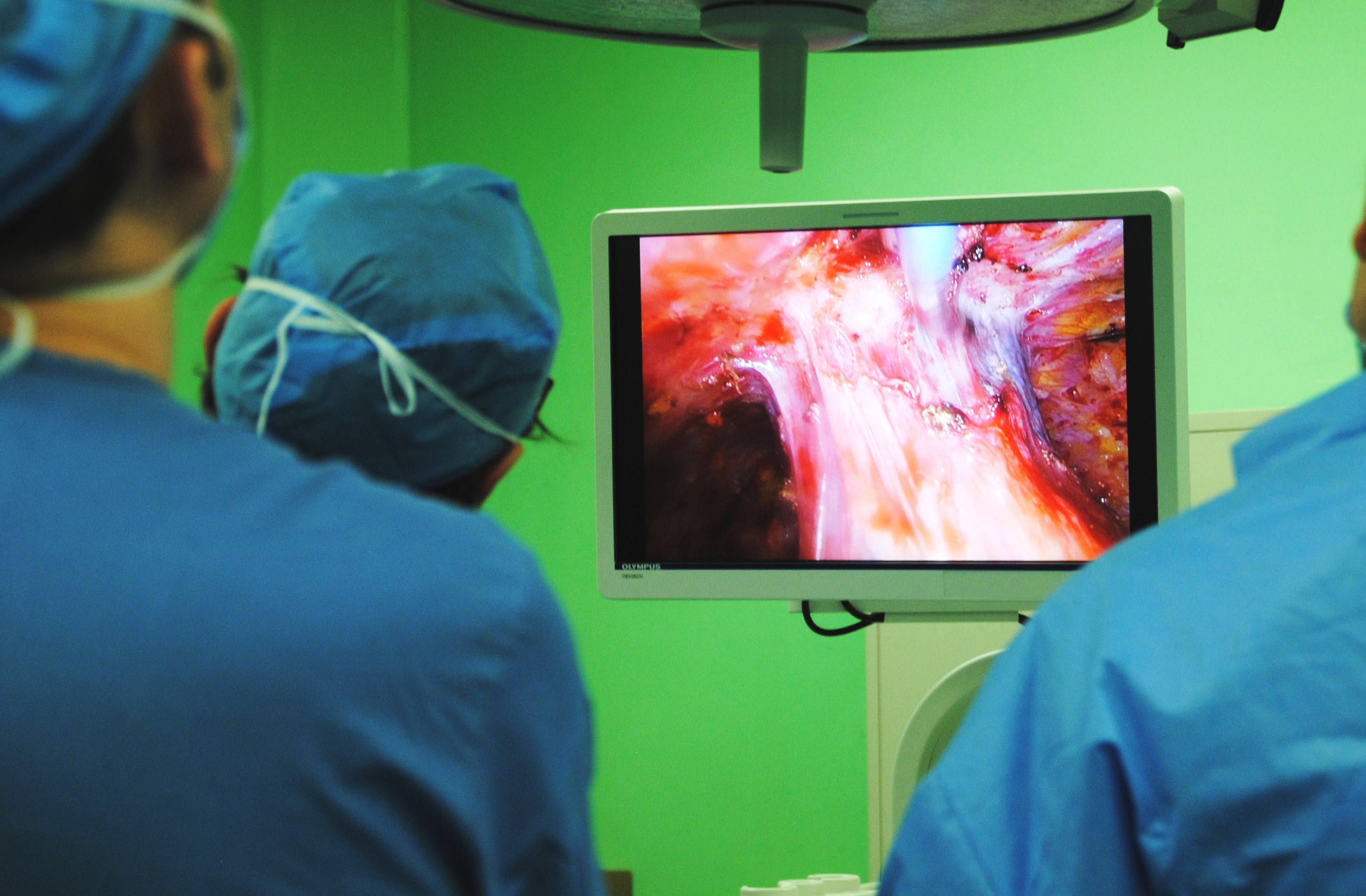
Gaziantep, Southeastern Anatolia
It’s been over a year since I first read Dr. Isik’s work on treating pleural mesothelioma. Since that time, Dr. Işik has continued his research into HITHOC and has now enrolled over 79 patients into the hyperthermic treatment group including one of the patients I met during my visit. (There are 29 surviving patients in the study, 13 in the mesothelioma group, the remainder are secondary pleural cancers.).
(If you are a patient seeking treatment, or would like more information about Dr. Isik (or Dr. Gonzalez Rivas, Dr. Sihoe or any of the other modern Masters of thoracic surgery), we are happy to assist you. Contact me at kristin@americanphysiciansnetwork.org
First impressions are deceiving
I don’t know what I expected Gaziantep to look like as one of the world’s oldest cities, but from the moment the airplane begins its descent into a beige dust cloud, to the desolate brush and dirt of the airport outside the city, it isn’t what I expected. Much of the antiquity of the biblical city of Antiochia has been replaced by a bustling modern city. Historic ruins and ancient Roman roads marking this as part of the original Silk Road are conspicuous, only by their scarcity.
modern Gaziantep is featureless at first glance
There are a handful of museums and monuments to the area’s rich history, but like the new name of Gaziantep (replacing Antep after the first world war), Turkey’s sixth largest city is modern; a collection of traffic and squat square buildings of post-modern architecture.
Kale
The city is also a mosaic of people. There are groups of foreign journalists in the lobby of our hotel, and convoys of United Nations vehicles cruising the streets. Crowds of Syrian children play in the park, calling out in Arabic to their parents resting on the benches nearby. There is a smattering of Americans and English speakers interspersed, many are college students and other foreign aid workers on humanitarian missions to help alleviate the strain caused by large numbers of people displaced by the Syrian civil war.
Gaziantep is famed for their copper work
But like a mosaic, there is always more to see, the closer you look. For me, as I look closer, I just want to see more. I feel the same about Dr. Elbeyli’s thoracic surgery department.
The closer you look, the more you see. photo courtesy of wiki-commons
The border (and the largest Syrian city of Aleppo) lies just to the south – and the impact of the Islāmic militants is felt throughout the region. No where is this more evident than at the local university hospital, where I meet Dr. Ahmet Işık and the Chief of Thoracic Surgery, Dr. Levent Elbeyli.
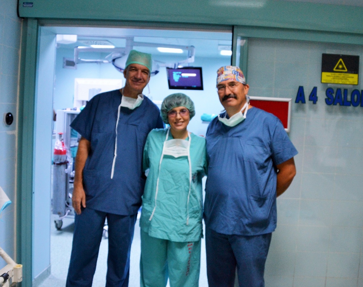
with Dr. Elbeyli (left) and Dr. Isik
Dr. Ahmet Feridun Işık
I like Dr. Işık immediately. He is friendly and appears genuinely interested by my visit. He’s from Giresun in the Black Sea region of northern Anatolia of Turkey. He attended medical school at Ankara University and completed his thoracic surgery training in Ankara before going to Adiyaman State Hospital in the bordering Turkish province of Adiyaman in southeastern Turkey.
He was an associate professor of thoracic surgery at Yuzuncu Yil University in the far eastern province of Van, Turkey before coming to Gaziantep in 2005. He became a full professor at the University of Gaziantep in 2013. In additional to authoring and contributing to his own publications, he also served as a reviewer for the Edorium series of open access journals.
It helps that his English is miles better than my non-existent Turkish. (Reading about the Turkish language in phrase books is one thing, pronouncing words correctly is another.)
He doesn’t seem to mind my questions tumbling out one after another. I’d like to be the cool, sophisticated visitor, but I’ve been waiting so long to ask some of these questions – and frankly, I am just excited to be there.
Dead-ends in medicine
There are a lot of “dead ends” in medicine – treatments that at first appear promising, but then end up being either impractical or ineffective. In fact, for the first ten years of HIPEC, most surgeons dismissed it as a ‘dead-end’ treatment; the surgery was too radical and mortality too high. But researchers kept trying experimental protocols; tweaking medications (less toxic) and procedures – and finding the right patients (not too frail prior to surgery) – and the literature shifted; from a largely useless ‘last ditch’ salvage procedure to a large, but potentially life-saving treatment. HITHOC is HIPEC in another color…
So I fire away –
Since our last post about Dr. Işık – he has performed several more cases of HITHOC on patients with pleural mesothelioma, pleural based cancers and advanced lung cancers. He now has 79 patients in the HITHOC treatment group. He has been receiving patients from all over Turkey, including Istanbul to be evaluated for eligibility for this procedure. While the majority of patients are referred by their oncologists, others come to Gaziantep after reading about Dr. Işık on the internet.
None of the original patients (from 2009) are still alive, but their survival still exceeded all expectations, with 13 patients (of 14 HITHOC patients) living 24 to 36 months after the procedure. (I don’t mean to be vague – but I was asking some of these questions in the operating room and I forgot to stuff my little notebook in my scrub pocket.)
While much of the literature surrounding the procedure cites renal failure as one of the major complications of the procedure, Dr. Işık has had one case of renal failure requiring dialysis. Any other instances of elevated creatinine were mild and transient. He doesn’t use any chemical renal prophylaxis but he does use fluid rehydration to limit nephrotoxicity.
He reports that while many surgeons consider sarcomas to be a contraindication to this procedure, he has had good outcomes with these patients.
He does state that diaphragmatic involvement in mesothelioma is an absolute contraindication because while the diaphragm can be resected / patched etc, it is almost impossible to guarantee or absolutely prevent the seeding of microscopic cancer cells from the diaphragm to the abdominal cavity – which increases the risk of disseminated disease.
He still uses Cisplatin – since that is what the original HITHOC researchers were using, but he uses a slightly higher dose of 300mg. He’d like to do some prospective studies utilizing HITHOC (these have all been retrospective in nature – comparing today’s patients with past patients that received PDD and pleurodesis for similar conditions). Prospective studies would allow him to better match his patients and to compare treatments head to head. It would also allow him to compare different techniques or chemotherapeutic agents.
Unfortunately, as he explained, many of these types of studies of ineligible for government funding in Turkey because the government doesn’t want to pay for experimental / unproven treatments for patients even if there are few or no alternatives for treatment. He is hoping to appeal this regulation so that he can continue his research since there is such a high rate of mesothelioma, that disproportionately affects rural Turkish patients.
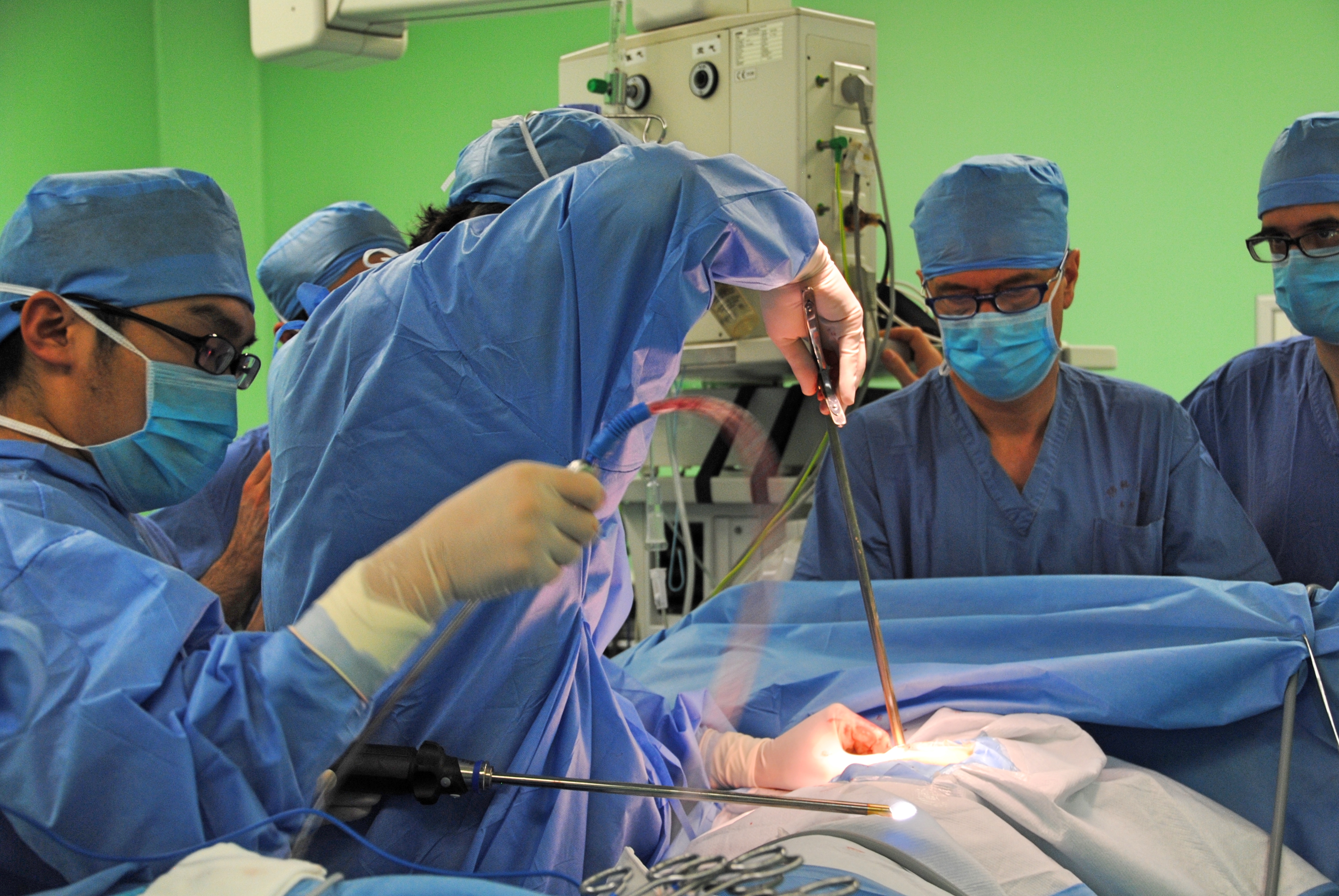
The University Hospital is one of several hospitals in Gaziantep. The academic institution has over 900 beds and 20 operating rooms spread out over three floors. There is a large 24 bed surgical ICU which includes 4 dedicated thoracic surgery beds.
Thoracic surgery may not be the advertised superstar of the hospital but it is the backbone of patient care. There are three full-time professors of surgery; Dr. Ahmet Isik, Dr. Levent Elbeyli and Dr. Bulent Tunçözgür, along with an associate professor, Dr. Maruf Sanli, several thoracic surgery fellows and research assistants. Together the thoracic surgery team performs over 1000 cases a year.
Dr. Levent Elbeyli is the driving force for thoracic surgery. A Gaziantep native, he founded the department in 1992, and has seen it grow from a few scattered beds to a full-fledged program with a full-time clinic, 2 dedicated operating rooms, 4 ICU beds and 15 to 20 cases a week.
Dr. Levent Elbeyli (in loupes) in the operating room
For the thoracic nurse, the department of Thoracic Surgery is a dream come true; tracheal cases, surgical resections, esophagectomies, thoracic trauma – all of the bread and butter that makes our hearts go pitter-pat. But then there is also plenty of pediatric cases, pectus repair, foreign body removal (oro-esophageal) and on-going surgical research. They do a large amount of pediatric and infant bronchoscopies (for foreign body obstructions, tracheal malformations etc).
There is the slightly exotic hydatid cysts and the more mundane (but my personal favorite) empyema thoracis to be treated. Cancers to be staged, and chest wall resections to undertake. I feel almost overwhelmed in my own petite version of a candy store; everywhere I turn I see opportunities to learn, case reports to write and new things to see.
Dr. Levent Elbeyli operates as Dr. Isik observes.
My non-medical readers might be slightly repulsed by my glee – but it is this intellectual interest that keeps me captivated, engaged and enamored with thoracic surgery and caring for thoracic surgery patients. And then there is the HITHOC program. With a large volume of mesothelioma and pleural based cancers due to endemic environmental asbestos in rural regions of Turkey, there is an opportunity to bring hope and alleviate suffering on a larger level. (Dr. Isik sees more cases here in his clinic in one year than I have seen in my entire career).
In this study, Dr. Isik and hs team looked at 73 patients with malignant pleural mesothelioma (MPM) who were in three different treatment groups. Group 1 received surgery only (extrapleural pneumonectomy). Group 2 received palliative treatment only. Group 3 received lung sparing surgery with hyperthermic chemotherapy (HITHOC). Lung sparing surgery included pleural decortication.
While the treatment groups are small, the results show a clear survival benefit to the patients receiving HITHOC. Surprisingly, the palliative group lived longer than the surgery alone group.
Survival based on treatment modality:
Surgery only: 5 months average surgery. 15% survival at 2 years
Palliative treatment only: 6 months average survival 17.6% at 2 years
HITHOC group: 27 months average survival 56.5% at 2 years
Selected Bibliography for Dr. Işık
Işık AF, Sanlı M, Yılmaz M, Meteroğlu F, Dikensoy O, Sevinç A, Camcı C, Tunçözgür B, Elbeyli L (2013). Intrapleural hyperthermicperfusion chemotherapy in subjects with metastatic pleural malignancies. Respir Med. 2013 May;107(5):762-7. doi: 10.1016/j.rmed.2013.01.010. Epub 2013 Feb 23. The article that brought me to Turkey, and part of our series of articles on the evolving research behind HITHOC.
Isik AF, Tuncozgur B, Elbeyli L, Akar E. (2007). Congenital chest wall deformities: a modified surgical technique. Acta Chir Belg. 2007 Jun;107(3):313-6.
Er M, Işik AF, Kurnaz M, Cobanoğlu U, Sağay S, Yalçinkaya I. (2003). Clinical results of four hundred and twenty-four cases with chest trauma. Ulus Travma Acil Cerrahi Derg. 2003 Oct;9(4):267-74. Turkish.
Sanli M, Arslan E, Isik AF, Tuncozgur B, Elbeyli L. (2013). Carinal sleeve pneumonectomy for lung cancer. Acta Chir Belg. 2013 Jul-Aug;113(4):258-62.
Sanli M, Isik AF, Zincirkeser S, Elbek O, Mete A, Tuncozgur B, Elbeyli L. (2009). The reliability of mediastinoscopic frozen sections in deciding on oncological surgery in bronchogenic carcinoma. J Thorac Cardiovasc Surg. 2009 Nov;138(5):1200-5. doi: 10.1016/j.jtcvs.2009.03.035. Epub 2009 Jun 18.
Sanli M, Işik AF, Tunçözgür B, Arslan E, Elbeyli L. (2009). Resection via median sternotomy in patients with lung cancer invading the main pulmonary artery. Acta Chir Belg. 2009 Jul-Aug;109(4):484-8.
Sanli M, Isik AF, Tuncozgur B, Elbeyli L. (2010). Successful repair in a child with traumatic complex bronchial rupture. Pediatr Int. 2010 Feb;52(1):e26-8. doi: 10.1111/j.1442-200X.2009.03000.x
Sanli M, Işik AF, Tunçözgür B, Meteroğlu F, Elbeyli L. (2009). Diagnosis that should be remembered during evaluation of trauma patients: diaphragmatic rupture]. Ulus Travma Acil Cerrahi Derg. 2009 Jan;15(1):71-6. Turkish.
Talking about the roles of traditional VATS, single port surgery and robots in modern thoracic surgery.
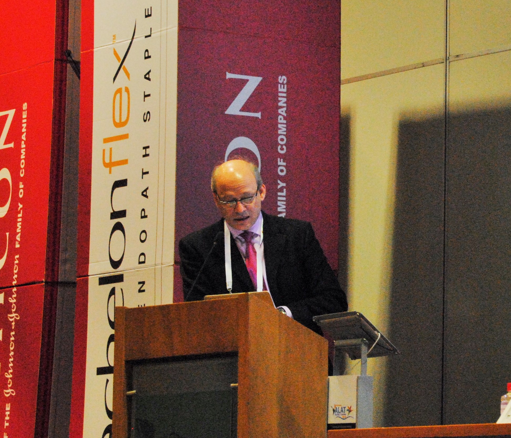
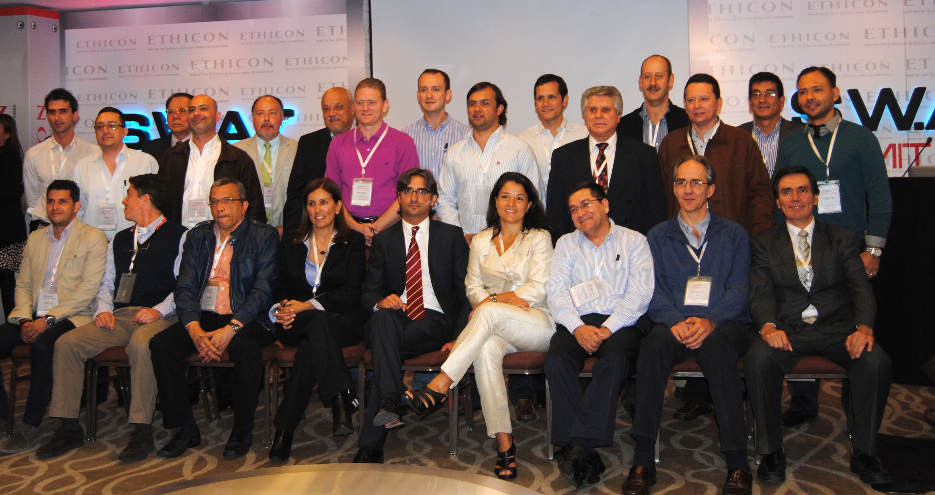
The Ethicon (Johnson & Johnson) sponsored session was by far, the best of the conference – and an excellent overview of modern technologies in thoracic surgery.
starting with Dr. Ricardo Buitrago (purple tie), Dr. Diego Gonzalez Rivas (blue tie) and Dr. Mario Ghefter (pink tie) are changing the future of thoracic surgery
Dr. Diego Gonzalez Rivas
“Is uni-port surgery feasible for advanced cancers?” Short answer: Yes.
The first speaker, was Dr. Diego Gonzalez Rivas of Coruna, Spain. He is a world-renown thoracic surgeon and innovator of uni-port thoracoscopic surgery. He discussed the evolution of single port surgery as well as the most recent developments with this technique, including more advanced and technically challenging cases such as chest wall resections (2013), sleeve resections/ reconstructions (2013), pulmonary artery reconstructions (2013) and surgery on non-intubated, awake patients (2014).
Experience and Management of bleeding
The biggest challenges to surgeons learning this technique is management of bleeding. But as he explained in previous lectures, this can be overcome with a direct approach. (these lectures and YouTube videos, Dr. Gonzalez explains the best ways to manage intra-operative bleeding.) In the vast majority of cases – this did not require deviation or conversion from the uni-port technique.)
As surgeons gain proficiency with this technique which mirrors open surgery, the only contra-indications for surgical resection of cancerous tissue (by single port) are tumors of great size, and surgeon discomfort with the technique.
Dr. Mario Ghefter
My favorite lecture of the series was given by Dr. Mario Ghefter of Sao Paolo, Brazil. While his lecture was ostensibly about video-assisted thoracoscopy (VATS), it was more of a retrospective vision and discussion of the modern history of thoracic surgery as seen through the eyes of a 22 year veteran surgeon.
He talked about the beginnings of VATS surgery and the contributions from such legends as Cefolio and D’Amico, including the 2005 paper – and modern-day thoracic bible, “Troubleshooting video-assisted thoracoscopic lobectomy (Demmy, James, Swanson, McKenna and D’Amico).
Dr. Ghefter also talked about how improved imaging and diagnostic procedures such as PET-CT and EBUS have been able to provide additional diagnostic information pre-operatively that helps surgeons to plan their procedures and treatment strategies more effectively.
Dr. Mario Ghefter
As a counterpoint to both Dr. Gonzalez and Dr. Buitrago, Dr. Ghefter acquitted himself admirably. He reminded audience members that even the newer technologies have some drawbacks – both as procedures and for the surgeons themselves.
He also successfully argued (in my opinion) that while the popularity of procedures such as multiple port VATS and even open thoracotomies have dropped drastically as thoracic surgeons embrace newer technologies, there will always be a place and time for these more traditional procedures.
Dr. Mario Ghefter is the Director of Thoracic Surgery at Hospital do Servidor Público Estadual – Sāo Paulo and on staff at the Hospital Alemão Oswaldo Cruz.
Dr. Ricardo Buitrago
Native Colombian (and my former professor), Dr. Ricardo Buitrago is acknowledged as one of the foremost experts in robotic thoracic surgery in Latin America.
During his presentation, he discussed the principles and basics of use of robotic techniques in thoracic surgery. He reviewed the existing literature surrounding the use of robotic surgery, and comparisons of outcomes between thoracic surgery and traditional lobectomy.
He reviewed several recent robotic surgery cases and the use of robotics as a training tool for residents and fellows.
While he mentioned some of previously discussed limitations of robotic surgery (namely cost of equipment) he cited recent studies demonstrating significant cost savings due to decreased length of stay and a reduced incidence of surgical complications.
He also discussed recent studies (by pioneering surgeons such as Dr. Dylewski) demonstrated short operating times of around 90 minutes.
the Babe Ruth of thoracic surgery continues his winning streak; and Dr. Benny Weksler heads south to the University of Tennessee. Kudos to both of these fine surgeons!
It’s another home run for Dr. Gonzalez Rivas as he and his team perform a single port (uni-port) thoracoscopic lobectomy with under local anesthesia, as reported by a recent story, “Operan un tumor e pulmón con una sola incisión y anestesia local” by Raul Romar in La Voz de Galicia.
Dr. Gonzalez Rivas demonstrates uniportal VATS
The answer is International collaboration and sharing of ideas
Dr. Gonzalez Rivas is used to sharing his ideas. After all, he spends a considerable amount of time traveling the world doing just that; sharing information about and teaching surgeons how to perform the single port thoracoscopic technique. But that doesn’t mean that he does find time to learn from his peers during his travels.
The article above highlights the importance of this international collaboration as it details how Dr. Gonzalez Rivas began to consider applying a local anesthesia approach to the single port surgical technique after talking (and visiting) surgeons in Taiwan and China.
Once he found the perfect candidate, he was ready to implement local anesthesia into his single port approach.. The rest, as they say – is now headed for the Annals of Thoracic Surgery.
Click here for English translation (note translation is not exact).
Dr. Benny Weksler, our own American (via Brazil) superstar surgeon recently made the move to the University of Tennessee. Dr. Weksler made the move in November of 2013 and is now settling in to his new position as Chief of Thoracic Surgery for the University of Tennessee (UT) Health Science Center and UT – Methodist.
Dr. Weksler, one of the United States most prominent thoracic surgeons, particularly in the area of esophageal surgery reports that he has big plans for the UT health system and the thoracic surgery department.
Big Plans for UT and the city of Memphis
These plans include a lung cancer screening program targeting vulnerable populations in Memphis including the uninsured/ underinsured, African-Americans (who are disproportionately affected) and smokers.
Related: Dr. Weksler talks about smoking cessation
Minimally invasive techniques for esophageal surgery
He has also started a new minimally invasive esophageal surgery program for esophageal cancer and reports “that there is almost an epidemics of squamous cell carcinoma of the esophagus” which is something tha was more rare in his previous practice in Pittsburgh, Pennsylvania.
Dr. Weksler and his colleagues are putting together a multi-disciplinary treatment plan to try to get these patients to a complete evaluation with a surgeon, an oncologist, and a radiation oncologist to provide patients with comprehensive, multi-faceted and coördinated care.
“The Surgeon Speaks” – Dr. Weksler talks about robotic surgery in this 2009 Jefferson University publication.
As a former Memphis resident, I want to say, “Welcome to the mid-south.. Hope you find time in your busy schedule to enjoy Beale Street, visit the Pink Palace and tour Graceland.. On behalf of all current Memphians, we are glad you are here.”
*This article was written by the author of this post.
the latest from Dr. Diego Gonzalez Rivas and the masters of thoracic surgery.
Dr. Gonzalez Rivas and the Thoracic Surgery Unit in Coruna, Spain are hosting the “International Symposium on Uniportal VATS” this week (February 26th to 28th, 2014).
Dr. Gonzalez Rivas demonstrates uniportal VATS
While the in-person, on-site event is limited to just 100 attendees, the event will be offering real-time live streaming surgery for viewers worldwide.
With registrations from around the world, Dr. Gonzalez Rivas estimates that thousands of pairs of eyes will be watching; from Australia to Saudi Arabia, Hong Kong to Colombia, Brazil to Russia, and the United States.
If you’ve ever wanted to learn more about single port VATS, this is the time to find out.
For more information:
Livethoracic.com – link to the event and on-line registration. Registration is 500 Euros.
Article at Examiner.com with more details on this event.
Discussing the classification and treatment of lung cancer according to the latest revisions (7th edition).
Medical City, Dallas, Texas USA
Sometimes location and timing is everything. Since I can’t attend all of the great thoracic surgery conferences and events, sometimes I just have to wait for something closer to home. But then again, “home” is a relative concept.
As a locum tenens provider, I travel around the country working in various hospital surgical programs on short-term contracts. It’s an interesting and always changing life but one that allows me to pursue my love of thoracic surgery to the fullest.
For the next few weeks, Medical City in Dallas, Texas is my home, as part of the cardiothoracic surgery service. It’s a return trip so it was nice to renew my acquaintance with the surgeons and staff of the CVICU and step-down units.
Today, as part of an ongoing continuing medical education program series, Dr. Mitchell Magee, of Southwest Cardiothoracic Surgeons gave an hour-long lecture entitled, “Lung cancer staging and evolving less invasive surgical treatment alternatives.” The focus of the talk was the changes in lung cancer classification and staging in the 7th edition guidelines. These revisions were proposed to replace previous versions which were based on a very small, select sample of patients at a single site. In comparison, the new revisions are based on over 100,000 patients worldwide.
T, N, M
T – tumor
N – nodes
M – metastasis
This classification system has been in use since the 1940’s and has been revised several times to reflect our growing knowledge. The latest revisions (7th edition) were released in 2012 after several years of research and debate. (For more on this process, see “The science behind the 7th edition Tumour, Node, Metastasis staging system for lung cancer” by Marshall et al, 2012).
Dr. Magee discussed the most recent revisions and how these changes affect both the treatment recommendations and prognoses for our patients. After reviewing these changes, he talked a bit about obtaining sufficient diagnostic information for accurate staging, including the role of EBUS, the new CT scan screening guidelines and the gold standard, mediastinoscopy. He also discussed some of the limitations of PET/CT and other non-invasive diagnostic imaging.
Upstaged/ Downstaged
As part of these changes in the subclassification of tumors, 10 stages have been downstaged (meaning that previously in-operable cases may now be eligible for resection) and seven classifications have been upstaged – meaning that the cancers are now considered more advanced.
For example, patients with two separate tumors in the same lobe of the lung has been upstaged to T3. Two different tumors in the same lung, but a different lobe is now T4 classification.
More specific
Some of the classifications have changed to make findings more specific. For example, T1 staging has now been subdivided into T1a and T1b.
Any invasion of the pleura, including microscopic – is now T2 staging.
He concluded the presentation with a short overview of the history of surgical resection for lung cancer, and the evolution of surgical techniques from open thoracotomies with pneumonectomies to lung sparing procedures utilizing more minimally invasive techniques.
Despite these changes, the hallmarks of a successful cancer operation remain unchanged – the right operation for the individual patient, and the need to respect oncological principles, like surgical margins, and a through lymph node dissection.
Lymph node dissection/ node sampling
Node sampling remains a crucial part of the cancer staging process despite the advent of less invasive imaging studies due to it’s infaliable accuracy. (There is either tumor tissue in the node or there isn’t, where as PET scan results can be false positive or false negative).
For this reason, tissue samples remain the gold standard of treatment and are the most accurate way to predict and prognosticate the extent of disease.
General rules regarding lymph node sampling are:
– More nodes are better. The minimum acceptable number of nodes for accurate staging is at least SIX for at least THREE different stations.
A good way to remember the relationship between node stations and node status is that bode stations are determined by distance from mediastinum; meaning that node station 14 is more peripheral that node 2.
N1 nodes are stations 10 – 14
N2 nodes are the single digit nodes (2, 4, 7 etc.)
Lymph nodes used for diagnosis and staging. Copyright Memorial Sloan-Kettering Cancer Center. Used with permission.
References and additional suggested readingBaltayiannis N, Chandrinos M, Anagnostopoulos D, Zarogoulidis P, Tsakiridis K, Mpakas A, Machairiotis N, Katsikogiannis N, Kougioumtzi I, Courcoutsakis N, Zarogoulidis K. (2013). Lung cancer surgery: an up to date. J Thorac Dis. 2013 Sep;5(Suppl 4):S425-S439. Review. Free pdf. Nice review article discussing the importance of staging for determining optimal treatment for lung cancer, as well as the impact of the latest revisions to the (7th edition) TNM classification system.
IASLC Staging Handbook in Thoracic Oncology – a site-specific guide on the new TNM classification of thoracic malignancies. This publication is published in coordination with the 7th editions of the TNM Classification of Malignant Tumors/UICC and AJCC Cancer Staging Manual.
Goldstraw P, Crowley J, Chansky K et al. (2007). The
IASLC lung cancer project: proposals for the revision of the
TNM stage groupings in the forthcoming (seventh) edition of
the TNM classification of malignant tumours. J Thorac Oncol
2007; 2: 706-714. Figure 1. Powerpoint slides TNM classification revisions for the 7th edition.
Quick and easy summary of the 7th edition classifications for Lung cancer staging – 7th edition Lung cancer staging pdf from the American Joint Commission on Cancer.
Dr. Mitchell Magee, thoracic surgeon at Medical City – Dallas, Texas
Dr. Mitchell Magee is Surgical Director of Thoracic Oncology and the Minimally Invasive Therapy Institute for Lung and Esophagus at Medical City Dallas. While his partner, Dr. Dewey focuses exclusively on cardiac surgeries like cardiac bypass, valve replacement, TAVR, LVADS and cardiac transplantation, Dr. Magee is the thoracic arm of the two surgeon Southwest Cardiothoracic Surgeons practice. This means Dr. Magee is able to devote his time to a sizable portion of all of the esophageal tears, empyemas, mediastinal masses and lung pathology that a city the size of Dallas has to offer.
Dr. Mitchell Magee & Amber Bethea, PA-C
Dr. Magee is also part of the CLEAR Clinic at Medical City – which is the lung cancer screening center at the Medical City Dallas facility.
in the operating room with Dr. Diego Gonzalez Rivas for single port thoracoscopic (uniportal) surgery.
Hilar mass resection using single port thoracoscopy with Dr. Diego Gonzalez – Rivas
K. Eckland & Andres M. Neira, MD
Instituto Nacional de Cancerlogia
Bogota, Colombia
Surgeon(s): Dr. Diego Gonzalez Rivas and Dr. Ricardo Buitrago
Dr. Diego Gonzalez Rivas demonstrates single port thoracoscopy
Case History:
59-year-old female with past medical history significant for recurrent mediastinal mass previously resectioned via right VATS. Additional past medical history included prior right-sided nephrectomy.
Pre-operative labs:
CBC: WBC 7230 Neu 73% Hgb:14.1 Hct 37 platelets 365000
Pt 12.1 / INR1.1 PTT: 28.3
Diagnostics:
Pre-operative CT scan: chest
edited to preserve patient privacy
Procedure: Single port thoracoscopy with resection of mediastinal mass and lymph node sampling
After review of relevant patient history including radiographs, patient was positioned for a right-sided procedure. After being prepped, and draped, surgery procedure in sterile fashion. A linear incision was made in the anterior chest – mid clavicular line at approximately the fifth intercostal space. A 10mm port was briefly inserted and the chest cavity inspected. The port was then removed, and the incision was expanded by an additional centimeter to allow for the passage of multiple instruments; including camera, grasper and suction catheter.
Dr. Gonzalez Rivas and Dr. Ricardo Buitrago at National Cancer Institute
The chest cavity, pleura and lung were inspected. The medial mediastinal mass was then identified.
As previously indicated on pre-operative CT scan, the mass was located adjacent and adherent to the vessels of the hilum. This area was carefully dissected free, in a painstaking fashion. After freeing the mediastinal mass from the hilum, the remaining surfaces of the mass were resected. The mass was fixed to the artery pulmonary and infiltrating it) . The mass was removed en-bloc. Care was then taken to identify, and sample the adjacent lymph nodes which were located at stations (4, 7 and 10).
Following removal of the tumor and lymph nodes, the area was re-inspected, and the lung was re-inflated. A 28 french chest tube was inserted in the original incision, with suturing of the fascia, subcutaneous and skin layers.
closing the single port incision
Hemostasis was maintained during the procedure with minimal blood loss.
Patient was hemodynamically stable throughout the case, and maintained appropriate oxygen saturations. Following surgery, the patient was awakened, extubated and transferred to the surgical intensive care unit.
Post-operative: Post-operative chest x-ray confirmed appropriate chest tube placement and no significant bleeding or pneumothorax.
Immediate post-operative film (chest tube visible)
Patient did well post-operatively. Chest tube was discontinued on POD#2 and discharged home.
PA & LAT films on post-operative day 2
Discussion: Since the initial published reports of single-port thoracoscopy, this procedure has been applied to an increasing range of cases. Dr. Gonzalez and his team have published reports demonstrating the safety and utility of the single-port technique for multiple procedures including lobectomies, sleeve resections, segmentectomies, pneumonectomies and mediastinal mass resections. Dr. Hanao Chen (Taiwan) has reported several successful esophagectomies using this technical, as well as bilateral pleural drainage using a unilateral single-port approach.
Contrary to popular perception, the use of a single-port versus traditional VATS procedures (three or more) is actually associated with better visibility and accessibility for surgeons. Surgeons using this technical have also reported better ergonomics with less operating fatigue related to awkward body positioning while operating.
The learn curve for this surgical approach is less than anticipated due to the reasons cited above, and has been cited at 5 to 20 cases by Dr. Gonzalez, the creator of this approach.
The main limitations for surgeons using this technique is often related to anticipated (but potentially unrealized) fears regarding the need for urgent conversion to open thoracotomy. In reality, many of the complications that may lead to urgent conversion, such as major bleeding, are manageable thoracoscopically once surgeons are experienced and comfortable with this approach.
Dr. Gonzalez and his colleagues have reported a conversion rate of less than 1% in their practice. Subsequent reports by Dr. Gonzalez and his colleagues have documented these findings.
Other barriers to adoption of this technique are surgeon-based, and may be related to the individual surgeon’s willingness or reluctance to adopt new techniques and technology. Many of these surgeons would be surprised by how this technique mimics open surgery.
The successful adoption of this technique by numerous thoracic surgery fellows shows the feasibility and ease of learning single-port thoracoscopy by surgeons interested in adopting and advancing their surgical proficiency in minimally invasive surgery.
The benefits for utilizing this technique include decreased length of stay, decreased patient discomfort and greater patient satisfaction.
References/ Additional Readings
Bertolaccini, L., Rocco, G., Viti, A. & Terzi, A. (2013). Surgical technique: Geometrical characteristics of uniportal VATS. J. Thorac Dis. 2013, Apr 07. Article from thoracic surgeons at the National Cancer Institute in Naples, Italy explains how the geometric advantages of uniportal VATS improves visibility and spatial perception over traditional VATS, and mimics open surgery.
Calvin, S. H. Ng (2013). Uniportal VATS in Asia. J Thorac Dis 2013 Jun 20. Article discussing the spread of uniportal techniques in Taiwan, China and other parts of Asia.
Chen, Chin-Hao, Lin, Wei-Sha, Chang, Ho, Lee, Shih-Yi, Tzu-Ti, Hung & Tai, Chih-Yin (2013). Treatment of bilateral empyema thoracis using unilateral single-port thoracoscopic approach. Ann Thorac Cardiovasc Surg 2013.
Gonzalez Rivas, D., Fieira, E., Delgado, M., Mendez, L., Fernandez, R. & De la Torre, M. (2013). Surgical technique: Uniportal video-assisted thoracoscopic lobectomy. J. Thorac Dis. 2013 July 4.
Gonzalez Rivas, D., Delgado, M., Fieira, E., Mendez, L. Fernandez, R. & De la Torre, M. (2013). Surgical technique: Uniportal video-assisted thoracoscopic pneumonectomy. J. Thorac Dis. 2013 July 4.
Rocco, G. (2013). VATS and uniportal VATS: a glimpse into the future. J. Thorac Dis. 2013 July 04. After coming across several articles by Dr. Gaetano Rocco, and actively pursuing several other publications by this Italian thoracic surgeon, I have become increasingly convinced that Gaetano Rocco, along with Dr. Gonzalez Rivas is one of the world’s leading innovators in thoracic surgery. Hopefully, cirugia de torax will be able to catch up to Dr. Rocco at some point for an in-depth discussion.
Rocco G. Single port video-assisted thoracic surgery (uniportal) in the routine general thoracic surgical practice. Op Tech (Society of Thoracic and Cardiovascular Surgeons). 2009;14:326–335.
Rocco G, Khalil M, Jutley R. Uniportal video-assisted thoracoscopic surgery wedge lung biopsy in the diagnosis of interstitial lung diseases. J Thorac Cardiovasc Surg. 2005;129:947–948.
5 / Video-assisted thoracic surgery lobectomy: 3-year initial experience with 200 cases. Gonzalez D, De la Torre M, Paradela M, Fernandez R, Delgado M, Garcia J,Fieira E, Mendez L. Eur J Cardiothorac Surg. 2011 40(1):e21-8.
6 / Single-port Video-Assisted Thoracoscopic Anatomical Resection: Initial Experience. Diego Gonzalez , Ricardo Fernandez, Mercedes De La Torre, Maria Delgado, Marina Paradela, Lucia Mendez. Innovations.Vol 6.Number 3. May/jun 2011. Page 165.
the 2013 S.W.A.T conference, presented by Johnson & Johnson. Featured presenters Dr. Diego Gonzalez Rivas and Dr. Paula Ugalde discuss single port thoracoscopy and topics in minimally invasive surgery
Very pleased that despite the initial difficulties, we are able to provide information regarding the recent conference.
Talking about Single-port surgery in Bogotá, Colombia – 2013 S.W.A.T. Summit
Dr. Diego Gonzalez Rivas and Dr. Paula Ugalde were the headliners at the recent Johnson and Johnson thoracic surgery summit on minimally invasive surgery. Both surgeons gave multiple presentations on several topics. They were joined at the lectern by several local Colombian surgeons including Dr. Stella Martinez Jaramillo (Bogotá), Dr. Luis Fernando Rueda (Barranquilla), Dr. Jose Maineri (Venezuela) Dr. Mario Lopez (Bogotá) and Dr. Pardo (Cartagena).
Thoracic surgeons at the 2013 S.W.A.T Summit in Bogota, Colombia. Drs. Gonzalez-Rivas and Dr. Paula Ugalde are center, front-row
Target audience missing from conference
The audience was made up of thirty Latin American surgeons from Colombia, Costa Rica and Venezuela. This surgeons were hand-picked for this invitation-only event. Unfortunately, while Johnson and Johnson organized and presented a lovely event; their apparent lack of knowledge about the local (Colombian) thoracic surgery community resulted in the exclusion of several key surgeons including Dr. Mauricio Velasquez, one of Colombia’s earliest adopters of single-port thoracoscopy. Also excluded were the junior members of the community, including Dr. Castano, Dr. Carlos Carvajal, and current thoracic surgery fellows. It was an otherwise outstandingand informative event.
The Gonzalez – Rivas dissector, photo courtesy of Scanlon International
As discussed in multiple publications, previous posts as well as during the conference itself, it is these younger members who are more likely to adopt newer surgical techniques versus older, more experienced surgeons. More seasoned surgeons may be hesitant to change their practices since they are more comfortable and accustomed to open surgical procedures.
Despite their absence, it was an engaging and interesting conference which engendered lively discussion among the surgeons present.
Of course, the highlight of the conference actually occurred the day before, when Dr. Gonzalez- Rivas demonstrated his technique during two separate cases at the National Cancer Institute in Bogotá, Colombia. (Case report).
Dr. Gonzalez-Rivas and Dr. Ricardo Buitrago performing single port thoracoscopy at the National Cancer Institute
Featured presenters:
Dr. Diego Gonzalez – Rivas is a world-renown thoracic surgeon jointly credited (along with Dr. Gaetano Rocco) with the development of single-port thoracoscopic (uni-port) surgery. He and his colleagues at the Minimally Invasive Surgery Unit in La Coruna, Spain give classes and lectures on this technique internationally. Recent publications include three papers in July alone detailing the application of this surgical approach, as well as several YouTube videos demonstrating use of this technique for a wide variety of cases.
Dr. Gonzalez Rivas
Dr. Paula Ugalde, a Chilean-borne thoracic surgeon (from Brazil) who gave several presentations on minimally-invasive surgery topics. She is currently affiliated with a facility in Quebec, Canada.
Dr. Paula Ugalde
Refuting the folklore
Part of the conference focused on refuting the ‘folklore’ of minimally-invasive procedures. Some of these falsehoods have plagued minimally-invasive surgery since the beginning of VATS (in 1991), such as the belief that VATS should not be applied in oncology cases. The presenters also discussed how uniportal VATS actually provides improved visibility and spatial perception over traditional VATS (Bertolaccini et al. 2013).
However, Gonzalez-Rivas, Ugalde and the other surgeons in attendance presented a wealth of data, and publications to demonstrate:
– VATS is safe and feasible for surgical resection in patients with cancer. (Like all surgeries, oncological principles like obtaining clear margins, and performing a thorough lymph node dissection need to be maintained).
– Thorough and complete lymph node dissection is possible using minimally invasive techniques like single-port surgery. Multiple studies have demonstrated that on average, surgeons using this technique obtain more nodes than surgeons using more traditional methods.
– Large surgeries like pneumonectomies and sleeve resections are reasonable and feasible to perform with single-port thoracoscopy. Using these techniques may reduce morbidity, pain and length of stay in these patients.
– Rates of conversion to open surgery are very low (rare occurrence). In single-port surgery, “conversion” usually means adding another port – not making a larger incision.
– Learning curve fallacies: the learning curve varies with each individual surgeon – but in general, surgeons proficient in traditional VATS and younger surgeons (the “X box generation”) will readily adapt to single-port surgery.
– Bleeding, even significant bleeding can be managed using single-port thoracoscopy. Dr. Gonzalez Rivas gave a separate presentation using several operative videos to demonstrate methods of controlling bleeding during single-port surgery – since this is a common concern among surgeons hesitant to apply these advanced surgical techniques.
Additional References / Readings about Single-Port Thoracoscopy
Scanlon single-port thoracoscopy kits – informational brochure about specially designed instruments endorsed by Dr. Gonzalez Rivas.
Dr. Diego Gonzalez Rivas – YouTube channel : Dr. Gonzalez Rivas maintains an active YouTube channel with multiple videos demonstrating his surgical technique during a variety of cases. Below is a full-length video demonstrating the uniportal technique.
Additional posts at Cirugia de Torax about Dr. Diego Gonzalez- Rivas
Upcoming conference in Florida – information about registering for September conference for hands-on course in single-port thoracoscopic surgery with Dr. Gonzalez-Rivas
Youtube video for web conference on Single-port thoracoscopic surgery
Bertolaccini, L., Rocco, G., Viti, A. & Terzi, A. (2013). Surgical technique: Geometrical characteristics of uniportal VATS. J. Thorac Dis. 2013, Apr 07. Article from thoracic surgeons at the National Cancer Institute in Naples, Italy explains how the geometric advantages of uniportal VATS improves visibility and spatial perception over traditional VATS, and mimics open surgery.
Calvin, S. H. Ng (2013). Uniportal VATS in Asia.J Thorac Dis 2013 Jun 20. Article discussing the spread of uniportal techniques in Taiwan, China and other parts of Asia.
Rocco, G. (2013). VATS and uniportal VATS: a glimpse into the future.J. Thorac Dis. 2013 July 04. After coming across several articles by Dr. Gaetano Rocco, and actively pursuing several other publications by this Italian thoracic surgeon, I have become increasingly convinced that Gaetano Rocco, along with Dr. Gonzalez Rivas is one of the world’s leading innovators in thoracic surgery. Hopefully, cirugia de torax will be able to catch up to Dr. Rocco at some point for an in-depth discussion.
While I advance criticism of this event – it was a fantastic conference. My only reservations were to the exclusivity of the event. While this was certainly related to the costs of providing facilities and services for this event – hopefully, the next J & J thoracic event will be open to more interested individuals including young surgeons and nurses.
writing about Dr. Diego Gonzalez Rivas and the other living legends in thoracic surgery and connecting people to the world of thoracic surgery
Readers at Cirugia de Torax have certainly noticed that there are numerous articles regarding the work of Dr. Diego Gonzalez Rivas. This week in particular, after a recent thoracic surgery conference and an afternoon in the operating room – there is a lot to say about the Spanish surgeon.
It’s also hard to escape that fact that I regard him in considerable awe and esteem for his numerous contributions to thoracic surgery and prolific publications. I imagine that this is similar to how many people felt about Drs. Cooley, Pearson or Debakey during their prime.
Making thoracic surgery accessible
But the difference is Dr. Diego Gonzalez Rivas himself. Despite the international fame and critical surgical acclaim, he remains friendly and approachable. He has also been extremely supportive of my work, at a time when not many people in thoracic surgery see the necessity or utility of a nurse-run website.
After all, the internet is filled with other options for readers; CTSnet.org, multiple societies like the Society of Thoracic Surgeons (STS), and massive compilations like journal-based sites (Annals of Thoracic Surgery, Journal of Thoracic Disease, Interactive Journal of Cardiothoracic Surgery).
But the difference between Cirugia de Torax and those sites is like the difference between Dr. Gonzalez Rivas and many of the original masters of surgery: Approach-ability and accessibility.
This site is specifically designed for a wider range of appeal, for both professionals in thoracic surgery, and for our consumers – the patients and their families. Research, innovation, news and development matters to all of us, not just the professionals in the hallowed halls of academia. But sometimes it doesn’t feel that way.
Serving practicing surgeons
For practice-based clinicians, and international surgeons publication in an academia-based journal requires a significant effort. These surgeons usually don’t have research assistants, residents and government grants to support their efforts, collect their data and clean up their grammar. Often English is a second or third language. But that doesn’t mean that they don’t make valuable contributions to their patients and the practice of thoracic surgery. This is their platform, to bring their efforts to their peers and the world.
Heady aspirations
That may sound like a lofty goal, but we have readers from over a 110 countries, with hundreds of subscribers along with over 6,000 people with Cirugia de Torax directly on their smart phone. Each month, we attract more hits and more readers.
Every day, at least 200 people read “Blebs, Bullae and Spontanous Pneumothorax”. Why? Because it’s a concise article that explains what blebs are, how a pneumothorax occurs and how it’s treated. Another hundred people usually go on to read the accompanying case report about blebectomy, for similar reasons. There are links for more information, CT scans and intra-operative photos included, so that people can find exactly what they need with a minimum of effort.
Avoiding ‘Google overload’
With the massive volume of information available on the internet, high-quality, easily understood, applicable information has actually become even more difficult for patients to find than ever before. Patients spend hours upon hours browsing through academic jargon, commercial websites and biased materials while attempting to sift through the reams of information for pertinent and easily understandable information. There is also a lot of great material out there – so we provide links to reputable sites, recommend well-written articles and discuss related research.
Connecting patients to surgeons
We also provide patients with more information about the people they are entrusting their bodies, their hopes and their lives to. It’s important that they know about the Dr. Benny Wekslers, the Dr. Hanao Chens, and the Dr. Diego Gonzalez Rivas out there.
Update: June 2019
After multiple reader requests from this site, we have launched a service to assist readers in pursuiting minimally invasive thoracic surgery, uniportal surgery, HITHOC and other state-of-the-art thoracic surgery procedures with the modern masters of thoracic surgery. We won’t talk a lot about this on the site, but we do want readers to know that we are here to help you. If you are wondering what surgery costs like with one of the world’s experts – it’s often surprisingly affordable.
If you are interested in knowing more, please head to our sister site, www.americanphysiciansnetwork.org or send an email to kristin@americanphysiciansnetwork.org.
Keeping it ‘real’
Looking over the shoulder of Dr. Gonzalez Rivas in the operating room
As much as I may admire the work and the accomplishments of Dr. Gonzalez-Rivas – it’s important not to place him on a pedestal. He and his colleagues are real, practicing surgeons who operating on regular people, not just heads of state and celebrities. So when we interview these surgeons and head to the OR, it’s time to forget about the accolades, the published papers and the fancy titles. It’s time to focus on the operations, the techniques, the patients and the outcomes because ‘master of thoracic surgery’ or rural surgeon – the operation and patient are all that really matters.
Reviewing “Ten years experience on 644 patients undergoing single-port (uniportal) video-assisted” by Gaetano Rocco et al. at the National Cancer Institute in Naples, Italy
In this month’s issue of the Annals of Thoracic Surgery, Dr. Gaetano Rocco and his colleagues at the National Cancer Institute, Pascale Foundation in Naples, Italy reported their findings on ten year’s worth of single-port surgery in their institution.
Who: 644 patients; (334 males, 310 females)
Indications:
Annals of thoracic surgery – Rocco et. al (2013)
What: Outcomes and experiences in single port thoracic surgery over a ten-year period. All procedures performed by a single surgeon at this institution, and single-port VATS accounted for 27.7% of all surgeries performed during this time period.
When: data collected on thoracic surgery patients from January 2000 – December 2010.
Technical Notes:
Pre-operative CT scan was used for incision placement planning. Incision was up to 2.5 cm (1 inch) in length depending on indications for surgery.
Conversion rate to 2 or 3 port VATS: 2.2% (14 patients)
Conversion to mini-thoracotomy: 1.5% (10 patients)
Patients underwent conversion due to incomplete lung collapse (22 patients) and bleeding (2 patients).
There were no re-operations or “take backs”. The four patients with malignant effusions who died within the 30 day post-op period were re-admitted to the ICU.
Post-operatively:
Otherwise, all patients were admitted to either the floor or the step-down unit following surgery.
Pain management: post-operative pain was managed with a non-narcotic regimen consisting of a 24 hour IV infusion pump of ketorolac (20mg) and tramadol (100mg*). After the first 24 hours, patients were managed with oral analgesics such as paracetamol (acetaminophen).
Limitations: in this study, uni-port VATS was not used for major resections, as seen in the work of Dr. Diego Gonzalez and others. This may be due to the fact that uni-port VATS was an emerging technique at the initiation of this study.
Strengths: This is one of the largest studies examining the use of single-port thoracic surgery – and showed low morbidity and mortality. (Arguably, the 30 day mortality in this study was related to the patients’ underlying cancers, rather than the surgical procedure itself.)
*Intravenous tramadol is not available in the United States.
Rocco G. Single port video-assisted thoracic surgery (uniportal) in the routine general thoracic surgical practice. Op Tech (Society of Thoracic and Cardiovascular Surgeons). 2009;14:326–335.
Rocco G, Khalil M, Jutley R. Uniportal video-assisted thoracoscopic surgery wedge lung biopsy in the diagnosis of interstitial lung diseases. J Thorac Cardiovasc Surg. 2005;129:947–948.
What kind of pain should patients expect after thoracic surgery, and how long will it last? Also, is this normal? When should I call my doctor?
Like all posts here at Cirugia de Torax, this should serve as a guide for talking to your healthcare provider, and is not a substitute for medical advice.
Quite a few people have written in with questions about post-operative pain after thoracic surgery procedures so we will try to address those questions here.
1. What is a normal amount of pain after these procedures?
While no two people will experience pain the same, there are some general guidelines to consider. But to talk about this issue – we will need to refer to a basic pain scale which rates pain from 0 (no pain) to 10 – (excruciating, writhing pain, worst possible imaginable).
Unfortunately, for the majority of people who have thoracic surgery, there will be some pain and discomfort.
Pain depends on the procedure
In general, the intensity and duration of pain after thoracic procedures is related to the surgical approach – or the type of surgical incision used.
This open incision (with rib spreading) will hurt more.. Photo: advanced empyema requiring open thoracotomy for decortication
Pain will be much less with a single incision VATS surgery (now with chest tube) and specimen removed (photo courtesy of Dr. Chen)
Patients with larger incisions like a sternotomy, thoracotomy or clamshell incision will have more pain, for a longer period of time than patients that have minimally invasive procedures like VATS because there is more trauma to the surrounding tissues. People with larger incisions (from ‘open surgeries’) are also more likely to develop neuralgia symptoms as they recover.
. (I will post pictures of the various incisions once I return home to my collection of surgical images).
Many patients will require narcotics or strong analgesics for the first few days but most surgeons will try to transition patients to anti-inflammatories after surgery.
Post-operative surgical pain is often related to inflammation and surgical manipulation of the chest wall, particularly in procedures such as pleurodesis, decortication or pleurectomy. For many patients this pain will diminish gradually over time – but lasts about 4 to 6 weeks.
Anti-inflammatories
This pain is often better managed with over the counter medications such as ibuprofen than with stronger narcotics. That’s because the medication helps to relieve the inflammation in addition to relieving pain. Anti-inflammatory medications also avoid the risks of oversedation, drowsiness and severe constipation that often comes with narcotics.
Use with caution
However, even though these medications are available without a prescription be sure to talk to your local pharmacist about dosing because these medications can damage the kidneys. Also, be sure to keep hydrated while taking this medications.
People with high blood pressure should be particularly cautious when taking over the counter anti-inflammatories because many of these medications have drug interactions with blood pressure medications.
2. “I had surgery three weeks ago, and I recently developed a burning sensation near the incision”
Neuralgias after surgery
For many patients, the development of a neuralgia is a temporary effect and is part of the healing process. However, it can be quite disturbing if patients are unprepared. Neuralgic pain is often described as a burning or stinging sensation that extends across the chest wall from the initial incision area. Patients also describe it as a ‘pins and needles’ sensation or “like when your foot falls asleep”. This usually develops a few weeks after surgery as the nerves heal from the surgery itself.
It the discomfort is unmanageable, or persists beyond a few weeks, a return visit to your surgeon is warranted. He/She can prescribe medications like gabapentin which will soothe the irritated nerves and lessen the sensations. However, these medications may take some time to reach full effect.
Range of motion and exercise after surgery
Exercise limitations are related to the type of incision.
Sternotomy incisions/ sternotomy precautions
If you have a sternotomy incision – (an incision through the breast bone at the center of your chest), this incision requires strict precautions to prevent re-injury to the area. Since the sternal bone was cut, patients are usually restricted from lifting anything greater than 10 pounds for 6 to 12 weeks, and to avoid pushing, pulling or placing stress on the incision. Patients are also restricted from driving until bone healing is well underway. (Be sure to attend a rehab program or physical therapy program to learn the proper way to exercise during this time period).
Patients will also need to take care to prevent a surgical skin infection or something more serious like mediastinitis. The includes prohibitions against tub bathing/ soaking, swimming or over- aggressive cleaning of the incision with harsh abrasives like hydrogen peroxide or anti-bacterial soaps. These chemicals actually do more harm than good in most cases by destroying the newly healing tissue. A good rule of thumb to remember (unless your doctor says otherwise): No creams or lotions to your incisions until the scabs fall off.
Post-thoracotomy incisions
With a large thoracotomy incision, most patients will be restricted from lifting any items greater than 10 pounds on the surgery side for around two weeks. However, unlike sternotomy patients – we want you to use and exercise that arm daily – otherwise patients have a risk of developing a ‘disuse’ syndrome. One of the common exercises after a thoracotomy is called the spider crawl. This exercise helps the muscles to heal and prevent long-term disability or problems. The physiotherapist at your local hospital should have a list of several others that they can teach you to practice at home.
The spider crawl
In this example, the patient had a left thoracotomy:
1. Stand with your surgical side within arm’s length of the wall.
start with your hand at waist level
Now, use your hand to “walk” up the wall, similar to a spider crawling.
‘walking’ the hand up the wall
Continue to walk your hand up the wall until your arm is fully extended.
continue until arm fully extended
Perform this exercise (or similar ones) for several minutes 5-6 times a day. As you can see – it is fast and easy to do.
VATS
For patients with minimally invasive procedures – there are very few exercise restrictions, except no heavy lifting for 2 to 3 weeks (this is not the time to help your neighbor move his television.)
General incision care guidelines are similar to that for sternotomy patients – no soaking or bathing (showering is usually okay), no creams or lotions and no anti-bacterial soaps/ hydrogen peroxide/ harsh cleaners.
Whats NOT normal – when it’s time to call your surgeon
– dramatic increase in pain not associated with activity (i.e. lifting or reaching). If your pain has been a “4” for several days and suddenly increases to an “8”
– If the quality of the pain changes – ie. if it was a dull ache and becomes a stabbing pain.
– any breathlessness, shortness of breath or difficulty breathing
– Any increase in redness, or swelling around your incisions. Incisions may be pink and swollen for the first 2-3 days, but any increase after that warrants a ‘wound check’ by your surgeon
– Any fresh bleeding – bright red blood. A small amount of drainage (from chest tube sites) that is light pink, clear or yellow in nature may be normal for the first few days.
– Drainage from the other sites (not chest tube sites) such as your primary incision is not normal and may be a sign of a developing infection.
– Fever, particularly fever greater than 101.5 – may be a sign of an infection.
– If you are diabetic, and your blood sugars become elevated at home, this may also be a sign of infection. (Elevation in the first few days is normal, and is often treated with insulin – particularly if you are in the hospital.
– Pain that persists beyond 3 months may be a sign of nerve damage (and you will need additional medications / therapies).
Discussing Dr. Joseph Coselli and ‘the cowboys of cardiac surgery’ along with some of our own heros of thoracic surgery here at Cirugia de Torax.
There’s a great article in this month’s Annals of Thoracic Surgery, by Dr. Joseph Coselli, from Texas Heart Institute and the Michael DeBakey Department of Surgery at Baylor. His article, entitled,” My heros have always been cowboys” is more than just a title torn from the song sheets of Willie Nelson. It’s a look back at both the pioneers of cardiac surgery and his own experiences as a cardiac surgeon. He also discusses the role of surgeons, and medical practitioners in American society in general and the promises we make to both society at large and our patients.
Here at Cirugia de Torax, I’d like to take a moment to look back at the surgeons that inspired and encouraged me in this and all of my endeavors. Some of these surgeons knew me, and some of them didn’t – but their encouragement and kindnesses have spurred a career and life that have brought immense personal and professional satisfaction.
Like Dr. Coselli, I too, took inspiration from the likes of Dr. Denton Cooley. But our stories diverge greatly from there. I never met Dr. Cooley and I probably never will. But it was a related story, from my former boss (and cardiothoracic surgeon), Dr. Richard Embrey that led to an email to Dr. Cooley himself. My boss had too trained under Dr. Cooley, Dr. Debakey and the Texas Heart Institute, the citadel of American heart surgery. Then, somehow, along the way – Dr. Embrey stopped to work at our little rural Virginia hospital. We were the remnants of a larger Duke cardiothoracic program but we were a country hospital all the same.
While I learned the ins and outs of surgery from Dr. Embrey (and Dr. Geoffrey Graeber at West Virginia University) on a day-to-day basis, I was also weaned on the folklore of cardiothoracic surgery – stories of the giants of history, like the ones mentioned in Dr. Coselli’s article, as well as local Duke legends who occasionally roamed the halls of our tiny ICU and our two cardiothoracic OR suites; Dr. Duane Davis, Dr. Shu S. Lin and Dr. Peter Smith. While never working side-by-side, Dr. D’Amico’s name was almost as familiar as my own. As the sole nurse practitioner in this facility, without residents or fellows, there was no buffer, and little social divide in our daily practice. Certainly, this changed me – and my perceptions. I asked the ‘stupid’ questions but received intelligent and insightful answers. I asked even more questions, and learned even more..
These opportunities fed my mind, and nurtured my ambitions. Not to be a physician or a doctor, but to learn as much as possible about my specialty; to be the best nurse possible in my field. It also nurtured a desire to share these experiences, and this knowledge with my peers, my patients and everyone else who ever had an interest.
It was that tiny little email, a gracious three-line reply from Dr. Cooley himself that made me realize that I didn’t have to rely on folklore and second-hand stories to hear more. That’s critical; because as we’ve seen (here at Cirugia de Torax) there are a quite of few of “Masters of thoracic surgery” or perhaps future giants that haven’t had their stories told. Dr. Coselli and his fellow writers haven’t written about them yet.. So I will.
Sometimes I interview famous (or semi-famous) surgeons here, but other times, I interview lesser-known but equally talented/ innovative or promising surgeons. All of them share similar traits; dedication and love for the profession, immense surgical talent and proficiency and sincere belief in the future of technology of surgery.
So, let’s hope that it won’t take forty more years for these surgeons to be recognized for their contributions to thoracic surgery in the way that Cooley, DeBakey and Crawford are heralded in cardiac surgery.
a record number of surgeons fail to pass the American thoracic surgery certification exam, in the midst of a deepening shortage of surgeons.
A new report from the (American) Board of Thoracic Surgery shows a growing number of eligible surgeons are failing the thoracic surgery certification examination.
Record Failure Rate
As stated in the article published at Family Practice News, the failure rate has doubled to 28% in just a few short years. This comes at a critical period in American medicine as shortages in specialty surgeons have emerged around the country due to an aging workforce. This shortage is not confined to the United States – and has been echoed in Canada, the UK and several other industrialized nations.
Decrease in resident hours = decreased surgical knowledge
This record failure rate comes in the wake of recent reforms to resident surgical education – including several reductions in resident training hours, and the push for a condensed 6 year residency program.
Rapidly evolving surgical technology
At the same time, rapidly evolving surgical technology and research in thoracic surgery may actually require significant curriculum changes and increased length of specialty training, according to this report at Thoracic Surgery News.
But, as previously reported, the extensive training requirements for cardiothoracic surgery have led to fewer residents and widespread vacancies in residency programs as fewer and fewer surgical residents elect to devote themselves to cardiothoracic surgery due to concerns about diminishing financial returns, reduced economic opportunities, excessive student loan burdens and concerns related to the hardships of the ‘cardiothoracic lifestyle’.
Solo Cardiac, General Thoracic tracks may trump combined “Cardiothoracic”
Alternatively, North American surgeons may need to follow the example of many of their international peers and diverge into two separate tracks: cardiac surgery and general thoracic to maintain surgical proficiency without excessive education burden in an era of rapidly evolving surgical knowledge.
Awake epidural anesthesia for thoracoscopic pleurodesis: A prospective cohort study. a new publication from Dr. Mauricio Velasquez and his surgical team reviewing results from their 36 month study
On the heels of a recent announcement on CTSnet.org soliciting surgeon input on their experiences with non-general anesthesia for thoracic surgery procedures, Cirugia de torax is revisiting one of the surgeons we interviewed last year, Dr. Mauricio Velasquez at Fundacion Valle de Lili in Cali, Colombia.
Dr. Velasquez in the operating room with Lina Caicedo Quintero (nurse) Valle de Lili, Cali, Colombia
The trip to Cali was primarily to discuss Dr. Velasquez’s Thoracic Surgery Registry, and to observe him performing several single port surgery cases. However, during the trip, Dr. Velasquez also spoke about several other aspects of his current practice including some of his recent cases, and the thoracic surgery program at Fundacion Valle de Lili.
Dr. Mauricio Velasquez after another successful case
We also talked with his wife, (and lead author), the talented Dra. Cujiño, an anesthesiologist who subspecializes in thoracic anesthesia. Together, they have successfully performed several thoracic cases using thoracic epidural anesthesia on awake patients.
By chance, they published articles in both Revista Colombianas de anesthesia and Neumologia y cirugía de torax in the last few weeks.
Revista Colombianas de anesthesia
Patients receiving epidural anesthesia received a small dose of midazolam prior to insertion of epidural needle at the T3 – T4 intervertebral space. During the case, patients received bolus administration via epidural of 0.5% bupivacaine on a prn basis.
Short surgeries, single port approach
All patients, regardless of anesthesia type underwent single port thoracoscopic surgery for the talc pleurodesis procedure. Surgery times were brief, averaging 30 to 35 minutes for all cases (range 25 – 45 minutes) with the epidural patient cases being slightly shorter.
Dr. Mauricio Velasquez performing single port thorascopic surgery
Dramatic reduction in length of stay
In their study, patients receiving awake anesthesia had an average length of stay of four days compared with ten days for the general anesthesia group.
Decreased incidence of post-operative complications
There was a marked reduction in the incidence of post-operative respiratory complications (19 in general anesthesia group) versus 3 patients in the awake anesthesia group. Post-operative mortality was also decreased (six in general anesthesia) versus two deaths in the awake anesthesia group. However, the mortality statistics may also be impacted by the overall poor prognosis and median survival time of patients presenting with malignant effusions.
Post-operative pain
Study patients also self-reported less post-operative pain in the awake anesthesia group – with only one patient reporting severe pain versus seven patients in the general anesthesia group.
Conclusions
Cujiño, Velasquez and their team found awake thoracic epidural anesthesia (ATEA) was a safe and effective method for intra-operative anesthesia and was associated with a decreased post-operative pain, decreased length of stay (LOS) and decreased incidence of post-operative complications.
Notes
This study was funded by the authors with no relevant disclosures or outside financial support.
Gonzalez Rivas, D. (2013). Evolving thoracic surgery: from open surgery to single port thoracoscopic surgery and future robotic. Chinese Journal of Cancer Research, 25 (1) 4-6. Editorial pdf download.
the latest predictions on the impending shortage of surgeons in the United States
Unsurprisingly – rural area hospitals face additional challenges in attracting and retaining specialty surgeons in comparison to big cities/ metropolitan areas. However, as reported by Patrice Welding at Thoracic Surgery News in a report on the annual meeting of the Central Surgical Association, this may be viewed as a boon for the surgeons themselves as hospitals may devise new and enhanced incentives to attract surgeons to their facilities. The surgical specialties most likely to benefit from this strategy include (as previously reported), obstetrics and gynecology, orthopedic surgery, general surgery, otolaryngology, urology, neurosurgery, and thoracic surgery.
The article which quotes Dr. Thomas E. Williams, Jr. predicts that hospitals and institutions may break out into a ‘bidding war’ over surgeons.
While this is dire news for rural hospitals and the estimated 56 million patients served by these facilities, it comes as a relief for current thoracic surgery fellows and new thoracic surgeons who have faced an increasingly bleak economic landscape over the last few years.
Of course, more sanguine experts note that the impact of the impending shortage has been reported for several years – with little impact on the current job market for new graduates.
Dr. Thomas E. Williams Jr. is one of the main researchers on the impending shortage in the United States and published a book based on his findings in 2009, entitled, “The coming shortage of surgeons: why they are disappearing and what that means for our health“. (Praeger, ISBN #978-0313380709). His work has also be published in multiple journals, and presented in meetings and conferences across the country.
Interested in learning more about single port thoracoscopy, or talking to the inventors of this technique? This March – head to the 1st Asian single port surgery conference in Hong Kong.
It doesn’t look like Cirugia de Torax will be in attendance for this conference, but it’s another opportunity for practicing thoracic surgeons and thoracic surgery fellows to learn more about single port thoracoscopic surgery.
This March (7th – 8th), the Chinese University of Hong Kong, along with the Minimally Invasive Thoracic Surgery Unit (Coruna, Spain), and Duke University are presenting the 1st Asian Single Port Symposium and Live Surgery conference in Hong Kong.
This is your chance to meet the experts – and the inventors of this technique (such as Dr. Diego Gonzalez – Rivas, one of the new masters frequently featured here at Cirugia de Torax.)
what is the future of thoracic surgery education? A new American study asks the if it is time to separate the specialties of cardiac and thoracic surgery.
A new study by Cooke & Wisner performed at a large medical center in California (UC Davis) and published in the Annals of Thoracic Surgery provides additional weight to the idea that Thoracic Surgery has increasingly developed into it’s own subspecialty away from the traditional cardiothoracic surgery model (seen in the United States and several other countries.)
In an article published in Medical News Today, the authors of the study explained that the increased complexity of (noncardiac) thoracic surgery procedures for general thoracic conditions has led to increased referrals and utilization of general thoracic surgeons (versus cardiac or general surgeons). This shows a reversal in a previous trend away from specialists – with more patients now receiving “complex” thoracic surgery procedures from specialty trained, board-certified thoracic surgeons. Previously up to 75% of all thoracic surgery procedures were performed by general surgeons.
With lung cancer rates expected to climb dramatically in North America and Europe, particularly in women – along with esophageal cancer, and long waits already common, support and on-going discussion about the evolution of resident and fellow education is desperately needed.
Reference
Cooke, D. T. & Wisner, D. H. (2012). Who performs complex noncardiac thoracic Surgery in United States Academic Medical Centers? Ann Thorac Surg 2012;94:1060-1064. doi:10.1016/j.athoracsur.2012.04.018
a day in the operating room with one of Colombia’s New Masters of Thoracic Surgery
Cali, Colombia
Dr. Mauricio Velasquez is probably one of the most famous thoracic surgeons that you’ve never heard of. His thoracic surgery program at the internationally ranked Fundacion Valle del Lili in Cali, Colombia is one of just a handful of programs in the world to offer single port thoracic surgery. Dr. Velasquez has also single-handedly created a surgical registry for thoracic surgeons all over Colombia and recently gave a presentation on the registry at a national conference. This registry allows surgeons to track their surgical data and outcomes, in order to create specifically targeted programs for continued innovation and improvement in surgery (similar to the STS database for American surgeons).
Dr. Mauricio Velasquez after another successful case
Dr. Velasquez is also part of a team at Fundacion Valle del Lili which aims to add lung transplant to the repertoire of services available to the citizens of Cali and surrounding communities.
He is friendly, and enthusiastic about his work but humble and apparently unaware of his growing reputation as one of Colombia’s finest surgeons.
Education and training
After completing medical school at Universidad Pontificia Bolivariana in Medellin in 1997, he completed his general surgery residency at the Universidad del Valle in 2006, followed by his thoracic surgery fellowship at El Bosque in Bogotá.
The Colombia native has also trained with thoracic surgery greats such as Dr. Thomas D’Amico at Duke University in Durham, North Carolina, and single port surgery pioneer, Dr. Diego Gonzalez Rivas in Coruna, Spain. He is also planning to receive additional training in lung transplantation at the Cleveland Clinic, in Cleveland, Ohio this summer.
Single port surgery
Presently, Dr. Velasquez is just one of a very small handful of surgeons performing single port surgery. This surgery is an adaptation of a type of minimally invasive surgery called video-assisted thoracoscopy. This technique allows Dr. Velasquez to perform complex thoracic surgery techniques such as lobectomies and lung resections for lung cancer through a small 2 – 3 cm incision. Previously, surgeons performed these operations using either three small incisions or one large (10 to 20cm) incision called a thoracotomy.
By using a tiny single incision, much of the trauma, pain and lengthy hospitalization of a major lung surgery are avoided. Patients are able to recovery and return to their lives much sooner. The small incision size, and lack of rib spreading means less pain, less dependence on narcotics and a reduced incidence of post-operative pneumonia and other complications caused by prolonged immobilization and poor inspiratory effort.
However, this procedure is not just limited to the treatment of lung cancer, but can also be used to treat lung infections such as empyema, and large mediastinal masses or tumors like thymomas and thyroid cancers.
Dr. Velasquez in the operating room with Lina Caicedo Quintero (nurse)
Team approach
Part of his success in due in no small part to Dr. Velasquez’s surgical skill, another important asset to his surgical practice is his wife, Dr. Indira Cujiño, an anesthesiologist specializing in thoracic anesthesia. She trained for an additional year in Spain, in order to be able to provide specialized anesthesia for her husband’s patients, including in special circumstances, conscious sedation. This allows her husband to operate on critically ill patients who cannot tolerate general anesthesia. While Dr. Cujiño does not perform anesthesia for all of Dr. Velasquez’s cases, she is always available for the more complex cases or more critically ill patients.
In the operating room with Dr. Velasquez
I spent the day in the operating room with Dr. Velasquez for several cases and was immediate struck by the ease and adeptness of the single port approach. (While I’ve written quite a bit about the literature and surgeons using this technique, prior to this, I’ve had only limited exposure to the technique intra-operatively.) Visibility and maneuverability of surgical instruments was vastly superior to multi-port approaches. The technique also had the advantage that it added no time, or complexity to the procedure (unlike robotic surgery).
Dr. Velasquez performing single port thoracoscopy
Cases proceeded rapidly; with no complications.
close up view
Note to readers – some of the content, and information obtained during interviews, conversations etc. with Dr. Velasquez may be used on additional websites aimed at Colombia-based readers.
Dr. Chen discusses single port thoracoscopy – and specimen size.
Single port thoracoscopy for wedge resection – does size matter? Dr. Chih-Hao Chen, Thoracic Surgeon, Mackay Memorial Hospital, Taiwan Correspondence: musclenet2003@yahoo.com.tw
Case presentation and discussion
A 77 y/o woman was found to have a neoplasm in RML during routine health exam. (Full case presentation here.)
The incision was 2.5 cm. The specimen was removed successfully; patient experienced no complications and was discharged from the hospital without incident. However, when discussing this case with surgeon colleagues a specific question arose.
preparing to remove the specimen through the port
Does size matter? How big is too big to remove through a port incision?
An important issue is the theoretically smallest (and effective) incision size. The issue is very challenging for most surgeons. In the past, other surgeons have questioned me :
“The specimen to be removed is more than 12-15 cm. How can you remove the specimen from a 3.5 cm incision?”
BUT, the fact is “the smallest size depends mostly on the solid part of the neoplasm “.
The lung is soft in nature. In my experience, a 3.5 cm port is usually enough for any lobe. The only problem is that if the solid part is huge, pull-out of the specimen may be difficult.
Therefore, the solid part size is probably the smallest incision size we have to make, usually in the last step when dissection completed , if needed.
successful removal of lung specimen thru small port
Port with chest tube in place
Using endo-bags to avoid larger incisions
There is an exception. I have never do this but I think it may be possible. We usually avoid opening the tumor (or cutting tumor tissue into half ) within the pleural space in order to prevent metastasis from spillage of cancerous tissue.
What if we can protect the tumor from metastasis while we cut it into smaller pieces? For example, into much smaller portions that will easily pass through the small port incisions?
My idea is to use double-layer or even triple layers of endo-bag to contain the specimen. If it is huge, with perfect protection, we cut it into smaller parts within the bag. Then the huge specimen can be removed through a very tiny incision. This is possible.
We still have to avoid “fragmentations” since this might interfere or confuse the pathologist for accurate cancer staging. Therefore, in theory, we can cut it into smaller parts but not into fragments. However this idea is worthy of consideration, and I welcome debate from my fellow surgeons and medical colleagues.
Robotic (thoracic) surgery comes to Clinica de Marly in Bogota, Colombia
A year and a half ago, I interviewed and spent some time with Dr. Ricardo Buitrago at the National Cancer Institute, and Clinica de Marly while doing research for a book about thoracic surgeons. At that time, Dr. Buitrago stated he was interested in starting a robotic surgery program – and was planning to study robot-assisted thoracic surgery with Dr. Mark Dylewski.
Dr. Ricardo Buitrago in the operating room, April 2011
Fast forward 1 year – when I received a quick little email from Dr. Buitrago telling me about his first robotic surgery at the Clinica de Marly. At that point, I sent Dr. Buitrago an email asking if I could come to Colombia and see his robotic surgery program to learn more about it. We had several phone conversations about it and I also outlined a research proposal to gather data on thoracic surgery patients and outcomes at high altitude, to which he enthusiastically offered to assist with. Thus began my current endeavor, in Bogota, studying with Dr. Buitrago.
Now – after completing a proctoring period with Dr. Dylewski, Dr. Buitrago has more than a dozen independent robotic surgeries under his belt. He has successfully used the robot for lobectomies, mediastinal mass resections and several other surgeries.
As part of my studies with Dr. Buitrago – I’ve made a video for other people who may be interested in robotic surgery with the DaVinci robot and what it entails.
Notes from the day’s lectures at the XVI Congreso Boliviana de Cirugia Cardiaca, Toracica u Vascular in Santa Cruz de la Sierra, Bolivia
This afternoon’s thoracic surgery offerings were provided in a more relaxed, round-table style discussion.
Relaxed roundtable discussion – Latin American surgeons. (Dr. Fernando Bello is the second from the left)
Dr. Edwin Crespo Mendoza, thoracic surgeon, of Santa Cruz, Bolivia led the discussion on diaphragmatic hernia repair and reminded the audience that over 50% of traumatic diaphragmatic hernias go undiagnosed at the time of initial presentation after trauma. To illustrate this, Dr. Crespo presented several cases including a case of post-traumatic hernia diagnosed 13 years after initial auto accident.
successful diaphragmatic hernia repair – click to enlarge
Dr. Fernando E. Jemio Ojara, MD (cardiothoracic surgeon) here in Santa Cruz at the Clinica Folanini presented a fascinating case of bilateral lung injury after blunt trauma. In this case, the patient was preparing to undergo urgent repair of a right-sided bronchial tear but during attempted intubation saturations dropped dramatically to 60%. At that time, the patient was placed on ECMO by venous cannulation to maintain oxygenation during the case. The surgeons proceeded with a right posteriolateral thoracotomy. Patient had a short successful ECMO run of 85 minutes, with extubated within 36 hours of surgery, and had no further problems post-operatively,
Dr. Ojara also discussed the mechanism of these type of acceleration – deceleration injuries that most commonly affect the right middle lobe, and how stabilization with ECMO is an effective strategy to repair what is essentially a functional pneumonectomy (in this particularly patient).
Dr. Fidel Silva Julio, Thoracic Surgeon also talked on a similar theme in his overview of closed chest trauma. He reminded the audience that 75-85% of all closed chest trauma patients need some sort of surgical management from chest tube placement to urgent surgery. He reviewed the classic presentations and radiographic findings in some of the most common conditions after chest trauma such as tension pnuemothorax/ sucking chest wounds, flail chest, pneumomediastinum, cardiac tamponade and pulmonary contusions. There were several medical students in the audience, taking notes – so I have included links to the radiology signs mentioned in his lecture, as well as a basic radiology primer.
He also highlighted the need to prevent the typical trauma pitfall of massive volume resuscitation which can prove extremely detrimental in these patients.
the XXIX Congreso Latinoamericano de cirugia vascular y angiologia in Santa Cruz, Bolivia this October.
The Sociedad Boliviana de Cirugia Cardiaca, Toracica y Vascular has an upcoming conference this October, the XXIX Congreso Latinoamericano de cirugia vascular y angiologia (ALCVA). While much of the conference focuses on cardiovascular topics, there is one day reserved for thoracic surgery topics.
I am attending in hopes of recruiting some of La Paz’s thoracic surgeons into our high altitude project, and will be giving a presentation entitled, “Las verdades esenciales y falsedades sobre el manejo del paciente diabético” on October 6, 2012.
As part of this, I will be bringing readers coverage of this event. If you are going to be Santa Cruz, and you want to talk thoracics -contact me.
In Jupiter, Florida talking about robots, lung cancer screening and solitary pulmonary nodules with Dr. K. Adam Lee, thoracic surgeon
Jupiter, Florida
Dr. K. Adam Lee, MD and Dawn Bitgood, FNP
All my prepared questions fly out of my mind as I greet Dr. Lee and his team. It’s been several months since I first contacted Dr. Lee to ask about his new thoracic surgery program at Jupiter Medical Center in coastal Florida, but it has taken this long for me to find a way to Florida. After nine months here, Dr. Lee is well-settled into his new position as medical director of the thoracic surgery and lung center.
Detecting and treating lung cancer
We talk about the regional differences in thoracic surgery, with Dr. Lee confirming that the majority of his practice is surgical oncology; including diagnosed lung cancer and solitary pulmonary nodules. In fact, since coming to Jupiter, Dr. Lee has started a lung cancer screening program based on the newly released CT scan guidelines for the early detection of lung cancer, as well as a lung nodule clinic for the evaluation of lung nodules.
Minimally invasive surgery
With Dr. Lee, “minimally-invasive’ is the theme. “I want patients to ask, ‘do I have to have a thoracotomy?” he states. “I want patients to know that there are minimally invasive options,” he continues as he talks about the advantages of minimally invasive techniques such as robotic-assisted thoracic surgery (RATS) and video-assisted thoracoscopic surgery (VATS). “Why should patients have all the pain [associated with large surgical incisions] if there is no reason not to do minimally invasive surgery?”
Dr. Lee should know; he’s been performing robotic surgery since 2003.
Dr. Lee, performing surgery with the DaVinci robot
Teaching others
As part of his commitment to advancing technologies, he has recently paired with Ethicon Endo-surgery to be able to provide training in minimally invasive surgery to thoracic surgery colleagues all over the world. Twice a month, he travels to other facilities to demonstrate these techniques for other surgeons. The operating rooms at the hospital here have recently been outfitted for web-based broadcasting for the remainder of the time, so that surgeons, regardless of location are able to watch these demonstrations[1].
He reports that learning to perform robotic surgery is easier for surgeons to learn than video-assisted thoracoscopic surgery, explaining that while the instrumentation is different (using robotic arms to perform surgery), the tissue manipulation and techniques are closer to open surgery [compared to VATS], and thus more familiar to conventionally trained surgeons.
I didn’t get to see Dr. Lee in the operating room – but soon, everyone will be able to.
[1] Surgeons interested in learning more can contact either Ethicon Endo-surgery or Dr. Lee directly.
* I was surprised to find out that the lung cancer screening program (CT scan, radiology interpretation/ consultation and a consultation with a thoracic surgeon) is under 300.00 USD. In an age of exorbitant medical fees, this is an affordable option for early detection of lung cancer.
Case reports, which are one of the oldest form of medical literature are by and large becoming a lost art. Considered observational, and low-level evidence in a time of ‘pay-for-performance,’ CMS guidelines and ‘evidence-based practice,’ the utility and educational nature of case reports is often overlooked. To many in the medical publishing industry, as the meta-analysis dominates the scientific landscape, the traditional case report is rapidly becoming a thing of the past (Kasim et. al. 2009).
However, to the practitioners, the case report remains an invaluable tool. These anecdotal stories stay with us, and may facilitate recognition of signs and symptoms that would otherwise go unnoticed, particularly by junior medical providers. Also the case report is often the first entry into a career punctuated with scholarly writing.
As such, in honor of the historical, humble case report, we have dedicated this post to providing assistance in authoring high quality case reports. Below are a list of resources to help get you started in your case report writing efforts.
Many readers have also noticed that here at Cirugia de Torax, we have our own section for Case Reports – a practice area, if you will, for budding writers, and others to present cases as we polish our writing skills, and a forum to share information with the thoracic community in a more relaxed and informal way. So collect your films and stories and send them our way.
Talking to the interesting and entertaining Dr. Marcelo Argote Green, of Mexico City at the LXXI Congreso for Sociedad Mexicana de Neumologia y Cirugia de Torax
Dr. Argote Green has been practicing in Mexico City for four years, since completing his fellowship in thoracic surgery at Brigham Women’s & Children’s Hospital in Boston, Massachusetts. He trained under the guidance of several of the most prominent American thoracic surgeons including the ever elusive* Dr. David Sugarbaker.
Dr. Argote maintains an active, and diverse practice as he is currently operating in several facilities within the supercity, including that National Institute of Medical Science. This gives him a wide range of exposure and experience to patients from all across the city, and across all demographic lines. As a surgeon at one of the countries more prominent public facilities, he also receives patients from around the country – particularly the more challenging or complex cases. He reports that this along with the high incidence of HIV and other autoimmune conditions such as scleroderma and lupus make his day-to-day practice different from the average small town physicians practice, or even the West Roxbury Veteran’s facility where he completed some of this training. “I saw maybe two or three cases with patients with this level of complex co-morbidities when I was training, but here I see it everyday.” He enjoys this aspect of his work which gives him a deeper level of experience than he might otherwise have at this stage in his career.
He has also embraced minimally invasive technologies such as VATS, and RATS, and currently performs uni-port lung biopsies, and VATS lobectomies. He also particularly enjoys treating patients with mediastinal masses, and uses a unilateral VATS approach for many of these tumor resections versus the traditional median sternotomy.
During our in-depth conversation, we also discussed some of the differences I had observed during my time in Mexico – particularly the inter-collegial relationships between pulmonologists and thoracic surgeons. He explained that this is due in part to a shared history, and that in Mexico – thoracic surgery was a outbranching of pulmonary medicine, as pulmonologists initially sought additional training in more invasive procedures, including surgery. While there is now more delineation between the specialties, there remains a sense of commonality often spoken of during this conference by the participants which is sometimes striking to outsiders like myself.
* Long-time readers know of our ongoing attempts to contact Dr. Sugarbaker for an interview.
Talking with Sandra Ogawa, ACNP about ‘What to do when the purple pill fails?”
Phoenix, Arizona
During my visit with Dr. Bremner at St. Joseph’s Hospital, I had the pleasure of meeting several members of the thoracic surgery team, including Sandy Ogawa.
Ms. Ogawa is an acute care nurse practitioner specializing in Thoracic Surgery. She initially began working with Dr. Bremner at USC as a nurse coordinator, and has been working with Dr. Bremner since he was a thoracic surgery fellow. After returning to school for her master’s degree – Ms. Ogawa became a nurse practitioner in thoracic surgery.
Since then she has taken on a wide range of duties and responsibilities caring for thoracic surgery patients, with a keen interest in anti-reflux procedures such as the Nissen fundaplication and the Toupet procedure.
One of the things we talked about was her upcoming presentation on proper patient selection and patient referral, or as Ms. Ogawa states, “What to do when the purple pill fails?”
Who should consider surgery for reflux?
The best patients for surgical treatment of reflux are patients who have failed first-line medical treatments such as Nexium (or other proton pump inhibitors.) Patients should explore these options as well as standard medical recommendations such as weight loss, and dietary modification prior to seeking the advice of a surgeon.
Symptoms & Complications of Reflux
Symptoms of GERD are varied and can range from simple heartburn to dysphagia (difficulty swallowing), chest pain, respiratory infections and dental erosion. Uncontrolled gastric reflux has been shown to negatively impact the patient’s quality of life; through interrupted sleep, impaired eating and other activities of daily living.
At St. Joseph’s, Dr Bremner and his colleagues specialize in both of these procedures (Nissen fundaplication and Toupet procedure) as well as re-do procedures for patients with re-current symptoms or re-current hernias after surgery.
Pre-surgical Evaluation: Diagnosis & Testing
Having ‘heartburn’ alone isn’t the only factor to consider prior to undergoing an anti-reflux procedure. The are multiple physiological factors that also help surgeons determine whether surgery is an appropriate treatment, and which surgical procedure is the best surgical option.
As part of their anti-reflux program, all pre-operative evaluation procedures (endoscopy with four quadrant biopsies, barium esophagrams, and manometry are performed in-house. In fact, the department has their own manometry lab, where they read all of their studies (versus sending patients to multiple departments). These tests help determine whether reflux is related to different conditions such as the presence of a hiatal hernia, or a malfunctioning esophageal sphincter. It is also important to rule out other causes of symptoms such as dysphagia such as an esophageal stricture since this condition is treated differently. If there is acid damaged tissue (tissue changes in the esophagus and stomach due to acid erosion), biopsies of the tissue will be taken to rule out Barrett’s esophagus or esophageal cancer.
Guess we’ll have to wait for the rest of Ms. Ogawa’s presentation to hear more.
Additional Resources: Anti-reflux procedures, GERD and treatment strategies
Talking with Dr. Ross Bremner, Chief of Thoracic Surgery and Chair of Thoracic Disease & Transplant at St. Joseph’s Hospital in Phoenix, Arizona.
Dr. Ross Bremner
St. Joseph’s Hospital
After talking to Dr. Bremner of the phone, I felt compelled to come down to Phoenix and meet him in person. I am glad I did. While St. Joseph’s is a large 607 bed hospital – it’s just one of many large healthcare facilities in the Phoenix area. The same can not be said of their robust thoracic surgery program. They have a surprising range of thoracic surgery subspecialties, and sub-specialty programs including transplant, anti-reflux surgery, minimally invasive surgery, esophageal surgery program and robotic surgery. As you can imagine, I felt a bit like a kid in a candy store – so overwhelmed by the array of services, that my mind was just bursting with questions. (I rounded with the group and got to see the full spectrum of patients – including four recent post-transplant patients.) They also have a pediatric thoracic surgery program and plan to start a pediatric transplant program soon.
Dr. Ross Bremner & Dr. Mike Smith, Heart & Lung Institute
The head of the program, Dr. Ross Bremner is one of five thoracic surgeons at the Heart & Lung Institute of St. Joseph’s Hospital in Phoenix, Arizona which is currently the state’s largest thoracic surgery program. A native of Johannesburg, Dr. Bremner maintains international ties to his home country by staying active in the South African Cardiothoracic Surgery Society. He began his thoracic surgery career at University of Southern California (USC) where he met and recruited both Dr. Michael Smith, MD and Sandra Ogawa, ACNP.
As Arizona has grown, so has thoracic surgery. Despite the relatively small population of Arizona overall, both the esophageal surgery program and the lung transplant program maintain volumes that are competitive with the big-name east coast institutions.
With over 45 lung transplants last year – and the University of Arizona currently out of the running, Dr. Bremner* and his team are set to boost those numbers this year. They have already done ten transplants here in the first quarter of 2012, and anticipate doing fifty to sixty this year. (If you remember from our previous posts about lung transplantation – even very large institutions are not doing huge numbers of transplants. In fact, you can check the numbers at the Organ Procurement and Transplant Network if you’re interested*.)
St. Joseph’s also has an esophageal surgery program which maintains the high volumes of esophagectomies needed for optimal outcomes. Dr. Bremner reports that they perform on average 50 – 60 esophagectomies for esophageal cancer ever year in addition to their benign esophageal surgery program. (As we discussed with Dr. Molena, ‘benign’ is a bit of a misnomer for esophageal conditions since achalasia, esophageal strictures and other non-cancerous conditions of the esophagus may have a huge negative impact on the individual’s quality of life.)
The Heart & Lung Institute also offers training courses for surgeons and residents in minimally invasive surgery – in fact, they are teaching a course the weekend of my visit.
As a practicing surgeon in Phoenix, Arizona, Dr. Bremner also sees numerous cases of Coccidoidomycosis** (or Valley Fever) which is endemic to this area of the country. In fact, Maricopa county, which encompasses the city of Phoenix sees more cases annually than the California valley the disease was originally named for.
* At the site, you can create data reports by organ, region, outcome, waiting period, etc.. For example – using this data table – we can see that there were a total 1,516 isolated lung transplants in the United States in 2011 which is actually a decrease from 2010 and 2009.
** Readers can anticipate a future article on this topic
Dr. Bremner is a genial gentleman and a ready conversationalist. Our interview was relaxed, but informative. He welcomed my questions on a variety of topics and was generous with his time. In fact, I had ready access to multiple members of his team, and spent the entire afternoon with the department of thoracic surgery. It was an engaging afternoon, and highlighted one of the reasons I pursue interviews and opportunities to speak to my colleagues within thoracics; it was an opportunity to learn more about the specialty, and the care of thoracic surgery patients.
Dr. Bremner is a board-certified thoracic surgeon. After obtaining his baccalaureate degree and medical school training at Witwatersrand University in South Africa, he continued his education in the United States.
He completed his general surgery residency, PhD research and thoracic surgery residency at the University of Southern California. He was the Director of the Hastings Thoracic Oncology Research Laboratory on the USC campus. At this lab, surgeons along with researchers from multiple disciplines conduct research on the diagnosis and treatment of lung cancer including research in gene therapies prior to coming to Arizona.
He has several YouTube videos talking about his current research projects at St. Joseph’s.
He also has an informational series for patients about Lung Transplant over at EmpowHer.com
Dr. Ross M. Bremner, MD, PhD
Chief of Thoracic Surgery
Chair of the Center for Thoracic Disease & Transplantation
Heart & Lung Institute – St. Joseph’s Hospital and Medical Center
500 W. Thomas Road, Suite 500
Phoenix, Arizona 85013
Tele: (602) 406 4000
Fax: (602) 406 3090
Selected publications (not a full listing)
Jacobs JV, Hodges TN, Bremner RM, Walia R, Huang J, Smith MA. (2011). Hardware preservation after sternal wound infection in a lung transplant recipient. Ann Thorac Surg. 2011 Aug;92(2):718-20. [no free text available].
Felton VM, Inge LJ, Willis BC, Bremner RM, Smith MA. (2011). Immunosuppression-induced bronchial epithelial-mesenchymal transition: a potential contributor to obliterative bronchiolitis. J Thorac Cardiovasc Surg. 2011 Feb;141(2):523-30. [no free text available].
Backhus LM, Sievers E, Lin GY, Castanos R, Bart RD, Starnes VA, Bremner RM. (2006). Perioperative cyclooxygenase 2 inhibition to reduce tumor cell adhesion and metastatic potential of circulating tumor cells in non-small cell lung cancer. J Thorac Cardiovasc Surg. 2006 Aug;132(2):297-303. [no free full-text available].
Backhus LM, Sievers EM, Schenkel FA, Barr ML, Cohen RG, Smith MA, Starnes VA, Bremner RM. (2005). Pleural space problems after living lobar transplantation. J Heart Lung Transplant. 2005 Dec;24(12):2086-90. [no free text available].
Backhus LM, Petasis NA, Uddin J, Schönthal AH, Bart RD, Lin Y, Starnes VA, Bremner RM. (2005). Dimethyl celecoxib as a novel non-cyclooxygenase 2 therapy in the treatment of non-small cell lung cancer. J Thorac Cardiovasc Surg. 2005 Nov;130(5):1406-12. [no free full-text available].
Sievers EM, Bart RD, Backhus LM, Lin Y, Starnes M, Castanos R, Starnes VA, Bremner RM. (2005). Evaluation of cyclooxygenase-2 inhibition in an orthotopic murine model of lung cancer for dose-dependent effect. J Thorac Cardiovasc Surg. 2005 Jun;129(6):1242-9. [no free full-text available].
Bowdish ME, Barr ML, Schenkel FA, Woo MS, Bremner RM, Horn MV, Baker CJ, Barbers RG, Wells WJ, Starnes VA. (2004). A decade of living lobar lung transplantation: perioperative complications after 253 donor lobectomies. Am J Transplant. 2004 Aug;4(8):1283-8. [no free full-text available].
Cirugia de Torax and the role of social media in the promotion of specialty practice.
“Using social media & technology to promote specialty practice” is the title of the abstract submitted (and accepted) for presentation at the American Academy of Nurse Practitioners (AANP) national conference in June 2012. As part of this presentation, we will be talking about and presenting information about the Cirugia de Torax website and associated social media. We will be presenting information about the evolution from the first post last spring, to the development of our first (and basic) Android app to more sophisticated applications such as the STS General database application.
We will also be presenting statistics – website visits, numbers of subscribers, numbers of applications downloaded, emails received as well as where our readers come from. So I wanted to take a moment to thank everyone who has made this project a success; to all of the surgeons and thoracic surgery specialists (nurse practitioners, physician’s assistants, nurses, etc.) that invited me into their operating rooms, made time in their schedules for interviews, phone calls, and answered my many emails.
Thank you to all of my readers – especially the medical / nursing students and patients out there that requested or suggested topics or articles. (I never knew how fascinated we all are with RATS (robot-assisted thoracoscopic surgery) until I started receiving all of those emails. So thank you for the gracious and generous feedback.
Lastly, if you are in Orlando, Florida area this June – I’d like to invite you to stop by and introduce yourself. (I’ll be posting more details as the date nears.)
K. Eckland
Update: 23 June 2012
27th AANP conference
Social Media Handouts – with information about Cirugia de Torax and other web blogs, websites, and social media by health care professionals (primarily nurse practitioners).
a selection of full text references talking about robotics and thoracic surgery.
Continuing on our theme of robotic assisted thoracic surgery, here’s a selection of full-text case reports and published reports on thoracic surgery. Some of these stories discuss the more technical aspects of this approach, such as optimal patient positioning, and equipment placement.
Robotic assisted lung resection – an interesting italian/ american study looking at 38 cases (single surgeon experience) from 2001 – 2009. Nicely illustrated. It’s a fascinating study, so I’ve written to the lead author (and surgeon) for more information.
Where do thoracic surgeons go to learn robotic surgery techniques?
Along with minimally invasive surgery (such as single port surgery), robotic surgery is one of the evolving therapies in thoracic surgery. While the first-generation robots were unweldly, and awkward to position and use in the operating room, newer models and increased collective and individual experience has addressed some of the initial problems as well as the steep learning curve.
The most well known of these devices is the da Vinci robot. While the robot has been used for several years, there still aren’t many thoracic surgeons using this technique. (I have emailed a couple thoracic surgeons who practicing robotic techniques – so hopefully I can bring more information about their programs soon.)
But where do interested, practicing thoracic surgeons go for training? What kind of training is available? There are several programs nationwide, and more coming.
an in-depth look at video-assisted thoracoscopy for decortication of advanced empyema.
In a previous series of posts discussing a recent paper presented by a group of Australian pulmonologists, we debated the use of VATS for decortication of advanced empyemas versus medical treatments. Today, I would like to talk more about the VATS decortication procedure itself. This procedure is performed to remove infected material (pus) from the thoracic cavity so the lung can re-expand.
when fully encapsulated as seen in this ct scan may be difficult to distinguish from lung abscess – but note the compressed lung, which is a characteristic of empyema.
In advanced empyema, a tough, fibrous layer (or peel) forms around the lung and prevents full re-expansion. (This peel has the appearance and texture of rubbery chicken skin.)
thick pleural removed during decortication
In these cases, decortication (or peel removal) is necessary for full recovery.
VATS decortication of a loculated empyema
If the peel isn’t removed, the lung will remain compressed and infection can easily recur. In VATS surgery, several ports are used (small 2cm incisions) versus a larger thoracotomy incision. This isn’t always possible; if the infection is severe, or surgeons are unable to free the lung through the smaller incisions. Sometimes surgeons have to convert to open surgery intra-operatively. However, VATS is preferable for patients, (if possible). Smaller incisions mean less injury, less pain leading to fasting healing, and a shorter hospital stay.
advanced empyema requiring open thoracotomy for decortication
Asian Cardiovasc Thorac Ann 2010;18:337–43. Thoracic empyema in high-risk patients: conservative management or surgery? Bar I, Stav D, Fink G, Peer A, Lazarovitch T, Papiashvilli M. Limited study of 119 patients showing benefit in both groups of patients with surgery used as primary management strategy in clinically unstable patients. (Increased mortality in this limited study of surgical interventions versus medical management can be attributed to the fact that surgery was used as a last resort in the sicker, more debilitated patients by the authors descriptions).
Metin M, Yeginsu A, Sayar A, Alzafer S, Solak O, Ozgul A, Erkorkmaz U, Gürses A. Treatment of multiloculated empyema thoracis. Singapore Med J. 2010, Mar 51(3): 242-6. Comparison of VATS, open surgery and conventional treatment for empyema. Authors recommend VATS for first line treatment.
second in a series of articles questioning whether thoracic surgery remains a relevant treatment for pleural diseases – as discussed in an article by Davies et. al.
In a previous post, we presented an article by Australian pulmonologists that challenged several of the current surgical treatments utilized in thoracic surgery for different pleural conditions. In today’s post we will discuss Davies, Rosenstengel & Lee’s contention that fibrinolytics and thoracostomy drainage are superior versus surgical decortication and evacuation for treatment of empyema. (An empyema is a collection of purulent material or pus from a lung infection that collects in the pleural space. Additional references and information on this condition are listed below.)
Unfortunately, Davies et.al are operating on a faulty premise – that all empyemas are currently managed with surgery or that current treatment theories support the use of surgery for uncomplicated empyemas. For the most part, in early, and uncomplicated empyemas (stage I) – thoracostomy (chest tube placement) and antibiotics are the most common first line treatment. In fact, Na, Dikensoy & Light at Vanderbilt (2008) attributed the high mortality rates in this condition to the failure to pursue more aggressive( surgical) treatment after early evidence of treatment failure (with antibiotics, and thoracentesis.) They, along with most of the thoracic surgery community, advocate surgery as treatment once initial conservative measures fail (as discussed in this article by Barbetakis et. al(2011).
Davies also contends that thoracentesis is an effective measure noting that thoracostomy catheter size is not an issue, stating “Empyema fails to drain most commonly because of multiple septations, a hurdle which large drains will not overcome; increasing numbers of studies now show that larger drain size does not increase efficacy, even in empyema” as an argument against surgery – however – it is this very condition (septations) that is best served by surgery, where surgeons can physically break up and remove these pocketed areas of infectious material.
.
Photo courtesy of CTSnet – CT scan showing loculatations
While the Austrailian authors argue that the use of antibiotics has changed the treatment regimen of empyema in recent times, a look back at a previous article by our guest commentator shows this too, to be a dated approach.
Surgery is usually reserved for advanced empyemas, with patients presenting in septic conditions, or failure of conservative measures (antibiotics & chest tube treatment) or residual trapped lung following attempted drainage. (However, multiple authors content that the problem with the treatment of empyema is that surgery is not utilized early enough).
As commonly reported in the literature, advanced empyema (stage II or III) or empyema with septic presentation is a serious condition with patient mortality approaching or exceeding THIRTY percent. In these cases, it can easily be argued that more aggressive (and rapid) treatment of these critically ill patients is warranted. Many of these patients have already failed multiple rounds of antibiotics. Evacuation of the infected fluid is key to survival in these patients – and VATS decortication is the most effective way to remove the fibrinous material (that causes loculations and trapped lung.) In these patients – treatment failures lead to rapid reaccumulation of purulent material (pus), and worsening of patient condition.
Another factor to be considered – is that many of these patients initially present to hospitals with later stage empyemas due to delayed diagnosis in outpatient settings. These patients have loculations and evidence of trapped lung on initial CT evaluation. Given the gravity of this condition, and the relative ease (and safety) of modern-day surgery by VATS – surgical intervention at this time is not unreasonable. If we take practical issues into consideration – the risk of hemorrhage and bleeding with fibrinolytics not withstanding – VATS allows for direct visualization and manipulation within the pleural space.
Surgeons can physically and manually remove purulent material and necrotic tissue, and free compressed lung. (in comparison – fibrinolytics such as t-Pa are injected blindly into the pleural space in an attempt to chemically dissolve fibrous tissue.) These medications (which are also known as ‘clot busters’) can cause severe bleeding – particularly in these patients which often have very friable (or delicate) pleural tissue due to the extensive infection.
Conclusion: A review of existing literature and available studies shows mixed results – making Davies et.al.’s conclusions premature at best – and potentially harmful to this fragile subset of patients. For patients with advanced empyema, or empyema complicated by sepsis – surgical intervention remains the best course of treatment to reduce morbidity and mortality.
Additional references: (limited selection of more recent literature)
Tuberk Toraks. 2008;56(1):113-20. New trends in the diagnosis and treatment in parapneumonic effusion and empyema. Na MJ, Dikensoy O, Light RW
Rahman et. al (2011) reported modest results in their double-blind randominzed study using fibrolytics versus placebo in “Intrapleural Use of Tissue Plasminogen Activator and DNase in Pleural Infection,” with use of a combination of agents showing modest decrease in hospital stays and surgical referrals. No benefit was seen with a single agent alone versus placebo. There was no difference in the incidence of adverse outcomes in the treatment group versus placebo.
Curr Opin Pulm Med. 2011 Jul;17(4):255-9. Comparison of video-assisted thoracoscopic surgery and open surgery in the management of primary empyema. Zahid I, Nagendran M, Routledge T, Scarci M. (no free full text available.) In comparison to Davies et. al, Zahid et. al, contend that current evidence supports the use of early VATS decortication rather than conservative measures in this article, published in the same issue of Current Opinions in Pulmonary Medicine.
Clin Med Insights Circ Respir Pulm Med. 2010 Jun 17;4:1-8. Empyema thoracis. Ahmed AE, Yacoub TE. While the authors cite fibrinolytics and thoracostomy for first line treatment in children (who rarely have underlying co-morbidities) – the authors readily concede that VATS decortication is the treatment of choice in adults.
Monaldi Arch Chest Dis. 2010 Sep;73(3):124-9. Practical management of pleural empyema. Tassi GF, Marchetti GP, Pinelli V, Chiari S. (No free full text available). The authors in this review of the literature acknowledge the effectiveness of VATS decortication for the treatment of empyema but recommend additional consideration of medical manangement particularly in more fragile patients.
Asian Cardiovasc Thorac Ann 2010;18:337–43. Thoracic empyema in high-risk patients: conservative management or surgery? Bar I, Stav D, Fink G, Peer A, Lazarovitch T, Papiashvilli M. Limited study of 119 patients showing benefit in both groups of patients with surgery used as primary management strategy in clinically unstable patients. (Increased mortality in this limited study of surgical interventions versus medical management can be easily attributed to the fact that surgery was used as a last resort in the sicker, more debilitated patients by the authors descriptions).
a new article published in Cancer, and summarized at Medscape talks about the importance of Lung Resection for long-term survival in Lung Cancer.
Re-posting an article on the benefits of early surgical intervention on elderly patients with early stage lung cancers from Medscape.com. This is a nice article summarizing the research study conducted by Dr. Nancy Keating at Harvard Medical School in Boston, MA. A link to the original research abstract is here, but no free full-text available.
This article that highlights the importance of surgery – even for patients that primary care physicians and others may not immediately think of as great surgical candidates (frail elderly, COPD, other illnesses.)
Unfortunately, they didn’t address WHO was doing the surgeries – was it thoracic surgeons in high resection geographic areas (on the higher risk patients) as is often the case? Were surgeries in the areas with lower resection rates more likely to be done by general surgeons who are less experienced in operating on more frail thoracic patients? [all thoracic patients are frail to some decrease given the nature of the condition – so specialty trained thoracic surgeons are usually much more experienced in caring for these patients]. It would have been nice to know.
Surgery Rates tied to Lung Cancer Outcomes in the Elderly
David Douglas (Medscape)
NEW YORK (Reuters Health) Aug 24 – People with early non-small cell lung cancer (NSCLC) live longer if they’re in regions of the U.S. where doctors perform more surgeries for that indication, according to a new study.
The link between higher surgery rates and better survival held true even for frailer patients.
“We found that areas with high rates of surgery tended to operate on older and sicker patients, yet still had better outcomes for early-stage lung cancer than areas with lower use of surgery,” said senior investigator Dr. Nancy L. Keating in an email to Reuters Health.
“These data suggest that areas with lower surgery rates may benefit from higher rates of surgery,” she said.
Dr. Keating, from Harvard Medical School in Boston, said, “Resection has by far the highest chance of cure.”
But, she noted, “It may be that fear of harm (surgeons being concerned about causing poor outcomes) may be leading to relative underuse of this effective treatment.”
“While there are some patients for whom the risks certainly outweigh the benefits,” she added, “those patients may be fewer than some physicians recognize.”
Dr. Keating and colleagues studied a population-based cohort of more than 17,000 Medicare beneficiaries at least 66 years old who were diagnosed with stage I or II NSCLC during 2001 to 2005.
Using Surveillance, Epidemiology, and End Results (SEER) data, they compared areas with high and low rates of curative surgery for early stage lung cancer.
Fewer than 63% of patients had operations in low-surgery areas, whereas more than 79% did in high-surgery areas, according to a July 28th online paper in Cancer.
The high-surgery areas saw more operations on older patients and in those with chronic obstructive pulmonary disease (COPD).
The one-year lung-cancer-specific mortality rate was 12% in the high-surgery regions and 17% in low-surgery. The adjusted odds ratio for each 10% increase in the surgery rate was 0.86. There were similar findings for all-cause mortality.
Original article reference information:
Cancer. 2011 Jul 28. doi: 10.1002/cncr.26363. [Epub ahead of print]. Improved outcomes associated with higher surgery rates for older patients with early stage nonsmall cell lung cancer. Gray SW, Landrum MB, Lamont EB, McNeil BJ, Jaklitsch MT, Keating NL.
Interview with Dr. Juan Carlos Garzon Ramirez in Bogota, Colombia
During a recent trip to Colombia, I stopped in to re-visit* renown Bogotá thoracic surgeon, Dr. Juan Carlos Garzon Ramirez. He’s tired from a long night with three back to back urgent cases – ending at 3 am but as always, he is charming, well-spoken and engaged in our discussions on thoracic surgery, and Bogotá, his home.
Dr. Juan Carlos Garzon during a VATS procedure
Dr. Juan Carlos Garzon is a dynamic, innovative young surgeon and was recently named one of Bogotá’s Hottest Young Surgeons by Adriaan Alsema of Colombia Reports for his efforts (April 2011**). (This article highlights several young surgeons contributing to the advancement of the Colombian medical community.) He currently works at several facilities including Fundacion Cardioinfantil, Clinica del Country, Clinica Colombia (and other Colsanitas facilities).
After completing his thoracic surgery training at the El Bosque in Bogotá, he went to Hong Kong for additional thoracoscopy training. He now trains other surgeons in these surgical techniques
During my visit, we talked about what he sees as the future of thoracic surgery (more minimally invasive surgery) the role of thoracic surgery in the medical tourism phenomenon and the potential role of Bogotá surgeons in this growing trend.
We also discussed his reaction to The Bogotá Surgeons which examines the interplay and dynamics among the twenty practicing thoracic surgeons in Bogotá, as well as the upcoming Thoracic Surgery conference this October (which Dr. Garzon chairs.) This year’s featured speaker, is Dr. Shu S. Lin, noted lung transplant surgeon (previously interviewed here.)
Dr. Juan Carlos Garzon
*Dr. Juan Carlos Garzon, is thoracic surgeon practicing in Bogotá, Colombia. He specializes in minimally invasive procedures such as VATS (video-assisted thoracoscopic surgery.) He was gracious enough during a recent visit to Bogotá to agree to several interviews as part of a separate project and is featured in both Bogotá! A Hidden Gem Guide to Surgical Tourism and The Thoracic Surgeons: Bogotá.*
More information about Dr. Garzon, and his surgical practice is available at his website, www.toracoscopica.com/
He also has several YouTube films, discussing surgical procedures (in Spanish)
**this report was based in part by information provided during an interview with Adriaan Alsema in April 2011, Medellin, Colombia.
Promoting the thoracic surgery specialty during an interview with Ilene Little, founder and writer for Traveling4Health.com
I was recently interviewed by a long-time journalist, Ilene Little. Ms. Little, a former reporter for the Seattle Times, who founded and maintains the Traveling 4 Health website, an on-line site created to provide savvy senior citizens with more information about overseas retirement and health care options.
Ilene Little and I discussed the role of nurse practitioners in surgical specialities, as well as the need to educate the public for continued patient safety. As part of this, I discussed the role and mission of Cirugia de Torax.org in providing patient education on surgical topics, and promoting the international thoracic surgery specialty among lay people. We also talked about the necessity of providing a worldview versus a country-specific (or United States dominated) discourse in this era of increased globalization.
Ms. Little’s recently published an article based in part of the interview and content from our site. It is available at her site, Traveling4health.com
Creation of a new ‘regional thoracic surgery center’ in Hamilton, Ontario highlights some of the issues we’ve talked about here before: high volume centers, optimizing outcomes, decreasing wait times, and quality/ consistency of care.
Since this center is in Canada – it is also important to note that this change will decrease waiting times (initial presentation to treatment) for patients. For people unfamiliar with socialized medicine – these waiting periods can be significant. The article phrases this differently, stating time of initial presentation to diagnosis – which can have a different meaning – (or if the diagnosis is made from surgical tissue, essentially the same.) However, that time was 95 DAYS (or over three months) and has now been reduced to 35 days.
In other Thoracic Surgery news – I am currently researching articles on high-altitude lung surgery, so there may be a gap in between postings as I continue to review the existing data.
Single lung transplant with Dr. R. Duane Davis, famous cardiothoracic surgeon, and chief of the Lung Tranplant program at Duke. Part of a series.
(Part of a series about the Lung Transplant Program at Duke University, in Durham, North Carolina).
3:40 am.. the surgical team keeps working steadily in the operating room as they wait for Dr. Reddy to arrive.. One diseased lung is being readied for removal to make room for the new organ. The anesthesia team maintains the patient’s oxygenation and blood pressure carefully, a delicate balance, using only one heavily damaged lung. The perfusionist stands by, to assist with CPB* (cardio-pulmonary bypass, also known as the heart-lung machine) if needed. [for this particular case – I was not present to witness this portion of the procedure]
at 03:45 we arrive, Dr. Reddy bringing in a wheeled cooler containing the organ. Quickly, Dr. Davis and another surgeon (one of the graduating thoracic surgery fellows) begins preparing the new lung for implantation.
The clock is ticking, and has been running since the moment the organ was removed, in that OR several states away. This is cold ischemia time – time when the organ is chilled, prior to implantation. The cold lessens the tissue damage, but doesn’t halt it completely. This is why organ procurement is such an orchestrated process, and why Dr. Davis is here, operating at 3 in the morning. Once the lung is removed from the icy solution and placed into the chest to start the implantation, the real clock (a wall mounted clock) is started, to keep track of warm ischemia time.
Warm ischemia time is the period during which the lung is being implanted into the recipient. This is when the most damage occurs to the harvested organ, and surgeons use several methods to limit the amount of warm ischemia that occurs. One of the ways they do this by infusing cold solutions intermittently while attaching the lung to the pulmonary arteries and veins (anastomosis) to re-establish blood flow prior to re-attaching the bronchus (the airway). The operating room clock is used to record the amount of warm ischemia so that the surgeons know when to re-infuse the solution and to keep track of the total ischemic time during the operation.
Once the lung is reattached by re-establishing both the blood supply from the recipient to the donor lung, and by connecting the airway – it’s time to re-inflate the lung. This is the most tense period of the operation – as surgeons wait to see if the new lung will function as intended. If not, the patient will be connected to ECMO (see below for more information on ECMO) to supply the lung with oxygen rich blood to prevent further injury. Often patients require ECMO for a short period of time after implantation.
One of the problems with lung transplantation is reperfusion injury, which begins at this point.
“Re-perfusion injury” itself is a generic term describing the injury that occurs to tissue once blood flow is re-established (whether this occurs during a treatment of a heart attack, stroke, organ transplantation or other disease process). A good way to think about re-perfusion injury is that this process occurs as part of the body’s (misguided) attempts to heal itself. Unfortunately, as the name implies – this sequelae of biochemical events results in more damage to the organ(s) itself. Medications are also used during this process to limit the amount of reperfusion injury.
Once the lung is functioning (breathing) and the patient is able to maintain oxygenation, and other vital signs (blood pressure and heart rate) the incisions are closed, and the patient is taken to the intensive care unit. From there, if everything goes well, the patient will be extubated (breathing tube removed and ventilator turned off) and out of bed and walking by the next day. (In lung transplant, like all lung surgeries, early ambulation is critical.)
Watching Dr. Davis operating is an experience in itself – among lung transplant surgeons, he is a legend along with Dr. Joel Cooper and Dr. Ken McCurry. Dr. Davis, locally known as “Dr. McDreamy” for his classic good looks; with silver blond hair and piercing blue eyes is affable and kind. He was patient with my multiple questions, which is important; as in lung transplant it seems like every answer you discover just leads to another layer of questions.
We’d met before, on one of his visits to the hospital in Virginia where I worked, but I didn’t know him as well as Dr. Shu Lin, who initially extended the invitation for me to visit the Lung Transplant service when I’d expressed interest.
Now here in the operating room, performing one of his many transplants (he’s done around 750 to 800 lung transplants), Dr. Davis was focused, but confident. He remained in tune to his patient underneath all those drapes, noticing every change in respiration, heart rate or blood pressure before anesthesia could bring it to his attention, even during the more delicate portions of the operation. I’d come into this part of the procedure after a long day myself, starting at 2am the previous day in Virginia, then meeting with Dr. Lin, rounding on patients, witnessing several cardiac operations before flying off with Dr. Reddy. It all culminated in the operating room with Dr. Davis more than 24 hours later – watching him operate, and watching the lung struggle to take its first breaths in its new home. Somehow, all tiredness vanishes at moments like this [though it returns with a vengeance.]
In this case – the lung struggled and needed a very short period of ECMO before recovering in the operating room*. During all of this, Dr. Davis was calm, and in control. After a few tense minutes, the lung recovered and ECMO was discontinued. The remainder of the case proceeded uneventfully, and I stumbled home to get a few hours sleep before my formal interview with Dr. Davis the next day. It was, all told – another successful surgery for Dr. R. Duane Davis, the patient and the Duke Lung Transplant Program.
* This is related to the physiological function of the donor lung, and the patient response – and not due to technical aspects of the surgery.
Additional References: (to be updated)
Dr. Joel Cooper – thoracic surgeon who performed first ‘successful’ single and double lung transplants, known as the “Father of Lung Transplant“. The first actual lung transplant was performed in 1963 by Dr. James D. Hardy at the University of Mississippi in Jackson, MS.
New project here on Cirugia de Torax.org: to compile a list of thoracic surgeons and thoracic surgery programs that are investigating and performing HITHOC procedures, but we need your help. Includes clinical trial information.
Since I’ve had enough web traffic and emails to see that I am not the only person that is interested in more news and research in the area of HITHOC, I have started contacting thoracic surgeons and programs that are doing research and treatment using hyperthermic intrathoracic chemotherapy. (I have identified thoracic surgeons through published literature.)
I’ve already contacted several (by email) and hope to hear back soon – so I can pass it on to interested readers. If you are currently researching this treatment, or know of a thoracic surgery program, please contact me via the site with more specific details.
HITHOC programs – Cytoreductive surgery with Hyperthermic intrathoracic chemotherapy
1. University Medical Center (Department of Cardiothoracic Surgery) and at the Barmherzige Brüder Regensburg (Department of Thoracic Surgery) – Regensburg, Germany (more details pending).
1 July 2011
Running into some roadblocks on this project – having a hard time contacting (and receiving replies) from authors researching HITHOC. Hopefully, I’ll get some more leads soon.
Other Research Programs:
HITHOC (Cytoreductive surgery + hyperthermic chemotherapy
new research results from the University of Pennsylvania on the treatment of pleural mesothelioma.
The University of Pennsylvania reports the latest results of a small study involving 28 patients with pleural mesothelioma.
This limited study compared combination treatment using photodynamic therapy along with a lesser lung surgery (14 patients) in comparison to extrapleural pneumonectomy alone (14 patients). 22 of the 28 patients also received chemotherapy.
Patient population: 28 patients – 12 /14 patients in either group with advanced (stage III/IV ) disease
Results: Extrapleural pneumonectomy group had a median survival of 8.9 months. The combination photodynamic/ surgery group median survival exceeded two years (when the study ended).
Take away message for readers: It’s too early, and the study groups are far too small for us to generalize these findings. However, these preliminary results are encouraging and should prompt more, larger scale studies / trials looking at photodynamic therapy as adjuvant therapy along with thoracic surgery for pleural mesothelioma.
Update: 06/15/2011:
the mesothelioma study from PA just got picked up by a major wire service, so expect to read and hear a lot more about it.
Update: 08/15/2014: Mesothelioma.net has asked that I link with their site. They offer some informational services for people facing mesothelioma. Please let me know if this site is spam-plagued or otherwise dubious and I will remove the link (the site is a bit ‘shiny’ and circular for my taste.)
A brief description of pulmonary metastasectomy (lung resection for metastatic disease) with a limited review of recent literature.
Pulmonary metastasectomy is a medical term used to indicate surgical resection or removal of a metastatic lung lesion. This terminology reflects the presence of an underlying non-lung primary cancer. This terminology can sometimes be confusing for patients; particularly as the surgical procedure itself is unchanged (from lung procedures for other lung lesions.)
In lay person’s terms; this is also sometimes called “cherry-picking[1]”.
The Procedure: Lung Resection
Usually, patients undergo the standard VATs or open wedge resection used for other primary lung lesions, to remove the cancerous tissue. The amount and type of surgery depends on the location and size of the lung lesion, so in some cases patients have bigger procedures such as lobectomies or pneumonectomies for this condition.
The reason for delineating a difference in terminology is related to long-term outcomes and adjuvant treatment strategies. This means that the accompanying treatments such as chemotherapy or radiation are different for different underlying diseases (ie. primary lung tumor versus metastatic disease from different area.)
For example:
Patient A has a wedge resection for a primary lung tumor, which turns out to be a bronchiogenic adenocarcinoma during intra-operative pathologic reporting (the lesion is sent to the pathologist during surgery & we wait for his report before completing the rest of the surgery.) The best treatment for this is a lobectomy, which is completed while the patient is still in the operating room.
Patient X has a history of colon cancer which was previously treated with surgical resection of the colon, and chemotherapy. X has been doing well but a recent CT scan shows a lesion in the right lower lobe of his lung, with no other lesions seen. Since Mr. X has a history of colon cancer – this lesion may be a metastatic colon lesion – and the adjuvant treatment, as well as the post-operative prognosis is very different.
How do we know who would benefit from pulmonary metastasectomy? (A brief look at the published literature)
1. The International Registry of Lung Metastases (IRLM): (the link is to a nice article explaining more about the history of registry and initial results). The registry was first started in 1990, and as the name suggests, this is an international registry that was created to track the outcomes of patients with lung metastases. By tracking this data, we are able to better understand which treatment therapies are useful/ life-saving and which treatments aren’t, according to patient disease characteristics (i.e patient with breast cancer and a lung lesion may fare differently than a patient X from our example above.)
The initial data from the registry actually came from fifty years of chart reviews, starting from 1945 to 1995. This study, by Pastorino, is considered the Hallmark for pulmonary metastases. All other studies build on this body of work, to either confirm, refute or expand on their findings.
Other researchers have looked at this as well: (this is just a small sample of recent research findings)
2. Zabaleta et. al (April 2011) published an article in Cirugia Espanola, “Review and update of prognostic factors in lung metastasis surgery” which nicely explains their findings. Zabaleta and his team compiled data on ten years worth of patients (146 total) and determined that the most important factors for predicting outcomes were: age of patient, disease free interval (after initial disease treated), the number of lung nodules and the size of these nodules. This study confirms the original findings.
Unsurprisingly, the patients that did the best (lived the longest and least or re-operations for more lesions) were the patients aged 41 – 79 who had long periods before the lesion appeared, with a solitary nodule less than one centimeter in size. Clear surgical margins were not determined to be statistically significant (due to low-frequency of positive margins in study population) but all of the patients with positive surgical margins failed to survive the study period (only nine cases with + margins).
Patient Population: The majority of the patients in this study (54.8%) had colo-rectal primaries, but there was a sizable sarcoma primary population
(12.3%) as well as several other primary types which adds to the generalizability of the study.
Patient outcomes: 38 patients (26%) developed recurrent lung lesions after lung surgery – with a mean time to recurrence of 18 months (range of 3 to 60 months). I would have liked to have known which primaries were responsible in the recurrence group, since certain cancers are more aggressive than others. Overall mean survival was 67 months, with a five-year survival rate of 52.4% While the authors mention the poor prognosis of sarcoma, it would have been nice if they could have broken down the survival statistics better by primary cancer type – as the authors attribute survivability by cancer factors rather than type (i.e. sarcoma usually has multiple mets).
3. A Japanese study published this winter in the Annals of Thoracic Surgery looking at colorectal patients with metastatic liver metastasis who underwent pulmonary metastasectomy (lung resection). Study population was small (19 patients) despite a long study period (1992 – 2006) but findings were interesting with a five-year survival rate of 60%.
Patient population: 77 patients who had 122 surgeries involving 273 nodules – this gives you a bit more of an idea how sarcoma can differ from other cancers (more lung mets – multiple nodules). Study period was a chart review of patients referred to thoracic surgery from 1990 to 2006.
Findings: number of metastatic lesions less important than resectability, meaning “Were they able to get it all?” This; [complete resectability] was the greatest predictor of survival – and 34.7 % of patients in this study survived 90 months (then investigators stopped looking). Mean survival was 36 months.
This is just a fraction of the literature out there, but all of these studies were well-written, and freely available without subscriptions. There are a couple of interesting studies that are awaiting journal publication – so I will try to update this article once they are published.
[1] The term cherry picking referred to the patients that are selected to undergo surgery for their metastatic lesions. Usually the patient has only one or two metastatic lesions – which can essentially be surgically removed or ‘plucked out’. This procedure is less feasible / successful in patients with multiple, bilateral lesions.
Interview with Dr. R. Duane Davis, the Chief of Lung Transplant at Duke University
(Part of a series on the lung transplant program at Duke University in Durham, North Carolina)
Dr. R. Duane Davis is the Chief of the Lung Transplant Program at Duke, which is the second largest lung transplant program in the United States (after Cleveland Clinic which performed 154 transplants last year to Duke’s 117*). During my visit, surgeons performed their 70 & 71st transplant this year, and will probably complete around 140 transplants before the year’s end. Much of Duke’s success at becoming a high volume transplant center lays at the feet of Dr. Davis, who took over the program in 1994, after training with the famed Dr. Joel Cooper. However, he credits many of his achievements to his training with several pulmonologists. In fact, he reports that it was his collaborative efforts with pulmonologists that led to his initial observations regarding the frequency of gastric reflux among transplant patients. (Later this was translated into the seminal work on the relationship between reflux / aspiration and graft failure**.) He cites this as one of the more important recent discoveries in the field of lung transplantation.
He estimates that during his career (thus far) he has performed around 750 – 800 lung transplants, making him one of the world’s more prolific lung transplant surgeons.
Dr. Davis is also responsible for several decisions that have distinguished Duke among transplant programs.
One of these decisions was the elimination of upper age restrictions for recipients. Unlike many facilities, Duke does not limit eligibility for transplantation solely based on age. (Many facilities limit transplantation to patients in their mid-sixties or below.) As part of this, he also rejects much of the ageism that is often implicit in transplant. “If we have two patients, otherwise equally matched but one is 18-years-old and the other is 53, then everyone always wants to give the transplant to the 18-year-old,” he explains. “It’s ageism to do that, particularly when research shows that the 18-year-old may not do as well [due to risk of noncompliance with anti-rejection meds. etc] so we need to carefully consider all of the factors before deciding,” he continues. “It makes us all feel good to give it to the 18-year-old, but that’s not always the right decision.”
One of the other decisions is something we talked about in a previous post – the use of organs that other facilities have turned down, sight unseen. He credits this decision with the short waiting periods for Duke patients compared with other patients nationwide.
Dr. Davis explains this policy along with other efforts to expand the donor pool. “There will always be a larger demand than the supply, but by expanding our eligibility criteria we increase our likelihood of finding an organ for our patients. As Dr. Davis explains, using traditional criteria, only about 10,000 donors are available every year, and in this donor pool, only about 17% of donations are lungs. He believes this number should be at least 40%, and that many useful organs that could have been transplanted are discarded. This is why Duke often sends their procuring surgeon on site to examine the lungs personally before deciding to reject organs for transplantation, if the lungs are otherwise a good match; (by size, blood type, etc.). In an attempt to meet demand and extend the lives of patients that might not otherwise receive transplants due to the shortage of donors, Duke surgeons recently implemented a program to accept donors from cardiac death patients (where the heart has stopped beating.) In traditional donor criteria, the patient’s heart is still beating at the time of organ retrieval. However, this criteria severely restricts the amount of donors available. As part of his efforts towards these ends, he reminds people of the value that organ donation has to help others, and encourages people to become organ donors.
During the interview, he also talks about breakthroughs in transplantation, such as the double transplantation of Laura Margaret, a 16-year-old with an immune deficiency disorder (bubble boy disease) who received a double lung transplant, followed by a bone marrow transplant from the same donor. In particular, he sees this particular case as a gateway to future discoveries in preventing graft (organ) rejection.
Like most Lung transplant surgeons, transplantation is only part of what Dr. Davis does, he is also an accomplished cardiothoracic surgeon, and performs cardiac surgery (such as bypass surgery/ valve replacement and surgery for atrial fibrillation) routinely. He states that he doesn’t have a favorite procedure, but he does have a favorite scenario. “I love the Christmas transplant,” he explains, when a patient receives a transplant on or around the holidays, and I get to come out to the family after the operation, and tell them Merry Christmas.” It really is a wonderful gift for the patient, he finishes.
** Surprisingly, even very large centers such as John Hopkins (24 last year), Massachusetts General (16) and Mayo Clinic (16) perform less lung transplants that we might expect. Even UCLA only performed 50 lung transplants in 2010. (Information from Organ Procurement and Transplant Network). Like many thoracic procedures, (as mentioned during discussions on esophagectomy and other lung surgeries) better surgical outcomes are linked to high-volume centers. Whether this is a result of ‘practice makes perfect’ or is due to the fact that high volume centers like Cleveland Clinic and Duke are usually large academic centers with a wealth of resources is not entirely clear. However, I would advise patients to use caution before seeking organ transplant at a center that does only a few transplants per year (less than 20- 30.) A recent study by Weiss et. al (2009) at John Hopkins places this number at 20, which as Dr. Mathew Hartwig noted during a recent lecture, is coincidentally,the three-year average [number of lung transplants] at that center.
A brief interview with Dr. Thomas D’Amico, Chief of Thoracic Surgery at Duke University Medical Center.
Dr. Thomas D’Amico is one of the first American thoracic surgeons I’ve had the privilege of interviewing for the website, after he was recommended to me by several other surgeons in Colombia. (Dr. D’Amico went to Medellin as an invited guest a few years ago and apparently made quite an impression.)
The irony in this scenario is unmistakable, since I worked for Duke (at another facility) for over three years – and knew of Dr. D’Amico, but had never met or spoken to him before.
Today, Dr. D’Amico took some time out of his busy schedule so we could talk about minimally invasive surgery, esophageal surgery programs and robots.
Dr. D’Amico is the Chief of Thoracic Surgery at Duke University Medical Center in Durham, North Carolina. Together with several other physicians that make up the thoracic surgery program; the surgeons at Duke perform 1600 – 1800 cases per year. This includes the entire spectrum of thoracic surgery procedures (thoracoscopic surgeries including lobectomies, wedge resections, mediastinal tumors, etc).
Last December, Duke started a minimally invasive esophageal surgery program, as well as a robotic thoracic surgery program. (Both of these concepts should be familiar to readers since we published articles on these very topics earlier this month, talking about the TIME trial in Europe, comparing outcomes between traditional and minimally invasive esophageal surgery, as well as previous post exploring the dearth of published literature on Robotic Thoracic Surgery. )
Since its inception six months ago, the program has done 80 -100 cases of minimally invasive esophageal surgery. Notably, Duke has an established esophageal cancer program – which performs about 70 – 80 esophagectomies a year. This doesn’t sound like a lot, but it actually distinguishes this program as a high volume center, which is important for reducing morbidity and mortality. Multiple studies have confirmed that esophageal surgery patients do better (less deaths, less complications) when they have surgery with thoracic surgeons at high volume centers.
The Robotics program, headed by Dr. Mark Onaitis is performing about 8 to 10 cases per month. The program is currently limited due to access to the Divinci robot. (Currently, thoracic surgery has use of the robot one day per week.) Dr. D’Amico reports that surgical case times have been increased on the robotic cases but states that much of this is robot maneuvering time as the robot is brought into position for surgery.
I’ve asked to observe a robotic case so I can bring you first hand observations (a la Bogotá Surgery style), as well as have a chance to look around the dedicated thoracic surgery unit at Duke hospital.
Pleural mesothelioma and related conditions are less well-defined within the Duke Thoracic surgery program. They only see about 20 or 25 cases per year, and don’t really have an established program for these patients. Dr. D’Amico reports they are not actively pursuing brachiotherapy or HITHOC (intrathoracic hyperthermic chemotherapy) options. The main focus of the program remains minimally invasive procedures, which is where Dr. D’Amico sees the future of thoracic surgery.
As for the surgeon himself, he is surprisingly closed lipped about his personal and professional life, and declined to answer any questions on the subject. He has a reputation around Duke as a shy, quiet and gentle man but my time with him was extremely limited, so I have no insights, or impressions to pass along to readers. Hopefully, I’ll get another chance to speak with him in the future, so I am able to give more details about these programs, and the surgeon behind it all.
An introduction to cytoreductive surgery with hyperthermic intrathoracic chemotherapy administrative for the treatment of malignant pleural mesothelioma.
Cytoreductive surgery with Intraoperative Hyperthermic Chemotherapy (HIPEC) has been used for over a decade now for abdominal cancers including metastatic colon cancer (peritoneal carcinomatosis) and malignant peritoneal mesothelioma. During this lengthy procedure, surgeons remove as much gross disease as possible, and then infuse heated chemotherapy agents directly into the abdominal cavity to kill any residual cancer cells. One of the benefits of this treatment is that by directly administering chemotherapy to the site of disease – the patient experiences less toxic side effects (versus intravenous or oral ingestion) and higher concentrations can be used, which are more effective at killing the malignant cells. Research findings have been encouraging, and have shown significant improvement in median survival in comparison to standard treatment.
In recent years, thoracic surgery has investigated and adopted some of this research for use and treatment of thoracic cancers, in a procedure known as HITHOC. In thoracic surgery, intrathoracic (inside the chest) administration of heated chemotherapy in the operating room has been used primarily to treat malignant thymomas and malignant pleural mesothelioma. Results of recent studies have been mixed – with the best results occurring in patients with thymomas. In patients with mesothelioma, prognosis is dependent on stage.
Rutgers and Bree et. al at the Netherlands Cancer Institute published several additional studies on the subject, looking at the effectiveness of different chemotherapeutic agents for HITHOC. Given their extensive experience and knowledge on the subject, I have contacted the researchers at the Netherlands Cancer Institute to invite the authors to submit a guest post. (I’d rather all of you hear from the experts!)
Additional References: (links when possible)
1. Dutch study using the Sugarbaker procedure for intrathoracic infusion for pleural thymomas and malignant pleural mesothelioma. Bree et. al (2000) from Chest. Small study with only 14 patients but a nice discussion of the procedure with graphics. Multiple other studies from these authors, as mentioned above.
2. A nice blog that explains the Sugarbaker procedure.
3. Very small Japanese study from 2003 – five patients. Notably, these patients had a different disease process – lung cancer with pleuritic carcinomatosis. 4 out of five patients demonstrated significant longevity after the procedure with no recurrence.
Extrapleural pneumonectomy for malignant mesothelioma
Extrapleural Pneumonectomy (EPP) is a radical operation in which the entire lung, and tissues of the lung space (pleura, diaphragm and pericardium) are removed. This is done as part of a cancer operation, often for an aggressive cancer called malignant mesothelioma. During cancer operations, surgeons have to remove all or as much cancer tissue as possible, including microscopic cells that are not visible to the surgeon at the time of surgery. Any tissue that is left behind may have cancer cells which will continue to grow, and spread. Due to the location of the cancer cells (in the lining), surgeons have to remove more tissue than if the cancer was centered in the lung itself. This includes replacing the diaphragm with a synthetic patch during surgery.
Mesothelioma is named after the cells it affects. These mesothelial cells make up the “linings” of the body cavities. For this reason, mesothelioma can affect other areas of the body, in the linings of the abdomen called the peritoneum, the pericardium (the lining around the heart), and most commonly, the pleura.
Inside the chest, coating the chest wall is a thin lining tissue called the pleura. The interior area of the rib cage and chest are thus called the pleural cavity. When mesothelial cancer cells invade this fragile tissue layer, it is called pleural mesothelioma, which is different from peritoneal mesothelioma. (Peritoneal mesothelioma affects the peritoneal cavity, or abdominal cavity.)
Not everyone is a candidate for this surgery. Due to the radical nature of this procedure, patients need to have good pulmonary function and overall functional status prior to surgery. (The patient is going to lose one whole lung during this procedure, so patients that are already oxygen dependent / bedridden or otherwise debilitated won’t be able to tolerate this procedure.) The best patients for this surgery (the patients who will have the best outcomes/ receive the most benefits from surgery) are patients with good functional status (able to perform normal activities of daily living) with earlier stages of the disease. In these patients – this surgery can extend their lives significantly. In patients with more advanced (stage III/ stage IV) disease, the surgery will make them live longer (months) but the quality of life may be worse post-operatively.
Prior to consideration for extrapleural pneumonectomy (or any other treatment) the doctors will want to definitively diagnose (prove the diagnosis through tissue biopsy) and do preliminary staging. (Final staging occurs after the operation when further tissue / lymph node biopsies are evaluated by the pathologist).
Preliminary staging and pre-operative evaluation is the process to try to figure out how much cancer is present (has it spread?) and whether the patient can tolerate a large operation. Mediastinoscopy; a surgical procedure to look at mediastinal lymph nodes (lymph nodes behind the sternum or breast bone), PET scans and blood for tumor markers help determine how much cancer is present. The tumor markers also help the oncologists figure out which chemotherapy drugs will work the best.
Pre-operative testing is looking at lung function, to see how well the patient will do with only one lung. Cardiac testing may be done as well since surgery can be stressful to the heart.
If the disease is controllable with surgery, and the surgeon thinks the patient can withstand surgery – the surgeon will consult with an oncologist about the timing of surgery and adjuvant treatments (chemotherapy/ radiation).
Even with radical surgery, the prognosis for malignant pleural mesothelioma is poor, but improves with combination therapies (surgery with chemotherapy/ radiation.) Currently, surgeons are investigating the use of cytoreductive hyperthermic chemotherapy (HIPEC/ Hithoc) for treatment of pleural based mesothelioma. (Previous studies by Dr. Paul Sugarbaker has shown this treatment to be effective with advanced abdominal cancers including malignant peritoneal mesothelioma.) We will discuss HIPEC further on a future post.
There are numerous studies looking at extrapleural pneumonectomy for treatment of malignant pleural mesothelioma. The links below is just a small sampling.
1. Extrapleural pneumonectomy for malignant pleural mesothelioma (2005) – Argote- Greene, Chang, and Sugarbaker. (Note: this article was co-authored by Dr. David Sugarbaker, Department of Thoracic surgery, Brigham Womens & Children’s, not Dr. Paul Sugarbaker, developer of HIPEC. I am going to attempt to contact Dr. Davis Sugarbaker for commentary for the site since he is the expert on this topic, so hopefully I’ll be able to update the site with his post in the future.
2. Radical surgery for malignant pleural mesothelioma – Japanese study comparing results for EPP versus pleurectomy / decortication. The main points to take away from this study is that stage of disease has a huge impact on prognosis, and outcomes after surgery. (The patients with stage I and II that underwent EPP did fairly well.)
3. Review of 83 cases of EPP – (2009) French study which unintentionally highlights the potential complications of surgery of this magnitude(almost 40% had major complications and had a re-operative rate of almost fifteen percent.)
Discussion of article by Bilimoria and the importance of high volume esophageal surgery programs for successful post-operative outcomes.
Here’s another study highlighting the importance of having esophagectomies (esophageal surgery) at high volume centers. It’s a topic we’ve talked about before, and as it’s something I feel very strongly about – something readers will probably see mentioned again. It also helps answer the question – “Do I need to travel to X for surgery or can I have it at the local hospital?” This was the main questions the researchers were looking at for this study in terms of costs, logistics and burdens on patient and family.
This article by Bilimoria et. al (2010), published in the Annals of Surgery, was actually comparing outcomes for multiple surgery types at small community hospital versus large specialty center, not just thoracic surgeries but the research findings are similar to what we have reported previously. The irony of this study is that the researchers were expressly trying to prove the opposite, that small hospitals are safe for high complexity, high morbidity/ mortality operations – as a way to cut costs, and save money by preventing additional patient shifting to larger institutions that may be at a considerable distance for patients. They demonstrated limited success in their results for other surgeries – but the need for high volume esophagectomy programs for successful surgical outcomes remains unchanged. (Some of this may be due to the fact that many of these esophagectomies at smaller hospitals are performed by general, not thoracic surgeons.)
The answer for patients with esophageal cancer is: Yes – you do need to go to the esophageal cancer center (not your local community hospital). This is regardless of classification of low or high risk (which is based on age, and a Charlson score – which is a score used to add up other risk factors). This is something I have had to address with patients in my own personal practice as both a referring provider (at a smaller facility) and as a receiving provider (when I was at a larger esophageal surgery center.)
I’ve reposted the abstract below, so you can read for yourself. (The article itself is several pages long). [Italics are mine..]
Bilimoria, et. al. (2010). Risk-based Selective Referral for Cancer Surgery: A Potential Strategy to Improve Perioperative Outcomes Annals of Surgery. 2010;251(4):708-716.
Abstract
Background: Studies have demonstrated volume-outcome relationships for numerous operations, providing an impetus for regionalization; however, volume-based regionalization may not be feasible or necessary. Our objective was to determine if low-risk patients undergoing surgery at Community Hospitals have perioperative mortality rates comparable with Specialized Centers.
Methods: From the National Cancer Data Base, 940,718 patients from ~1430 hospitals were identified who underwent resection for 1 of 15 cancers (2003–2005). Patients were stratified by preoperative risk according to age and comorbidities. Separately for each cancer, regression modeling stratified by high- and low-risk groups was used to compare 60-day mortality at Specialized Centers (National Cancer Institute-designated and/or highest-volume quintile institutions), Other Academic Institutions (lower-volume, non-National Cancer Institute), and Community Hospitals.
Results: Low-risk patients had statistically similar perioperative mortality rates at Specialized Centers and Community Hospitals for 13 of 15 operations. High-risk patients had significantly lower perioperative mortality rates at Specialized Centers compared with Community Hospitals for 9 of 15 cancers. Regardless of risk group, perioperative mortality rates were significantly lower for pancreatectomy and esophagectomy at Specialized Centers. Risk-based referral compared with volume-based regionalization of most patients would require fewer patients to change to Specialized Centers.
Conclusions: Perioperative mortality for low-risk patients was comparable at Specialized Centers and Community Hospitals for all cancers except esophageal and pancreatic, thus questioning volume-based regionalization of all patients. Rather, only high-risk patients may need to change hospitals. Mortality rates could be reduced if factors at Specialized Centers resulting in better outcomes for high-risk patients can be identified and transferred to other hospitals.
In and out of the operating room with Dr. Shu S. Lin, MD, PhD, cardiothoracic surgeon and member of the Duke lung transplant program.
Dr. Lin scrubs in preparation for surgery
As I noted at the beginning of this series of posts about the Duke Lung Transplant program, I have been fortunate enough to know and work with Dr. Lin during my career as a nurse practitioner. But, in all the time we spent rounding and caring for patients at a small community hospital in Virginia, I never scratched the surface of the multi-faceted, and multi-talented Dr. Shu Lin.
Like many talented surgeons (Dr. Thomas D’Amico, Dr. Rafael Beltran, Dr. Edgard Gutierrez are just a few other examples), Dr. Lin is a humble and down to earth person. He is quick to shrug off any praise or admiration of his talents. When receiving tearful thanks from a grateful family member of one of his patients after completing a multi-hour lung transplant, he gives a shy smile, and says “it’s no problem” before giving further details of the operation and the expected post-operative recovery.
At first glance, you expect this soft-spoken and gentle natured surgeon to be shy and retiring but you quickly learn to appreciate his keen wit and fine-tuned sense of humor.
The walls of his office are papered with just a sampling of the numerous awards and recognition he has garnered since childhood, as a concert master for the symphony orchestra (violin, high school), as an avid tennis player, research awards in both the fields of surgery and immunology. More awards undoubtably lay forgotten in a desk drawer somewhere, replaced by photos of his family.
Seeing all of this makes me reconsider the Shu Lin I thought I knew, and whom I call a friend. I always knew he was a gifted and talented surgeon (I always call him ‘quietly brilliant’ due to his unassuming nature) but now, even I am overwhelmed by it all. Yet, he remains unaffected; the gracious, caring surgeon I always knew. He looked surprised when I told him that his was the toughest interview I’d ever done; especially since I’d just come from interviewing one of his colleagues. But it’s true – I pride myself at trying to maintain an aura of objectivity, and to present information with a degree of clinical professionalism, but with Dr. Lin, it’s hard. Not because he isn’t a great surgeon, and a great clinician; but because he is. I worry that this previous working relationship colors my writing, but at the same time, it is these very qualities of patience, kindness and empathy coupled with surgical skill and clinical knowledge that are so important for patients to know about, and read about.
It’s a little awkward too, I think it’s difficult for Dr. Lin to see me in my new role of interviewer, and it’s the first time I’ve seen him in a year – since I left Virginia to embark as a medical writer in Bogotá, Colombia. But after a few minutes, we settle into our roles as he explains some of the issues in lung transplantation. It helps that he’s a great teacher [to the residents and fellows in cardiothoracic surgery].
“I haven’t updated my profile in a few years, I am no longer researching xenotransplantation” [using organs from other species] he says before we launch into a discussion on the effects of acid reflux on lung tissue and graft survival in patients post-transplant. This is one of the current projects at Duke (with Dr. Davis, and Dr. Mathew Hartwig, whom I hope to interview soon).
We review some of the history of lung transplantation, which is ‘still in its infancy’, and discuss the role of continued research to address the problems in transplant surgery; such as graft rejection. He also talks about the need to better delineate lung disease and effective treatment strategies. “Transplantation is always the final option for treatment, after medical therapies fail so it’s important that we know as much as we can about both the diseases and the effectiveness of current therapies.” This is one of the reasons Dr. Lin feels very strongly about the need for patients to participate in research studies – “the more we know, the more we can do to treat them.” This is also important when we discuss the future of lung transplantation.
Dr. Lin sees several evolving adjuvant treatments as essential for driving the future of lung transplant therapies:
– The development of mechanical devices similar to the newest ambulatory VAD (ventricular assist devices) used in heart failure, to allow patients to use mechanical lungs during daily living. Currently, ambulatory ECMO devices allow patients to walk (with assistance) while receiving therapy, which improves survival and prevents complications related to prolonged hospitalization. Dr. Lin foresees the development of more efficient devices (aka artificial lungs) that would resemble the newest VADs in functionality, and would allow patients to live longer, better quality of life while awaiting lung transplant.
– Tissue therapies, gene therapies – to prevent rejection of transplanted tissues,and prolong graft function.
Dr. Lin believes that the continuing development of all of these competing and adjuvant therapies will promote future research into lung transplantation, for better quality of life for patients, longevity and better post-transplant outcomes.
Dr. Lin still maintains an active cardiac surgery practice since by its nature, lung transplant surgery is sporadic. As a busy cardiac surgeon, Dr. Lin performs a full range of cardiac surgery procedures; most commonly coronary artery bypass surgery and valve repair/ replacement. So during my visit, after rounding on his transplant patients, and post-cardiac surgery patients, I observe him performing two bypass surgeries (coronary artery bypass grafting or CABG). This is familiar territory for me – but later, I see him in his role of transplant surgeon during a double lung transplant with Dr. Hartwig.
Lung Transplant Surgery with Dr. Shu S. Lin
In the operating room, Dr. Lin is deliberate and methodical in his actions, but surprisingly, appears more relaxed*. He tells jokes (clean ones) and stories while working, and appears more comfortable, and unguarded than I am used to seeing. It’s like he left all of the mundane problems of the world outside the operating room door. He works well, in tandem with Dr. Hartwig, who is the newest member of the thoracic surgery department and remains focused but completely aware of his surroundings. This time, I am able to witness the surgery entirely from the recipient’s angle – from the initial clamshell incision, and preparations for the removal of the first lung – to Dr. Reddy’s arrival and lung delivery, and the placement of both lungs to closure of the incisions.
This time, when the lungs are first inflated together, after completing the final anastomosis – there is no struggle, the lungs work perfectly. There were concerns when the donor network first called – that maybe one of the lungs had sustained injury, but as per department policy***, in these cases, Dr. Reddy investigates on site, and make the determination whether the lungs are useable after inspection (in telephone consultation with Dr. Lin).
The entire surgery takes less time than I expected, and by one am – we are in the waiting room, talking to the family.
*He’s not normally tense or uptight, but tends to be very serious during rounds, as part of his role of professor. [Dr. Lin is a professor of both surgery and immunology.]
*** This is one of the reasons Duke has the shortest waiting list time period of any transplant facility in the US. While many facilities reject offered ‘marginal’ appearing organs based on lab values, and the chest x-ray, the surgeons at Duke will often go to inspect the organs before rejecting them outright. As Dr. Davis explained during a separate interview, “sometimes they only give you information about the right or left lung, not both. Maybe the information they give you suggests the lung isn’t perfect, but not terrible either. In these cases, instead of turning the organs down, we would rather take a closer look – so a potentially useable organ doesn’t get wasted. A lot of times, when we actually examine the organs – at least one lung is actually in good condition [suitable for transplant]”.
By doing this – Duke does run the risk of having more ‘dry runs’ than other facilities, meaning that when they get to the donor, the organs aren’t useable, and the procurement team returns empty-handed, but they also get good, functional organs for their patients – that would have been lost. “We get more organs that other facilities,” Dr. Davis explains, “because UNOS/CDS (organ network/ carolina donor services) often call us after everyone else has turned them down.”
the health care crisis hits home: prolonged waiting times for patients with lung cancer results in the deaths of several patients in Canada – and this scenario is projected to be repeated in the USA and Europe due to surgeon shortages and limited access to health care.
In disturbing news from Canada, as reported by the Vancouver Sun in April 2011, as part of an ongoing court case, an estimated 250 lung cancer patients died awaiting surgery due to prolonged wait times. In this case, the thoracic surgeon, Dr. Ciaran McNamee had previously complained to hospital administrators at Capital Health in Alberta, Canada about the prolonged waiting times patients were experiencing due to insufficient operating room facilities. For his patient advocacy efforts, Dr. McNamee was fired, and slandered as experiencing ‘mental health issues.’ Dr. NcNamee also alleges that other doctors who complained about the problem were also punished or paid off to keep silent about the problem while their patients suffered.
While in this case, the prolonged wait times were caused by insufficient operating room facilities, in the future the problem may be more directly related to the lack of thoracic surgeons themselves.
May 13, 2011
I admire Dr. McNamee for his convictions and patient advocacy in the face of serious repercussions. I wrote to him at Brigham Womens & Childrens Hospital in Boston, where he is now a professor of surgery as part of the thoracic surgery program to extend an invitation to submit a guest post. (He specializes in esophagectomies along with VATS which are two subjects we always like to hear more about here at cirugia de torax.)
October 30, 2011 – the Calgary Herald updated this story among controversy over the original comments by Dr. McNamee and his successor, Dr. Tim Winton.
March 2, 2012 – The Vancouver Sun reports that the Canadian politicians continue to argue over the issue but do very little to address these allegations and the shortage of health care services affecting Alberta residents.
If you knew now that you were going to be in a horrible but completely unavoidable car accident in a few weeks -you’d do things differently, wouldn’t you? You’d make sure to be in a car with the maximum amount of safety features (we’d all be in Volvos) with 6 air bags, automatic assisted braking, five point seatbelts and helmets. You’d do all of this, to ensure your survival. You wouldn’t just hop into a pinto and drive off to work..
I’ve always considered esophagectomies (surgical treatment for esophageal cancer) to be the ‘open heart’ procedure of Thoracic Surgery. It’s a big surgery on precariously positioned patients, which often represents the only hope for definitive treatment or potential cure.
Due to the nature of the disease and it’s presentation, these patients are usually quite fragile pre-operatively. Early in my career, I was fortunate enough to work with Dr. Ronald Hill and Dr. Geoffrey Graeber, who stressed the absolute importance of early and aggressive pre-operative optimization and nutritional rehabilitation in these patients. I learned that albumin and pre-albumin (nutritional labs) were just as important that almost any other factor in predicting outcomes (independent of catastrophic bleeding or other surgical events).
This training, more than anything else, changed the way I practice – and changed the way I view surgery. Before working with esophageal cancer patients – I viewed surgery the way many people see surgery – as a treatment for a condition, a means to a desired outcome.
I understood physiological stress, and the stress response and all of these concepts – but I still viewed surgery as a treatment. Now I see surgery, particularly large surgeries such as this for what it really is – a profound, manmade injury. The benefits only come later – if your patient survives the initial injury and recovery phase.
This paradigm shift was absolutely essential for the continued health and well-being of my patients – and it’s something I really try to impart to my patients (without terrifying them). This paradigm makes us truly understand why all the advance planning is necessary, vital and absolutely essential.
If you knew now that you were going to be in a horrible but completely unavoidable car accident in a few weeks -you’d do things differently, wouldn’t you? You’d make sure to be in a car with the maximum amount of safety features (we’d all be in Volvos) with 6 air bags, automatic assisted braking, five point seatbelts and helmets. You’d do all of this, to ensure your survival. You wouldn’t just hop into a pinto and drive off to work..
Pre-optimization is giving your patient a helmet, a seatbelt, and array of airbags, and understanding that they are about to be in a severe roll-over crash.
When you do these things for your esophagectomy patients – you do this for all your patients – and take the time to explain and impart this knowledge to the patients, so they can be active participants in this process. This pre-operative training/ planning, in my experience is the one crucial factor; (more than surgical technique, surgeon* or hospital facility) in ultimately determining outcomes.
* All of the factors listed above have been postulated to predict post-operative outcomes. In particular, data shows that thoracic surgeons with greater than 12 – 25 cases (esophagectomies) per year have better outcomes than nonthoracic surgeons. Some argue that these successes are due to the widespread use of aggressive pre-operative strategies within the thoracic surgery specialty, and a better understanding of intra-operative factors; such as anatomy of the chest, leading to better understanding of tumors eligible for resection, and less intra-operative blood loss. For more information on the impact of thoracic surgery training on thoracic surgery outcomes, please see the post: Who is performing your thoracic surgery?
Additional Resources: Pre-operative management/ prevention of post-operative complications in patients undergoing esophagectomy
Akutsu, Y. & Matsubara, H. (2009) Perioperative Management for the Prevention of Postoperative Pneumonia with Esophageal Surgery. Ann Thorac Cardiovasc Surg. 2009 Oct;15(5):280-5. (free full text). This is a well written report by Japanese surgeons on several techniques to reduce post-operative pneumonia. Several of these items can be directly implemented by patients – such as pre-operative smoking cessation, pulmonary rehabilitation and good dental hygeine.
The majority of general thoracic surgical operations in the United States are performed by surgeons not specializing in thoracic surgery. [despite the fact that] Both general thoracic surgeons and cardiac surgeons achieve better outcomes than general surgeons.” Schipper et. al (2009).
Research has shown that speciality specific training contributes greatly to surgical outcomes, yet large numbers of surgeons persist in operating outside their area of expertise.
In fact, in the United States, the majority of thoracic surgery procedures are not performed by board-certified thoracic surgeons. Unfortunately, the majority of patients are uninformed about the different training and subspecialties among surgeons, and it appears that general surgeons are not hastening to inform them. While most patients are sophisticated enough to realize and understand that a general surgeon is not the best candidate to remove a large brain tumor, this does not apply to a lung tumor.
Why does this happen? As Wood & Farjah (2009) explain: (italics are mine)
“Thoracic surgeons are well aware of the apparent moral hazard that occurs in a community when a patient is referred to the local general surgeon for lung cancer resection but to the general thoracic surgeon if the patient is higher risk, is a “VIP” (health professional or relative, community or business leader), or if the patient demands specialist care. If high-risk or “important” patients benefit from operations done by thoracic surgeons, it seems likely that other patients will as well. This tacit understanding of the benefits of specialty care is obvious and is supported by research from Schipper and others, yet appears to be undermined by local factors that have yet to be confronted by hospitals, payers, patient advocacy groups, or policy makers.
Physicians referring patients requiring thoracic operations may prefer to direct a patient to a nonspecialist due to local politics and economics, potentially benefiting directly or indirectly if the patient is cared for within the same hospital or same medical group. Although many hospital credentials committees require specialty board certification to provide specialty care, this is often overlooked because of local traditions, reluctance to restrict or offend current medical staff, and concern about potential financial implications if lack of hospital “specialists” results in redirection of certain patients to a competing hospital.”
“National specialty societies representing surgeons are generally silent on the issue in an effort to avoid disenfranchising one or more of their constituencies. These well-intended but incongruous local incentives could be overcome by policy decisions by health care systems, payers, agencies evaluating quality, and government policy makers.”
Does local politics, local traditions and financial incentives to the referring physician seem like a good reason to refer a patient to an unqualified surgeon – when conclusive, and comprehensive data shows otherwise?
the development and application of single-port thoracoscopic surgery, (or the lack there of.)
Right now, single incision scopic surgery (laparoscopic, generally*) is in all the literature. This is a minimally invasinve technique using only one port (or incision) for access to the surgical area (usually the abdomen).
I’ve seen it performed by several general surgeons as part of my travels for BogotaSurgery.org and read the literature surrounding it, but hadn’t heard much about it’s close kin, single port thoracoscopic surgery, though I’d seen it performed during a trip to Cartagena early last year. At the time, I immediately noticed the difference in technique in the operating room (it’s not something you miss) but the surgeon performing the procedure just sort of shrugged, and went back to work, a “Yeah, well.. I do this all the time sort of thing.”
Since that trip, I’ve talked to several thoracic surgeons about this technique, and they all agreed; that due to limited visibility and maneverability, it was a procedure with “limited applications”. But it didn’t sound like any of them had attempted it, or knew much on the subject.
Since ‘limited applications’ describes many surgical techniques, I decided to go to the literature, and see what has been published on the topic.
Hmm.. Not much.
An article from two Spanish surgeons dating to 2009. It’s a well written article with a decent amount of subjects (24) for the treatment of spontaneous pneumothorax. They mention one of the adaptations required is use of the Coviden multi-station system to hold instruments – this is a silly piece of equipment that costs about a thousand dollars. I know that in general surgery, several surgeons have adapted a sterile surgical glove for the same purpose. Since use of this costly but specialized piece of rubber also requires an even bigger incision – I hope these surgeons have since moved on to the sterile glove technique. In this study, length of stay and amount of post-operative pain were not greatly reduced, which was a little surprising.
Jutley, Khalil and Rocco published a paper in 2005 in the European Journaol of Cardiothoracic Surgery on the same technique for spontaneous pneumothorax with 16 patients having uniport surgery (versus 19 in the standard three port group) with positive findings of reduced pain, and less residual neuralgias.
An Italian group reported similar positive findings (compared to Jutley, et. al) in 2008 on a similar sized group/ population (28 patients uniport versus 23 ‘traditional’ 3 port). They also reported a minimum of problems with the uniport technique.
So, three published studies (there are probably more, but this is what I could find over at Pubmed) with minimum of fuss or problems… So, why isn’t there more single incision thoracoscopic surgery? Where are the surgeons performing this technique? Maybe I’m just not talking to enough thoracic surgeons, or the right thoracic surgeons..
I’ll get back to you on this.
* This laparoscopic technique goes by the anacronym: SILS for single-incision laparoscopic surgery. It is also called uni-port (uniportal) laparoscopy and it has both it’s champions and detractors.
Today we are taking about one of my favorite topics, Fast Track Thoracic Surgery – which is a fancy name for trying to streamline the surgical experience to prevent complications and shorten the time to discharge. It’s an on-going process, and many of the things that were once just ideas “What if we extubated people in the operating room?” are now standard practice. But ten years ago, most people stayed intubated and on the ventilator overnight after surgery..
A lot of the techniques mentioned in the literature that we’ve included (links) here is now the current standard of treatment,(and these articles are just a few years old – which shows how quickly things can change) such as:
– VATS procedures versus open surgery,
– early extubation in the operating room,
and the one we are going to focus on,
–early mobilization (that’s walking, in plain English) but since all of this ‘early mobilization’ (or getting out of bed and walking right as early after surgery as possible) falls on you, the patient – it’s important that we explain why we are asking you to do all these things that are the absolutely the last thing you want to do after you’ve been operated on..
We’ve already discussed this another article, but since ‘early mobilization’ is one of those things that absolutely, truly makes a huge difference, but so often get missed; because the patient doesn’t feel receptive to the idea after surgery, the surgeon doesn’t mention it during his visits, and the nurses are too busy to encourage you (because it means more work for them anyway) – so we are going to revisit the concept.
So, you want me to get out of bed and walk around, just after surgery? But I am tired, sore, and I have all this stuff (IVs, chest tubes, urinary catheter) attached to me..
I know, I know – I wouldn’t want to do it either – but wanting to – and finding the strength and motivation to do it even when you really don’t want to – are two different things.. And you should know by now, I absolutely wouldn’t ask you to do it, if it wasn’t critically important.
But these low-tech things*, such as walking, and using an incentive spirometry really do make a huge difference – and yes, in some cases, a difference between life and death (from respiratory complications, etc.). So not only do I want you to walk – I want to you do it a couple times a day – at least three, and I want you to enlist your friends and family to help. (If I were there, I would be coming by to help you untangle all the equipment, make sure your behind is covered in your robe, and push your IV pole, while we chatted about other stuff – but in all likelihood – I’m not going to be there, so we better just get you moving anyway.)
And I want you to keep going – keep walking, even after discharge, when you get home. Don’t plop down on the couch or bed with the remote – keep doing all the walking, coughing/ deep breathing exercises, and using your incentive spirometer.. Keep it up until you see your doctor at your follow up appointment..
At that appointment – particularly if you had a lobectomy, or a large piece of lung taken out – be sure to ask him about a prescription of pulmonary rehabilitation, if you didn’t get one a few weeks before surgery, or when you were discharged from the hospital.
* I had a couple of patients in the past who expressed surprise that things like walking, not computer-based technology were the main driving force between rapid recovery and the development of complications. “There’s not some machine to do this?”
“Nope, just those legs you were born with..”
I’m not making fun, it’s just that it sounds far too simple for people to believe..which is why even though it sounds so obvious to you here, it’s one of the things I have to go over with people several times before and after surgery.
But, really, it is that simple.. Get up and walk like your life depended on it. It does.
Now in some of these articles, ambulation and pre-operative management get just a passing mention;- but remember, these articles are written by surgeons, not nurses.. They’d rather talk about surgery, not ‘physiotherapy’. But even so, they do take time to mention it – because it is important..
I’ll be updating this article with new references every so often.
How to maximize your chances before lung surgery to speed healing, post-operative recovery and reduce the incidence of complications.
As most of my patients from my native Virginia can attest; pre and post-operative surgical optimization is a critical component to a successful lung surgery. In most cases, lung surgery is performed on the very patients who are more likely to encounter pulmonary (lung) problems; either from underlying chronic diseases such as emphysema, or asthma or from the nature of the surgery itself.
Plainly speaking: the people who need lung surgery the most, are the people with bad lungs which makes surgery itself more risky.
During surgery, the surgeon has to operate using something called ‘unilung ventilation’. This means that while the surgeon is trying to get the tumor out – you, the patient, have to be able to tolerate using only one lung (so he can operate on the other.)
Pre-surgical optimization is akin to training for a marathon; it’s the process of enhancing a patient’s wellness prior to undergoing a surgical procedure. For diabetics, this means controlling blood sugars prior to surgery to prevent and reduce the risk of infection, and obtaining current vaccinations (flu and pneumonia) six weeks prior to surgery. For smokers, ideally it means stopping smoking 4 to 6 weeks prior to surgery.(1) It also means Pulmonary Rehabilitation.
Pulmonary Rehabilitation is a training program, available at most hospitals and rehabilitation centers that maximizes and builds lung capacity. Numerous studies have show the benefits of pre-surgical pulmonary rehabilitation programs for lung patients. Not only does pulmonary rehabilitation speed recovery, reduce the incidence of post-operative pneumonia,(2) and reduce the need for supplemental oxygen, it also may determine the aggressiveness of your treatment altogether.
In very simple terms, when talking about lung cancer; remember: “Better out than in.” This means patients that are able to have surgical resection (surgical removal) of their lung cancers do better, and live longer than patients who receive other forms of treatment such as chemotherapy or radiation.
If you are fortunate enough to have your lung cancer discovered at a point where it is possible to consider surgical excision – then we need you to take the next step, so you are eligible for the best surgery possible.
We need you to enhance your lung function through a supervised walking and lung exercise program so the surgeon can take as much lung as needed. In patients with marginal lung function,(3) the only option is for wedge resection of the tumor itself. This is a little pie slice taken out of the lung, with the tumor in it. This is better than chemotherapy or radiation, and is sometimes used with both – but it’s not the best cancer operation because there are often little, tiny, microscopic tumor cells left behind in the remaining lung tissue.
The best cancer operation is called a lobectomy, where the entire lobe containing the tumor is removed. (People have five lobes, so your lung function needs to be good enough for you to survive with only four.(4) This is the best chance to prevent a recurrence, because all of the surrounding tissue where tumors spread by direct extension is removed as well. Doctors also take all the surrounding lymph nodes, where cancer usually spreads to first. This is the best chance for five year survival, and by definition, cure. But since doctors are taking more lung, patients need to have better lung function , and this is where Pulmonary Rehab. comes in. In six weeks of dedicated pulmonary rehab – many patients who initially would not qualify for lobectomy, or for surgery at all – can improve their lung function to the point that surgery is possible.
Post-operatively, it is important to continue the principles of Pulmonary rehab with rapid extubation (from the ventilator), early ambulation (walking the hallways of the hospitals (5) and frequent ‘pulmonary toileting’ ie. coughing, deep breathing and incentive spirometry.
All of these things are important, where ever you have your surgery, but it’s particularly important here in Bogota due to the increased altitude.
One last thing for today:
a. Make sure to have post-pulmonary rehab Pulmonary Function Testing (PFTs, or spirometry) to measure your improvement to bring to your surgeon,
b. walk daily before surgery (training for a marathon, remember)
c. bring home (and use religiously!) the incentive spirometer provided by rehab.
ALL of the things mentioned here today, are things YOU can do to help yourself.
Footnotes:
1. Even after a diagnosis of lung cancer, stopping smoking 4 to 6 weeks before surgery will promote healing and speed recovery. Long term, it reduces the risk of developing new cancers.
2. Which can be fatal.
3. Lung function that permits only a small portion (or wedge section) to be removed
4. A gross measure of lung function is stair climbing; if you can climb three flights of stairs without stopping, you can probably tolerate a lobectomy.
5. This is why chest tube drainage systems have handles. (so get up and walk!)
and the snowball effect of atrial fibrillation after surgery. Discussion includes beta blockers and vitamin C as methods to reduce the incidence of post-operative atrial fibrillation with discussion of the literature supporting its use.
In previous posts, we’ve talked about prevention and management of respiratory complications of lung surgery. However, one of the more common complications of lung surgery, is atrial fibrillation, or an abnormal heart rate and rhythm. Most of the time, atrial fibrillation after surgery is temporary – but that does not make it a benign problem.
Developing atrial fibrillation is problematic for patients because increases length of stay (while we attempt to treat it) and increases the risk of other problems (such as stroke – particularly if we can’t get the heart rhythm to return to normal).
‘The Cootie Factor’ Length of stay is important for more than cost and convenience. One of the things I try to explain to my patients – is that hospitals are full of sick people, and in general, my surgery patients are not sick– they’ve had surgery..
But surgery increases their chance and susceptibility to contracting infections from other patients, and visitors. I call this ‘the cootie factor’. (Everyone laughs when you say cooties – but everyone knows exactly what you mean.) So the reason I am rushing my patients out the door is more than just for patient convenience and the comforts of home – it’s to prevent infection, and other serious complications that come from being hospitalized, in close quarters, with people who have may have some very bad cooties indeed (MRSA, resistant Klebsiella, VRE, Tuberculosis and other nasties.)
But besides, length of stay – atrial fibrillation, or a very rapid quivering of the atrial of the heart (250+ times per minute) increases the chance of clots forming within the atrial of the heart, and then being ejected by the ventricles straight up into central circulation – towards the brain – causing an embolic stroke.. Now that’s pretty nasty too..
Atrial fibrillation risk reduction
But there are some easy things we can do to reduce the chance of this happening..
One of the easiest ways to prevent / reduce the incidence of post-operative atrial fibrillation – to slow down the heart rate. We KNOW that just by slowing down the heart by 10 – 15 beats per minute, we can often prevent abnormal heart rhythms.
Most of the time we do this by pre-operative beta blockade, which is a fancy term for using a certain class of drugs, beta blockers (such as metoprolol, carvedilol, atenolol) to slow the heart rate, just a little bit before, during and after surgery.
In fact, this is so important – national/ and international criteria uses heart rate (and whether patients received these medications prior to surgery) as part of the ‘grading’ criteria for rating surgery/ surgeons/ and surgery programs. It’s part of both NSQIPs and the Surgical Apgar Scale – both of which are important tools for preventing intra-operative and post-operative problems..
The good thing is, most of these drugs are cheap (on the $4 plan), very safe, and easily tolerated by patients. Also, most patients only need to be on these medications for a few days before and after surgery – not forever.
Now, if you do develop atrial fibrillation (a. fib) after surgery – we will have to give you stronger (more expensive, more side effects) drugs such as amiodarone, or even digoxin (old, but effective) to try to control or convert your heart rhythm back to normal.
If you heart rhythm does not go back to normal in a day or two – we will have to start you on a blood thinner like warfarin to prevent the blood clots we talked about previously. (Then you may have to have another procedure – cardioversion, and more medicines, if it continues, so you can start to see why it’s so important to try to prevent it in the first place).
Research has also looked at statin drugs to prevent atrial fibrillation after surgery – results haven’t been encouraging, but if you are already on cholesterol medications prior to surgery, there are plenty of other reasons for us to continue statins during and after surgery.. (Now, since the literature is mixed on whether statins help prevent a. fib – I wouldn’t start them on patients having lung surgery, but that’s a different matter.)
Now Dr. Shu S. Lin, and some of the other cardiac surgeons did some studies down at Duke looking at pre-operative vitamin C (along with quite a few others) and the results have been interesting.. That doesn’t mean patients should go crazy with the supplements.. anything, even Vitamin C can harm you, if taken willy-nilly (though the risk with vitamin C is usually minimal).
In fact, the evidence was strong enough (and risk of adverse effects was low enough) that we always prescribed it to our pre-operative patients for both heart and lung surgery. (Heart patients are at high risk of atrial fibrillation too.) We prescribed 500mg twice a day for a week before surgery, until discharge – which is similar to several studies. I’ve included some of these studies before – please note most of them focus on atrial fibrillation after heart surgery.
Contrary to popular belief, performing a VATS procedure (versus open surgery) does not eliminate the risk of post-operative atrial fibrillation.
Now Dr. Onaitis, D’Amico and Harpole published some interesting results last year (and of course, as Duke Thoracic surgeons, I am partial) – but I can’t repost here since it’s limited access articles..
Discussion of treatment goals, and patient centered care for Malignant pleural effusions. This is the first in a series of articles on lung cancer, and lung surgery topics. Originally posted at our sister site.
Not all conditions are curable, and not all treatments are curative. Some treatments are based on improving quality of life, and alleviating symptoms. This is a hallmark of patient centered care – doing what we can to make the patient feel better even when we can’t ‘fix’ or cure the underlying disease. No where is this more evident than in the treatment of malignant effusions.
By definition, a Malignant Effusion is the development of fluid in the fluids related to an underlying (and sometimes previously undiagnosed) malignancy. Malignant effusions can be seen with several different kinds of cancers, most commonly lung and breast cancers. The development of a malignant effusion is a poor prognostic sign as it is an indicator of metastasis to the pleural tissue/ space.
The development of a malignant effusion usually presents with symptoms of shortness of breath, and difficulty breathing. While the treatment of the underlying cancer may vary, the primary goal of treatment of an effusion is palliative (or symptom relief). The best way to relieve symptoms is by removing the fluid.
This can be done several ways – but each has its own drawbacks.
Thoracentesis:
The fluid can be drawn out with a needle (thoracentesis) either bedside or under fluroscopy. This procedure is quick, and can be performed on an out-patient basis, in a doctor’s office, or in radiology.
The potential drawbacks with this treatment strategy are two-fold:
1. There is a chance that during the procedure, the needle will ‘poke’ or ‘pop’ the lung, causing a pneumothorax (or collapse of the lung). This then requires a chest tube to be placed so the lung can re-expand while it heals. However, if the procedure is performed uneventfully, (like it usually does) the patient can go home the same day.
2. The other complication – is rapid re – accumulation – since you haven’t treated the underlying cause, but have only removed the fluid. This also happens when the cause of the effusion (nonmalignant) is from congestive heart failure. This means the fluid (and symptoms of shortness of breath) may return quickly, requiring the patient to return to the hospital – which is hard of the patient and their family.
Video- Assisted Thoracoscopy: (VATs)
Malignant effusions can also be treated by VATS – this is a good option if we are uncertain of the etiology (or the reason) for the effusion. While all fluid removed is routinely sent for cytopathology (when removed during surgery, thoracentesis or chest tube placement) – but cytopathology can be notoriously inaccurate with false negative reports, because the diagnosis is dependent on the pathologist actually seeing cancer cells in the fluid. However, during the VATs procedure – the surgeon can take tissue samples, and photos along with fluid for diagnostic testing. This is important because I have had cases in the operating room (VATS) where the surgeon actually sees the tumor(s)** with the camera but the fluid comes back as negative.
** in these cases, we send biopsies of the tumor tissue – which is much more accurate and definitive.
But a VATS procedure requires an operation, chest tube placement and several days in the hospital.
Chest tube placement:
Another option is chest tube placement – which also requires several days in the hospital..
During both chest tube placement and VATS, a procedure called pleurodesis can be performed to try to prevent the fluid from re-accumulating.
But what if we know it’s a malignant effusion? What are the other options for treatment?
Catheter based treatments: (aka PleurX style catheter, or Heimlich valve)
(note: catheter means a small tube – a foley catheter is the type used to drain urine, but other types are used for many things – even an IV is a catheter.)
One of the options used in our practice was pleur X (brand) catheter placement. This catheter was a small flexible tube that could be placed under local anesthesia – either in the office or the operating room – as an ambulatory procedure. After some patient teaching, including a short video, most family members felt comfortable emptying the catheter every two or three days at home, to prevent fluid re -accumulation (and allowing the patient to continue normal activities, at home.)
PleurX catheter placement is preferred in many cases due to ease of use, and patient convenience. The Heimlich valve is messier – as it tends to leak, and harder for patients to hide under clothing.
Sometimes a visiting nurse would go out and empty the catheter, and in several cases, patients would come to the office, where I would do the same thing – it was a nice way to relieve the patient’s symptoms without requiring hospitalization, and several studies have shown that repeated drainage often caused spontaneous pleurodesis (fluid no longer accumulated.) We would then take the catheter out in the office.. Now, like any procedure, there is a chance for problems with this therapy as well, infection, catheter can clog, etc..
But here’s another study, showing that even frail patients benefit from home-based therapy – which is important when we go back and consider our original treatment goals:
-Improving quality of life
-Relieving symptoms
In the article, the authors used talc with the catheters and then applied a Heimlich valve, which is another technique very similar to pleurX catheter placement. (Sterile talc is used for the pleurodesis procedure – which we will talk about in more detail in the future.)