While working on a recent interview with one of the New Masters of Thoracic Surgery, I talked about one of his biggest contributions to his local community, which was establishing the first dedicated thoracic surgery program in that city. Then I realized that maybe readers wouldn’t know what that was important.. This article came from that interview
Memphis, Tennessee at night
Big hospitals, little hospitals. Major health systems and community facilities battle it out of our insurance dollars. Private wings, VIP suites, catered meals and fancy robots all try and lure patients in the doors. As a writer of several books based on the business of medical tourism – I’ve seen that the appeal of glistening marble floors, free fancy coffees and an aura of exclusivity can trump the principles of safe and effective patient care when it comes to attracting paying patients. This is acutely evident in the surgery wars; the wars to attract referrals between private practice and academic medicine (which usually, but not always – has less glamorous facilities**). But for a person facing a large, and possibly life-saving thoracic surgery, we need to explore the differences that are more than just skin-deep.
Subspecialty interest and skill
The difference between a true thoracic surgery program and a cardiothoracic surgery private practice group is often marked by the degree of continuing competence, subspecialty interest and skill in minimally invasive techniques. (For more about the overall differences between general thoracic and cardiothoracic surgery, read here.) This post is discussing the pitfalls of the private practice medical group and surgical referral patterns. Surgical partners in a lucrative practice don’t have continuing education requirements, but residencies do. In order to teach surgical residents, the attendings themselves need to be well-versed in the latest operating techniques and surgical outcomes research.
Where the patients come from
Private practice groups get their patients thru an ‘old boy network’ particularly in cities with few strong ties to university medical centers. Patients don’t just walk thru the door to see a thoracic surgeon – they are referred to one. Most people have never even heard of a thoracic surgeon before they or a loved one needs one.
As we talked about in one of our very first posts, “Who is performing your thoracic surgery?” – just because you need thoracic surgery, that doesn’t guarantee that a patient will see an actual board certified thoracic surgeon.
In a referral based system, patients are often not referred based on the skills or merits of the surgeon in the operating room, his rates of post-operative infection or even the health system affiliations – but by his charm, wit or connections on the social scene. In a city like Memphis, which is awash in old money, southern tradition and the Junior League, this means that patients are referred to the surgeon based on the friendships amongst wives, college fraternity friendships or 6 am tee-off times.
Cardiothoracic versusgeneralthoracic
Often times, the surgeon is not particularly gifted or even interested in modern lung or esophageal surgery techniques, meaning that the surgeon is most likely to revert to large thoracotomies or median sternotomies because that’s where his comfort lies. There is no standard or requirement to master minimally invasive techniques, so often these surgeons don’t. It’s not a criticism of cardiothoracic surgery, but a basic reality. A heart surgeon wants to be a heart surgeon. He doesn’t necessarily want to do lung or esophageal surgery, but he might not turn away these cases either, because everyone likes to make a living.
In comparison, a dedicated thoracic surgery program, particularly in an academic setting; is made up exclusively of thoracic surgeons who live and breathe general (noncardiac) thoracic surgery. This is what they do, this what they want to do, this is what they have always wanted to do. Academic settings also have more stringent requirements (in general) regarding maintaining clinical and educational competencies. These surgeons are learning or teaching the newer techniques, reading and writing the literature and actively pursuing advances in the field. This dedication is important for more than the most obvious reason – sure, you want your surgeon to be competent in the operating room – but you also want him to be knowledgeable and skilled outside of it.
Academic centers with general thoracic surgery programs are more likely to have a protocol based, formalized multidisciplinary approach to thoracic disease. This means that patients are treated by a team of specialists in a cooperative fashion. There are no conflicts between what the oncologist wants to do and what the surgeon wants. If the patient needs pre-operative radiation or chemotherapy, it’s coordinated in conjunction with surgery, so that the patient receives care in a timely and organized fashion based on the current treatment recommendations and clinical research**.
But American medical care is the best in the world, right?
Multidisciplinary approach, evidence-based practice, ongoing academic research and continuing surgical education: All of these themes don’t sound extraordinarily unusual to readers because I have been discussing and presenting surgeons who work within these types of programs for years here at Thoracics.org.
Not the norm
But it’s actually not the norm in the United States, which means that many American patients get woefully inadequate, outdated or just plain uncoordinated care. These patients have more pain, more suffering, longer lengths of stay, more complications and less quality of life than any of the patients who have been cared for by just about any surgeon ever mentioned on this site. Patients at the University of Pittsburgh, Duke, University of Virginia or John Hopkins were getting great care, but patients here in Memphis, Las Vegas or any of the other cities or regions without these types of specialized programs, weren’t and often still aren’t.
When added to the growing shortages in this specialty area, an appointment with a trained thoracic surgeon may become an elusive endeavor. Especially if patients don’t know to ask.
* A thoracic surgery program that focuses on diseases and conditions of the lungs, esophagus and mediastinum.
** There are several academic medicine facilities that have managed to boast their own celebrity style perks, like the VIP wings at John Hopkins.
aka, “Why we should be nice to plastic surgeons”. This case study highlights the need for close interdisciplinary partnerships among surgeons and also asks the question, “Are we addressing the emotional and psychosocial needs of our patients and their families?”
Bronchopleural fistula: an abnormal communication between the exterior environment and the pleural cavity, often caused entry of bacteria, fluids and other substances into the chest cavity by way of the bronchial tree, for example: bronchial stump breakdown. BPF most commonly occur after large thoracic surgeries such as pneumonectomy but can occur for other reasons such as infection or trauma.
Bronchopleural fistulas (BPF) are a dread complication of thoracic surgery that has (thankfully) become rare in most countries in the last few decades. Treatment of a large bronchopleural fistula can be massive undertaking requiring collaboration and cooperative from multiple specialties including radiology, infectious disease, pulmonology, wound management and plastic surgery.
Patients often endure several months of surgical and wound care treatments prior to undergoing definitive surgical management for this condition. This treatment includes the surgical creation of large open wounds to facilitate drainage of purulent materials, repair of the fistula tract and bronchial stump and debridement / revascularization for proper tissue healing. The case presented today illustrates the devastating emotional, physical and financial costs of bronchopleural fistula as well as the need for interdisciplinary collaboration for definitive surgical repair.
Surgical repair itself carries an elevated risk of morbidity and mortality primarily from respiratory complications, infections/ sepsis and hemorrhage.
More than physical consequences
Bronchopleural fistulas carries more than just the physical consequences of pain and disability for patients and their families. There are also devastating emotional and social effects. Patients can experience a myriad of psychosocial effects from this chronic wound and related treatment. The resultant deformity from many drainage and wound management techniques, in particular, can lead to depression and social ostracism. The development of a bronchopleural fistula can contribute to relationship and intimacy issues. Several of the surgeons interviewed including Dr. Boxiong specifically mentioned both divorce and suicide as being a risk in numerous cases[1].
Case Study
Dr. Boxiong Xie, thoracic surgeon
Dr. Dong Jiasheng & Dr. Zheng, Reconstructive/ Plastic Surgeons
Dr. Boxiong Xie, thoracic surgeon
The patient is a young male in his early forties who had undergone a right upper lobectomy for cancer several years prior at a facility in a far away province. He then presented with a large empyema. Initially, conservative treatments were attempted. The patient underwent several drainage procedures, by both open and closed methods. These measures along with attempts to repair the bronchial stump failed due to extensive infection and tissue destruction.
Following the failure of more conservative measures, the patient presented to this facility for specialty care. He had heard about this program, and travelled a long distance to be here at significant difficulty and expense. As his surgeon explained, “it’s his last chance at a normal life.”
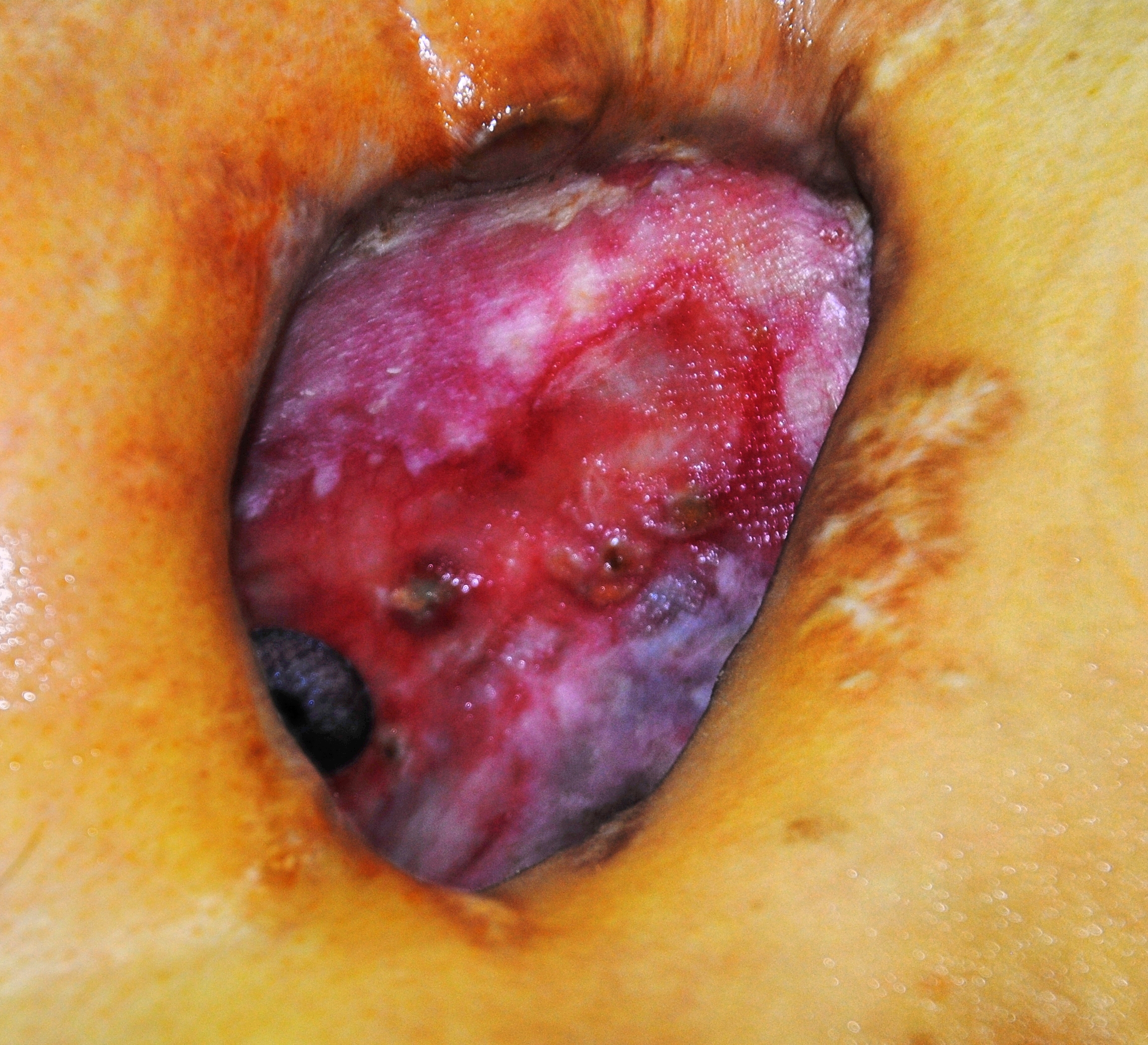
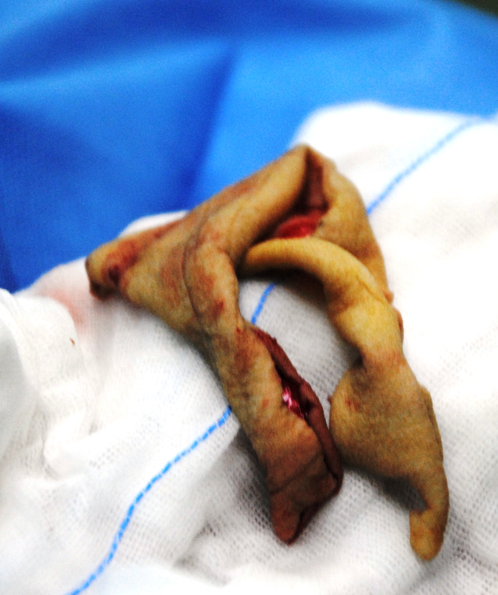
Over the continuing course of his treatment, a large opening on the anterior chest was created surgically. Due to the extent of necrotic tissue, this required the removal of anterior sections of ribs #2, 3, 4 and 5, leaving the patient with a very large open cavity, as seen in CT slices (pulmonary and tissue windows).
packing material can be seen in the right chest cavity.
tissue window showing extent of wound
This large cavity was left open for a period of around two years, while infected material was debrided and evacuated, and aggressive wound management was continued. At the time of his presentation to the operating room, the wound bed is dry and pink with a small amount of slough. An opening to the bronchus is visible (with bubbling on respiration at the site of the wound). The wound measures approximately 6 cm X 4 cm. As seen from the CT images above, the wound was also several centimeters in depth.
overview of wound – which tracks upwards several cm towards shoulder
The wound tracks up towards the shoulder, making it deeper and larger than it initially appears on gross visualization. There is a visible pulsation from the border of the cavity, (which may add to the patient and family’s distress).
Surgical procedure:
After the wound is cleaned and prepared with betadine solution, the anesthesiologist introduces a bronchoscope into the airway, for illumination and visualization of the airway. The light from the scope is immediately visible to observation within the chest. At that point, amplatzer patch was inserted into the bronchial stump.
Amplatzer patch visible in the chest cavity
After placement of the patch was confirmed, the patient was re-prepped, and draped. Dr. Boxiong expands the existing wound, and dissects down to healthy bleeding tissue, removing yellow eschar. The wound is lightly packed with moist gauze.
thoracic surgeons dissect down to healthy tissue (anterior wound site)
Then Dr. Dong and his assistant surgeon arrive, to start their portion of the operation. Dr. Dong starts another incision approximately 3 cm below the wound area. The incision is extended to the left side of the chest. The surgeon dissects down through skin, adipose and fascia to free the right internal mammary artery to use to ensure that the graft is well vascularized.
preparing the internal mammary artery for eventual anastomosis
Next step: Flap harvesting
Once the IMA was free, it was temporarily secured, and the wound was dressed. The patient was re-positioned, and re-prepped to allow access to the posterior aspect of the left chest. Due to muscle devascularization from the multiple previous surgeries on the right anterior chest, the surgeon harvests the left latissimus dorsi, using a large diamond-shaped incision.
Harvesting the myocutaneous flap from posterior chest
Once the flap was harvested, the patient was left with a large open defect, without enough surrounding skin to cover the area. The surgical site is dressed with a temporary dressing while Dr. Dong moves on to his next surgical site.
Next step: Skin Harvesting
Dr. Dong prepares to harvest skin for grafting
After preparing the patients right thigh, Dr. Dong applied a Padgett dermatome to shave off a thin layer of skin.
Harvesting skin from patient’s thigh
After multiple passes, the surgeons have enough skin to cover the defect from the flap site.
Skin harvested to cover flap site
Next step: Skin Grafting
Skin grafting at the myocutaneous flap site
The thin strips of skin were applied to the flap site and sutured into place.
suturing skin graft over flap harvest site (on back)
Once the sutures were completed, the wound was re-dressed and the patient was re-positioned for the last steps of the operation.
Next step: Anastomosis of mammary artery to flap
surgeons using the microscope to complete vascular anastamoses
Following re-positioning to supine position, the flap was placed within the right chest wound. The flap was loosely sutured into place to maintain a proper position while the painstaking vascular anastomoses were performed. Once the anastomoses were completed, the remaining incisions were carefully closed.
Total surgical time was greater than ten hours.
Discussion
As discussed by Lois and Noppen (2005), BPF management has traditionally been performed in a piece meal or stepwise fashion, with surgical interventions reserved as a last resort. Unfortunately, for some patients, this means that BPF becomes a chronic illness. As a chronic illness, (and all that chronic illness entails such as chronic malnutrition, chronic inflammation, long-term antibiotic therapy), the morbidity and mortality of this condition continues to increase for the duration of the illness. In the case study above, a relatively young, now cancer-free patient had now developed much of the disabilities associated with elderly patients due to the chronic nature of his illness (BPF after a lobectomy ten years prior). This certainly places the patient at significant risk for major complications once a large-scale definitive surgery is performed. Van Schill et al. (2014) notes that better understanding regarding the need for interdisciplinary management including aggressive physical therapy and nutritional support have reduced some of these complications.
While the impact of bronchopleural fistulas are usually discussed in terms of mortality, financial costs (surgical costs) and length of stay,and for this case, we would like to take a closer look at morbidity and quality of life issues raised by the development of this complication.
While BPF is rare, it truly can be a life-altering and destructive diagnosis. In addition to pain, physical debility, there may be gross deformity coupled with chronic wound care. Deformities caused by extensive tissue destruction and removal of several ribs can cause significant emotional and psychological anxiety and stress in both the patient and family members. The visible pulsation (cardiac movement) seen within the wound may exacerbate this anxiety. The stress of this wound combined with additional stressors related to this diagnosis have been observed to lead to a higher rate of marital discord and patient suicide. Patients may also feel a loss of sexuality and personal identity in the presence of this type of disfigurement, similar to some women after radical mastectomy (particularly in female patients).
To add insult to injury, unlike many conditions which can be readily corrected surgically, the creation of myocutaneous flap (and subsequent skin grafting) itself causes additional disfigurement. This patient required a lengthy (ten hour) surgery which resulted in the creation of three new surgical sites in addition to the patient’s original right-sided chest wound. While this is a drastic example, it does serve to highlight the on-going need to consider the psychological and emotional well-being of this patient (and all our patients).
BPF and professional relationships?
This case also reminds of the need for good interdisciplinary relationships. In thoracic surgery, cosmetic outcomes (other that pursuing minimally invasive options when possible) are not usually one of our primary considerations. This leaves us at a disadvantage when managing patients with such a drastic complication. We don’t always have a strong network or relationships with other surgical or medical disciplines outside of oncology or oncology-related fields. We need to take the opportunities available to become more familiar with our local reconstructive surgeons, as well as the latest techniques in reconstructive surgery. It’s not “good enough” to know the name of one of the plastic surgeons we brush elbows with in the surgical waiting lounge. It is not just about referrals and compensation. It is about having an open and free dialogue with surgical colleagues, so that when we do require their assistance, we can work together smoothly and coördinate care.
Consider the need to include social workers, psychologists and other counseling services in both the preoperative and postoperative care of our patients, when necessary for their long-term health and wellness. Unfortunately, due to social stigma, health care/ insurance or financial restrictions as well as provider hesitation**, not enough of our patients receive consultations or referrals to appropriate resources. We can’t change insurance regulations, but by becoming more familiar with our local resources and providers, we can overcome many of the other barriers to supporting our patients emotional health.
[1] I was unable to find literature that specifically cites BPF as a contributing factor to psychosocial complications such as divorce, depression or suicide but the impact of chronic wounds on emotional health, family life and other quality of life indicators are well documented. However, Okonta et. al (2015) and Lois & Noppen (2005) both cite QoL issues in patients with BPF.
** Provider hesitation is a nice term for all the reasons providers sometimes fail to seek mental health referrals for patients; such as fear of embarrassing our patients, believing that counseling is only needed for psychiatric emergencies, failure to understand local resources available, or our own discomfort with mental health “issues”.
References and Additional Readings
Arnold, P. G. & Pairolero, P. C. (1990). Intrathoracic muscle flaps: an account of their use in the management of 100 consecutive patients. Annals of Surgery, 1990; 211(6): 656-660. Study looking at one hundred cases from May 1977 and February 1988. In this potent reminder of the morbidity and mortality that is associated with patients requiring muscle flaps, as well as the advances in medicine over the last two decades, there were 16 operative deaths and 43 additional all-cause deaths in the operative survivors. Interestingly, one of these late-term deaths was due to suicide.
Goyal VD1, Gupta B2, Sharma S3 (2015). Intercostal muscle flap for repair of bronchopleural fistula. Lung India. 2015 Mar-Apr;32(2):152-4. doi: 10.4103/0970-2113.152628. Indian case study of patient presentation of BPF after treatment for spontaneous pneumothorax.
Van Schil PE1, Hendriks JM1, Lauwers P1 (2014). Focus on treatment complications and optimal management surgery. Transl Lung Cancer Res. 2014 Jun;3(3):181-6. doi: 10.3978/j.issn.2218-6751.2014.06.07. Belgian paper reviewing outcomes of 3,500 surgeries.
Levine, L. A. (2013). The clinical and psychosocial impact of Peyronie’s disease. Am J Manag Care. 2013 Mar;19(4 Suppl):S55-61. While unrelated to thoracic surgery, patients with Peyronie’s disease have many of the same emotional and psychological stressors as patients with other chronic wound conditions such as BPF.
Talking about the roles of traditional VATS, single port surgery and robots in modern thoracic surgery.
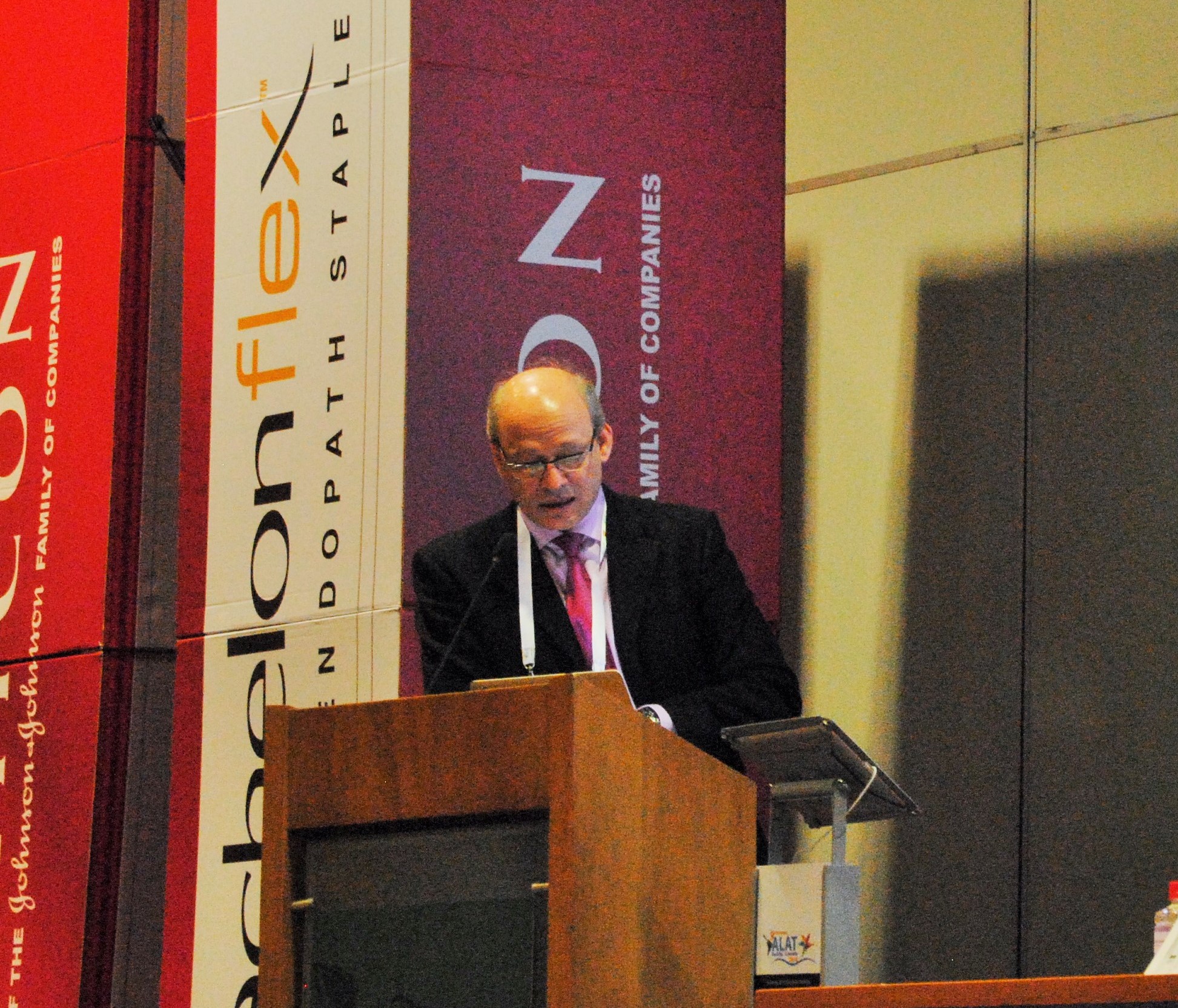
The Ethicon (Johnson & Johnson) sponsored session was by far, the best of the conference – and an excellent overview of modern technologies in thoracic surgery.
starting with Dr. Ricardo Buitrago (purple tie), Dr. Diego Gonzalez Rivas (blue tie) and Dr. Mario Ghefter (pink tie) are changing the future of thoracic surgery
Dr. Diego Gonzalez Rivas
“Is uni-port surgery feasible for advanced cancers?” Short answer: Yes.
The first speaker, was Dr. Diego Gonzalez Rivas of Coruna, Spain. He is a world-renown thoracic surgeon and innovator of uni-port thoracoscopic surgery. He discussed the evolution of single port surgery as well as the most recent developments with this technique, including more advanced and technically challenging cases such as chest wall resections (2013), sleeve resections/ reconstructions (2013), pulmonary artery reconstructions (2013) and surgery on non-intubated, awake patients (2014).
Experience and Management of bleeding
The biggest challenges to surgeons learning this technique is management of bleeding. But as he explained in previous lectures, this can be overcome with a direct approach. (these lectures and YouTube videos, Dr. Gonzalez explains the best ways to manage intra-operative bleeding.) In the vast majority of cases – this did not require deviation or conversion from the uni-port technique.)
As surgeons gain proficiency with this technique which mirrors open surgery, the only contra-indications for surgical resection of cancerous tissue (by single port) are tumors of great size, and surgeon discomfort with the technique.
Dr. Mario Ghefter
My favorite lecture of the series was given by Dr. Mario Ghefter of Sao Paolo, Brazil. While his lecture was ostensibly about video-assisted thoracoscopy (VATS), it was more of a retrospective vision and discussion of the modern history of thoracic surgery as seen through the eyes of a 22 year veteran surgeon.
He talked about the beginnings of VATS surgery and the contributions from such legends as Cefolio and D’Amico, including the 2005 paper – and modern-day thoracic bible, “Troubleshooting video-assisted thoracoscopic lobectomy (Demmy, James, Swanson, McKenna and D’Amico).
Dr. Ghefter also talked about how improved imaging and diagnostic procedures such as PET-CT and EBUS have been able to provide additional diagnostic information pre-operatively that helps surgeons to plan their procedures and treatment strategies more effectively.
Dr. Mario Ghefter
As a counterpoint to both Dr. Gonzalez and Dr. Buitrago, Dr. Ghefter acquitted himself admirably. He reminded audience members that even the newer technologies have some drawbacks – both as procedures and for the surgeons themselves.
He also successfully argued (in my opinion) that while the popularity of procedures such as multiple port VATS and even open thoracotomies have dropped drastically as thoracic surgeons embrace newer technologies, there will always be a place and time for these more traditional procedures.
Dr. Mario Ghefter is the Director of Thoracic Surgery at Hospital do Servidor Público Estadual – Sāo Paulo and on staff at the Hospital Alemão Oswaldo Cruz.
Dr. Ricardo Buitrago
Native Colombian (and my former professor), Dr. Ricardo Buitrago is acknowledged as one of the foremost experts in robotic thoracic surgery in Latin America.
During his presentation, he discussed the principles and basics of use of robotic techniques in thoracic surgery. He reviewed the existing literature surrounding the use of robotic surgery, and comparisons of outcomes between thoracic surgery and traditional lobectomy.
He reviewed several recent robotic surgery cases and the use of robotics as a training tool for residents and fellows.
While he mentioned some of previously discussed limitations of robotic surgery (namely cost of equipment) he cited recent studies demonstrating significant cost savings due to decreased length of stay and a reduced incidence of surgical complications.
He also discussed recent studies (by pioneering surgeons such as Dr. Dylewski) demonstrated short operating times of around 90 minutes.
in the operating room with Dr. Diego Gonzalez Rivas for single port thoracoscopic (uniportal) surgery.
Hilar mass resection using single port thoracoscopy with Dr. Diego Gonzalez – Rivas
K. Eckland & Andres M. Neira, MD
Instituto Nacional de Cancerlogia
Bogota, Colombia
Surgeon(s): Dr. Diego Gonzalez Rivas and Dr. Ricardo Buitrago
Dr. Diego Gonzalez Rivas demonstrates single port thoracoscopy
Case History:
59-year-old female with past medical history significant for recurrent mediastinal mass previously resectioned via right VATS. Additional past medical history included prior right-sided nephrectomy.
Pre-operative labs:
CBC: WBC 7230 Neu 73% Hgb:14.1 Hct 37 platelets 365000
Pt 12.1 / INR1.1 PTT: 28.3
Diagnostics:
Pre-operative CT scan: chest
edited to preserve patient privacy
Procedure: Single port thoracoscopy with resection of mediastinal mass and lymph node sampling
After review of relevant patient history including radiographs, patient was positioned for a right-sided procedure. After being prepped, and draped, surgery procedure in sterile fashion. A linear incision was made in the anterior chest – mid clavicular line at approximately the fifth intercostal space. A 10mm port was briefly inserted and the chest cavity inspected. The port was then removed, and the incision was expanded by an additional centimeter to allow for the passage of multiple instruments; including camera, grasper and suction catheter.
Dr. Gonzalez Rivas and Dr. Ricardo Buitrago at National Cancer Institute
The chest cavity, pleura and lung were inspected. The medial mediastinal mass was then identified.
As previously indicated on pre-operative CT scan, the mass was located adjacent and adherent to the vessels of the hilum. This area was carefully dissected free, in a painstaking fashion. After freeing the mediastinal mass from the hilum, the remaining surfaces of the mass were resected. The mass was fixed to the artery pulmonary and infiltrating it) . The mass was removed en-bloc. Care was then taken to identify, and sample the adjacent lymph nodes which were located at stations (4, 7 and 10).
Following removal of the tumor and lymph nodes, the area was re-inspected, and the lung was re-inflated. A 28 french chest tube was inserted in the original incision, with suturing of the fascia, subcutaneous and skin layers.
closing the single port incision
Hemostasis was maintained during the procedure with minimal blood loss.
Patient was hemodynamically stable throughout the case, and maintained appropriate oxygen saturations. Following surgery, the patient was awakened, extubated and transferred to the surgical intensive care unit.
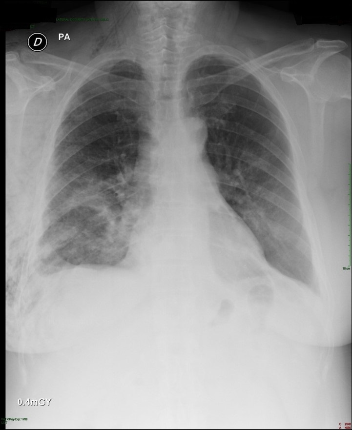
Post-operative: Post-operative chest x-ray confirmed appropriate chest tube placement and no significant bleeding or pneumothorax.
Immediate post-operative film (chest tube visible)
Patient did well post-operatively. Chest tube was discontinued on POD#2 and discharged home.
PA & LAT films on post-operative day 2
Discussion: Since the initial published reports of single-port thoracoscopy, this procedure has been applied to an increasing range of cases. Dr. Gonzalez and his team have published reports demonstrating the safety and utility of the single-port technique for multiple procedures including lobectomies, sleeve resections, segmentectomies, pneumonectomies and mediastinal mass resections. Dr. Hanao Chen (Taiwan) has reported several successful esophagectomies using this technical, as well as bilateral pleural drainage using a unilateral single-port approach.
Contrary to popular perception, the use of a single-port versus traditional VATS procedures (three or more) is actually associated with better visibility and accessibility for surgeons. Surgeons using this technical have also reported better ergonomics with less operating fatigue related to awkward body positioning while operating.
The learn curve for this surgical approach is less than anticipated due to the reasons cited above, and has been cited at 5 to 20 cases by Dr. Gonzalez, the creator of this approach.
The main limitations for surgeons using this technique is often related to anticipated (but potentially unrealized) fears regarding the need for urgent conversion to open thoracotomy. In reality, many of the complications that may lead to urgent conversion, such as major bleeding, are manageable thoracoscopically once surgeons are experienced and comfortable with this approach.
Dr. Gonzalez and his colleagues have reported a conversion rate of less than 1% in their practice. Subsequent reports by Dr. Gonzalez and his colleagues have documented these findings.
Other barriers to adoption of this technique are surgeon-based, and may be related to the individual surgeon’s willingness or reluctance to adopt new techniques and technology. Many of these surgeons would be surprised by how this technique mimics open surgery.
The successful adoption of this technique by numerous thoracic surgery fellows shows the feasibility and ease of learning single-port thoracoscopy by surgeons interested in adopting and advancing their surgical proficiency in minimally invasive surgery.
The benefits for utilizing this technique include decreased length of stay, decreased patient discomfort and greater patient satisfaction.
References/ Additional Readings
Bertolaccini, L., Rocco, G., Viti, A. & Terzi, A. (2013). Surgical technique: Geometrical characteristics of uniportal VATS. J. Thorac Dis. 2013, Apr 07. Article from thoracic surgeons at the National Cancer Institute in Naples, Italy explains how the geometric advantages of uniportal VATS improves visibility and spatial perception over traditional VATS, and mimics open surgery.
Calvin, S. H. Ng (2013). Uniportal VATS in Asia. J Thorac Dis 2013 Jun 20. Article discussing the spread of uniportal techniques in Taiwan, China and other parts of Asia.
Chen, Chin-Hao, Lin, Wei-Sha, Chang, Ho, Lee, Shih-Yi, Tzu-Ti, Hung & Tai, Chih-Yin (2013). Treatment of bilateral empyema thoracis using unilateral single-port thoracoscopic approach. Ann Thorac Cardiovasc Surg 2013.
Gonzalez Rivas, D., Fieira, E., Delgado, M., Mendez, L., Fernandez, R. & De la Torre, M. (2013). Surgical technique: Uniportal video-assisted thoracoscopic lobectomy. J. Thorac Dis. 2013 July 4.
Gonzalez Rivas, D., Delgado, M., Fieira, E., Mendez, L. Fernandez, R. & De la Torre, M. (2013). Surgical technique: Uniportal video-assisted thoracoscopic pneumonectomy. J. Thorac Dis. 2013 July 4.
Rocco, G. (2013). VATS and uniportal VATS: a glimpse into the future. J. Thorac Dis. 2013 July 04. After coming across several articles by Dr. Gaetano Rocco, and actively pursuing several other publications by this Italian thoracic surgeon, I have become increasingly convinced that Gaetano Rocco, along with Dr. Gonzalez Rivas is one of the world’s leading innovators in thoracic surgery. Hopefully, cirugia de torax will be able to catch up to Dr. Rocco at some point for an in-depth discussion.
Rocco G. Single port video-assisted thoracic surgery (uniportal) in the routine general thoracic surgical practice. Op Tech (Society of Thoracic and Cardiovascular Surgeons). 2009;14:326–335.
Rocco G, Khalil M, Jutley R. Uniportal video-assisted thoracoscopic surgery wedge lung biopsy in the diagnosis of interstitial lung diseases. J Thorac Cardiovasc Surg. 2005;129:947–948.
5 / Video-assisted thoracic surgery lobectomy: 3-year initial experience with 200 cases. Gonzalez D, De la Torre M, Paradela M, Fernandez R, Delgado M, Garcia J,Fieira E, Mendez L. Eur J Cardiothorac Surg. 2011 40(1):e21-8.
6 / Single-port Video-Assisted Thoracoscopic Anatomical Resection: Initial Experience. Diego Gonzalez , Ricardo Fernandez, Mercedes De La Torre, Maria Delgado, Marina Paradela, Lucia Mendez. Innovations.Vol 6.Number 3. May/jun 2011. Page 165.
the 2013 S.W.A.T conference, presented by Johnson & Johnson. Featured presenters Dr. Diego Gonzalez Rivas and Dr. Paula Ugalde discuss single port thoracoscopy and topics in minimally invasive surgery
Very pleased that despite the initial difficulties, we are able to provide information regarding the recent conference.
Talking about Single-port surgery in Bogotá, Colombia – 2013 S.W.A.T. Summit
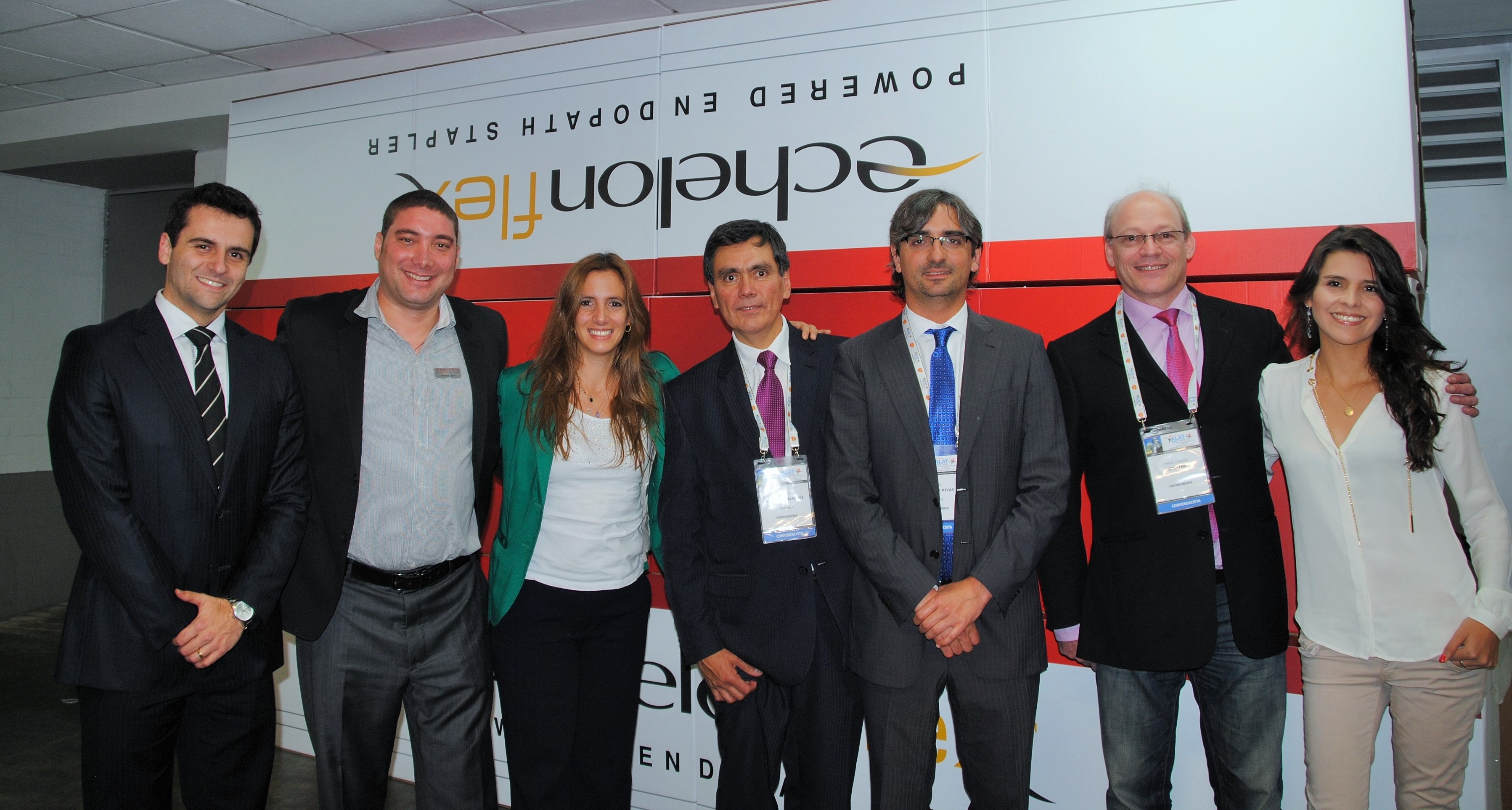
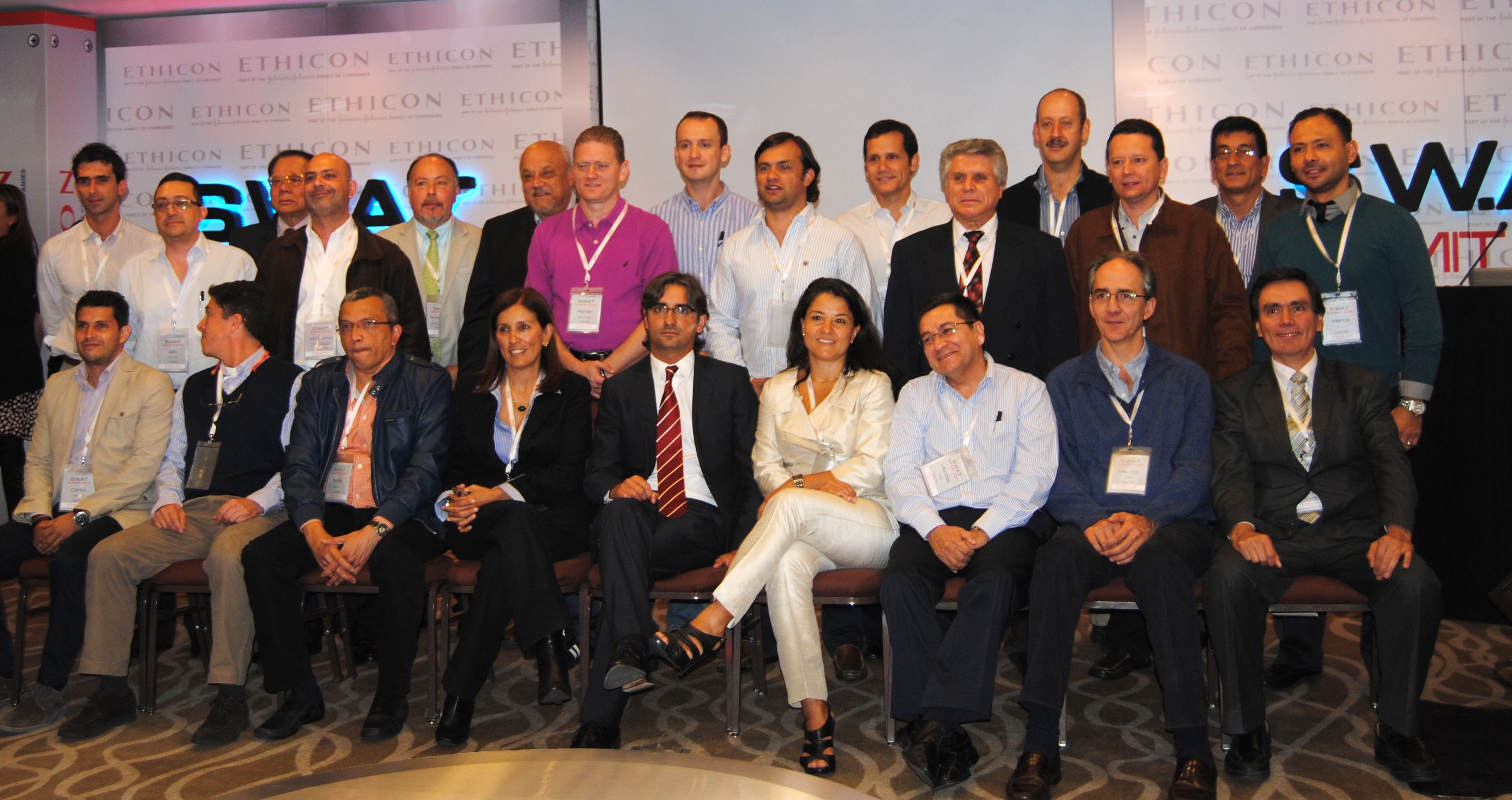
Dr. Diego Gonzalez Rivas and Dr. Paula Ugalde were the headliners at the recent Johnson and Johnson thoracic surgery summit on minimally invasive surgery. Both surgeons gave multiple presentations on several topics. They were joined at the lectern by several local Colombian surgeons including Dr. Stella Martinez Jaramillo (Bogotá), Dr. Luis Fernando Rueda (Barranquilla), Dr. Jose Maineri (Venezuela) Dr. Mario Lopez (Bogotá) and Dr. Pardo (Cartagena).
Thoracic surgeons at the 2013 S.W.A.T Summit in Bogota, Colombia. Drs. Gonzalez-Rivas and Dr. Paula Ugalde are center, front-row
Target audience missing from conference
The audience was made up of thirty Latin American surgeons from Colombia, Costa Rica and Venezuela. This surgeons were hand-picked for this invitation-only event. Unfortunately, while Johnson and Johnson organized and presented a lovely event; their apparent lack of knowledge about the local (Colombian) thoracic surgery community resulted in the exclusion of several key surgeons including Dr. Mauricio Velasquez, one of Colombia’s earliest adopters of single-port thoracoscopy. Also excluded were the junior members of the community, including Dr. Castano, Dr. Carlos Carvajal, and current thoracic surgery fellows. It was an otherwise outstandingand informative event.
The Gonzalez – Rivas dissector, photo courtesy of Scanlon International
As discussed in multiple publications, previous posts as well as during the conference itself, it is these younger members who are more likely to adopt newer surgical techniques versus older, more experienced surgeons. More seasoned surgeons may be hesitant to change their practices since they are more comfortable and accustomed to open surgical procedures.
Despite their absence, it was an engaging and interesting conference which engendered lively discussion among the surgeons present.
Of course, the highlight of the conference actually occurred the day before, when Dr. Gonzalez- Rivas demonstrated his technique during two separate cases at the National Cancer Institute in Bogotá, Colombia. (Case report).
Dr. Gonzalez-Rivas and Dr. Ricardo Buitrago performing single port thoracoscopy at the National Cancer Institute
Featured presenters:
Dr. Diego Gonzalez – Rivas is a world-renown thoracic surgeon jointly credited (along with Dr. Gaetano Rocco) with the development of single-port thoracoscopic (uni-port) surgery. He and his colleagues at the Minimally Invasive Surgery Unit in La Coruna, Spain give classes and lectures on this technique internationally. Recent publications include three papers in July alone detailing the application of this surgical approach, as well as several YouTube videos demonstrating use of this technique for a wide variety of cases.
Dr. Gonzalez Rivas
Dr. Paula Ugalde, a Chilean-borne thoracic surgeon (from Brazil) who gave several presentations on minimally-invasive surgery topics. She is currently affiliated with a facility in Quebec, Canada.
Dr. Paula Ugalde
Refuting the folklore
Part of the conference focused on refuting the ‘folklore’ of minimally-invasive procedures. Some of these falsehoods have plagued minimally-invasive surgery since the beginning of VATS (in 1991), such as the belief that VATS should not be applied in oncology cases. The presenters also discussed how uniportal VATS actually provides improved visibility and spatial perception over traditional VATS (Bertolaccini et al. 2013).
However, Gonzalez-Rivas, Ugalde and the other surgeons in attendance presented a wealth of data, and publications to demonstrate:
– VATS is safe and feasible for surgical resection in patients with cancer. (Like all surgeries, oncological principles like obtaining clear margins, and performing a thorough lymph node dissection need to be maintained).
– Thorough and complete lymph node dissection is possible using minimally invasive techniques like single-port surgery. Multiple studies have demonstrated that on average, surgeons using this technique obtain more nodes than surgeons using more traditional methods.
– Large surgeries like pneumonectomies and sleeve resections are reasonable and feasible to perform with single-port thoracoscopy. Using these techniques may reduce morbidity, pain and length of stay in these patients.
– Rates of conversion to open surgery are very low (rare occurrence). In single-port surgery, “conversion” usually means adding another port – not making a larger incision.
– Learning curve fallacies: the learning curve varies with each individual surgeon – but in general, surgeons proficient in traditional VATS and younger surgeons (the “X box generation”) will readily adapt to single-port surgery.
– Bleeding, even significant bleeding can be managed using single-port thoracoscopy. Dr. Gonzalez Rivas gave a separate presentation using several operative videos to demonstrate methods of controlling bleeding during single-port surgery – since this is a common concern among surgeons hesitant to apply these advanced surgical techniques.
Additional References / Readings about Single-Port Thoracoscopy
Scanlon single-port thoracoscopy kits – informational brochure about specially designed instruments endorsed by Dr. Gonzalez Rivas.
Dr. Diego Gonzalez Rivas – YouTube channel : Dr. Gonzalez Rivas maintains an active YouTube channel with multiple videos demonstrating his surgical technique during a variety of cases. Below is a full-length video demonstrating the uniportal technique.
Additional posts at Cirugia de Torax about Dr. Diego Gonzalez- Rivas
Upcoming conference in Florida – information about registering for September conference for hands-on course in single-port thoracoscopic surgery with Dr. Gonzalez-Rivas
Youtube video for web conference on Single-port thoracoscopic surgery
Bertolaccini, L., Rocco, G., Viti, A. & Terzi, A. (2013). Surgical technique: Geometrical characteristics of uniportal VATS. J. Thorac Dis. 2013, Apr 07. Article from thoracic surgeons at the National Cancer Institute in Naples, Italy explains how the geometric advantages of uniportal VATS improves visibility and spatial perception over traditional VATS, and mimics open surgery.
Calvin, S. H. Ng (2013). Uniportal VATS in Asia.J Thorac Dis 2013 Jun 20. Article discussing the spread of uniportal techniques in Taiwan, China and other parts of Asia.
Rocco, G. (2013). VATS and uniportal VATS: a glimpse into the future.J. Thorac Dis. 2013 July 04. After coming across several articles by Dr. Gaetano Rocco, and actively pursuing several other publications by this Italian thoracic surgeon, I have become increasingly convinced that Gaetano Rocco, along with Dr. Gonzalez Rivas is one of the world’s leading innovators in thoracic surgery. Hopefully, cirugia de torax will be able to catch up to Dr. Rocco at some point for an in-depth discussion.
While I advance criticism of this event – it was a fantastic conference. My only reservations were to the exclusivity of the event. While this was certainly related to the costs of providing facilities and services for this event – hopefully, the next J & J thoracic event will be open to more interested individuals including young surgeons and nurses.
Reviewing “Ten years experience on 644 patients undergoing single-port (uniportal) video-assisted” by Gaetano Rocco et al. at the National Cancer Institute in Naples, Italy
In this month’s issue of the Annals of Thoracic Surgery, Dr. Gaetano Rocco and his colleagues at the National Cancer Institute, Pascale Foundation in Naples, Italy reported their findings on ten year’s worth of single-port surgery in their institution.
Who: 644 patients; (334 males, 310 females)
Indications:
Annals of thoracic surgery – Rocco et. al (2013)
What: Outcomes and experiences in single port thoracic surgery over a ten-year period. All procedures performed by a single surgeon at this institution, and single-port VATS accounted for 27.7% of all surgeries performed during this time period.
When: data collected on thoracic surgery patients from January 2000 – December 2010.
Technical Notes:
Pre-operative CT scan was used for incision placement planning. Incision was up to 2.5 cm (1 inch) in length depending on indications for surgery.
Conversion rate to 2 or 3 port VATS: 2.2% (14 patients)
Conversion to mini-thoracotomy: 1.5% (10 patients)
Patients underwent conversion due to incomplete lung collapse (22 patients) and bleeding (2 patients).
There were no re-operations or “take backs”. The four patients with malignant effusions who died within the 30 day post-op period were re-admitted to the ICU.
Post-operatively:
Otherwise, all patients were admitted to either the floor or the step-down unit following surgery.
Pain management: post-operative pain was managed with a non-narcotic regimen consisting of a 24 hour IV infusion pump of ketorolac (20mg) and tramadol (100mg*). After the first 24 hours, patients were managed with oral analgesics such as paracetamol (acetaminophen).
Limitations: in this study, uni-port VATS was not used for major resections, as seen in the work of Dr. Diego Gonzalez and others. This may be due to the fact that uni-port VATS was an emerging technique at the initiation of this study.
Strengths: This is one of the largest studies examining the use of single-port thoracic surgery – and showed low morbidity and mortality. (Arguably, the 30 day mortality in this study was related to the patients’ underlying cancers, rather than the surgical procedure itself.)
*Intravenous tramadol is not available in the United States.
Rocco G. Single port video-assisted thoracic surgery (uniportal) in the routine general thoracic surgical practice. Op Tech (Society of Thoracic and Cardiovascular Surgeons). 2009;14:326–335.
Rocco G, Khalil M, Jutley R. Uniportal video-assisted thoracoscopic surgery wedge lung biopsy in the diagnosis of interstitial lung diseases. J Thorac Cardiovasc Surg. 2005;129:947–948.
Discussing Dr. Joseph Coselli and ‘the cowboys of cardiac surgery’ along with some of our own heros of thoracic surgery here at Cirugia de Torax.
There’s a great article in this month’s Annals of Thoracic Surgery, by Dr. Joseph Coselli, from Texas Heart Institute and the Michael DeBakey Department of Surgery at Baylor. His article, entitled,” My heros have always been cowboys” is more than just a title torn from the song sheets of Willie Nelson. It’s a look back at both the pioneers of cardiac surgery and his own experiences as a cardiac surgeon. He also discusses the role of surgeons, and medical practitioners in American society in general and the promises we make to both society at large and our patients.
Here at Cirugia de Torax, I’d like to take a moment to look back at the surgeons that inspired and encouraged me in this and all of my endeavors. Some of these surgeons knew me, and some of them didn’t – but their encouragement and kindnesses have spurred a career and life that have brought immense personal and professional satisfaction.
Like Dr. Coselli, I too, took inspiration from the likes of Dr. Denton Cooley. But our stories diverge greatly from there. I never met Dr. Cooley and I probably never will. But it was a related story, from my former boss (and cardiothoracic surgeon), Dr. Richard Embrey that led to an email to Dr. Cooley himself. My boss had too trained under Dr. Cooley, Dr. Debakey and the Texas Heart Institute, the citadel of American heart surgery. Then, somehow, along the way – Dr. Embrey stopped to work at our little rural Virginia hospital. We were the remnants of a larger Duke cardiothoracic program but we were a country hospital all the same.
While I learned the ins and outs of surgery from Dr. Embrey (and Dr. Geoffrey Graeber at West Virginia University) on a day-to-day basis, I was also weaned on the folklore of cardiothoracic surgery – stories of the giants of history, like the ones mentioned in Dr. Coselli’s article, as well as local Duke legends who occasionally roamed the halls of our tiny ICU and our two cardiothoracic OR suites; Dr. Duane Davis, Dr. Shu S. Lin and Dr. Peter Smith. While never working side-by-side, Dr. D’Amico’s name was almost as familiar as my own. As the sole nurse practitioner in this facility, without residents or fellows, there was no buffer, and little social divide in our daily practice. Certainly, this changed me – and my perceptions. I asked the ‘stupid’ questions but received intelligent and insightful answers. I asked even more questions, and learned even more..
These opportunities fed my mind, and nurtured my ambitions. Not to be a physician or a doctor, but to learn as much as possible about my specialty; to be the best nurse possible in my field. It also nurtured a desire to share these experiences, and this knowledge with my peers, my patients and everyone else who ever had an interest.
It was that tiny little email, a gracious three-line reply from Dr. Cooley himself that made me realize that I didn’t have to rely on folklore and second-hand stories to hear more. That’s critical; because as we’ve seen (here at Cirugia de Torax) there are a quite of few of “Masters of thoracic surgery” or perhaps future giants that haven’t had their stories told. Dr. Coselli and his fellow writers haven’t written about them yet.. So I will.
Sometimes I interview famous (or semi-famous) surgeons here, but other times, I interview lesser-known but equally talented/ innovative or promising surgeons. All of them share similar traits; dedication and love for the profession, immense surgical talent and proficiency and sincere belief in the future of technology of surgery.
So, let’s hope that it won’t take forty more years for these surgeons to be recognized for their contributions to thoracic surgery in the way that Cooley, DeBakey and Crawford are heralded in cardiac surgery.
A discussion of Meimarakis’ recently published article, “Prolonged overall survival after pulmonary metastatectomy in patients with breast cancer.”
As reported in the Society of Thoracic Surgeons, and multiple other outlets, a newly published study by several surgeons in Germany shows that surgical removal of metastatic breast cancer that has spread to the lungs may improve overall patient survival. The study, by Meimarakis et al. was published in the April 2013 issue of the Annals of Thoracic Surgery.
pulmonary metastatectomy in metastatic breast cancer
The Meimarakis study included 81 patients over a twenty-five year period. The study looked at the overall survival time in breast cancer patients with a pulmonary metastasis. The study began in 1992, and data was collected retrospectively to 1982.
Poor median survival despite advances in chemotherapy
Current survival time in these patients ranges from 12 to 24 months. However, the authors note that in up to 23% of these patients, the sole metastatic lesion is in the lung or pleural space. In these patients with pulmonary metastasis alone, the majority survived less than 22 months after diagnosis, despite chemotherapy. The 10 year survival has been previously reported as a dismal 9% in this population in prior studies conducted as M. D. Anderson (Meimarakis, et. al, 2013).
Role of pulmonary metastatectomy in advanced breast cancer
Unlike pulmonary metastatectomy for colon cancer, metastatectomy has been used sparingly in this population and with no clear-cut criteria to distinguish which breast cancer patients would benefit from surgery, surgery in addition to chemotherapy, versus chemotherapy alone.
Aim of study
The authors, at Ludwig-Maximilian University in Munich, Germany attempt to address this deficiency by investigating surgical, pathological and demographic factors that impact survival in this patient population to help determine which candidates would benefit the most from surgical intervention.
The authors looked at a multitude of factors such as presence and type of hormone receptor, histological type, size of both primary and metastatic lesions, the number of metastatic lesions, surgical grade/ resectability and the laterality of these lesions. They also collected and compared additional markers such as CEA, LDH and CA 15-3.
These factors and their impact on survival were analyzed using statistical analysis, Kaplan-Meier estimators, log-rank tests as well as matched pair analysis of 2 year survival (metastectomy vs. standard therapy only). These factors included data from pathological specimens and tumor typing (Meimarakis, 2013).
What makes this study particularly interesting and noteworthy, is the operative inclusions. While patients with local residual disease, additional (non-lung) metastases or recurrent primary breast tumors were excluded, patients with contralateral lung lesions were not.
Selected patient demographics
Total number of patients: 81
Median age: 58.2 (range 28.2 to 76.3)
Breast cancers: Histological types
64.1% invasive ductal carcinoma, 17.2 % with ductal carcinoma in situ? and 18.7% other breast cancer.
Number and size of metastatic lesions:
61 (75.3%) lesions were less than 3 cm in size.
20 (24.7%) of lesions were 3 cm or greater.
The majority (51 (63%) of patients presented with a solitary lung lesion, whereas 30 (37. %) presented with two or more lesions.
Operative procedures
Meimarakis et al. performed a total of 92 operations. These included 71 patients who underwent one procedure, 9 patients for two procedures and 1 patient with three procedures.
All of the patients undergoing more than one procedure had contralateral surgery for newly occurring metastases. (The authors re-operated on patients within 4 to 6 weeks for synchronous metastatic lung lesions.) This is important to remember when reviewing the primary article since the terminology ‘re-do’ operations and repeat operations can be confusing. However, after clarifying with the primary author, there were no completion procedures (i.e. wedge converted to lobectomy based on final pathology) and no returns to the operating room for surgery due to complications. There was no return to the operating room for any procedures on the same side as the original procedure. Thus for clarification, no “re-do” procedures.
All patients underwent resection via anterolateral thoracotomy. However, patients with peripheral, previously unbiopsied nodules were initially approached via VATS with conversion to anterolateral thoracotomy for positive intraoperative pathology.
67 operations were wedge resection, with an additional 10 segmental resections. The remainder of procedures included 7 lobectomies, 7 pneumonectomies and 1 bilobectomy.
Median operating room time was 83 minutes, with a fairly lengthy hospitalization stay (median 9 days, with a range of 3 – 63 days.) Complication rate was 7.6% (3 patients with pneumonia, 4 patients with atelectasis).
Limitations of Study
The median follow-up was only 27.2 months. At the end of this period, 27 of the 81 patients (33.3%) had died. While the published study was lengthy and detailed (10 pages with multiple charts and graphs) much of this was related to discussion regarding receptor status, and existing literature. A clearer, more streamlined algorithmic approach or scoring system utilize to their findings would be more helpful to readers in determining the likelihood of successful outcomes with surgical resection, and for encouraging replication of their research.
Results
Despite the limited number of patients with multiple metastatic lung lesions in this study, the underlying rules of surgical resection remain consistent. Patients who did the best, with the longest overall survival time were patients with complete surgical resection (R0). While patients with a completely resection of a single metastasis lived longer than patients with complete resection of multiple metastases, the R0 patients with multiple metastases had greater median survival than all patients with incomplete resection, regardless of the degree of residual (R1, R2) disease (microscopic or gross disease).
Receptor positive patients with better outcomes
As seen in multiple studies, tumor types were a crucial factor in long-term outcomes; whether estrogen receptor positive (ER+), human growth factor receptor 2 positive (HER2+), progesterone receptor+ (PR+).
Median survival of all patients after metastatectomy was 82.4 months with the greatest median survival time in the 31 patients with + hormone receptor tumors (HR+) at 127.4 months (range 33.2 to 221.6 months). In comparison, the 8 patients with HER+ had a mean survival of 66 months and only 27 months median survival for the 14 triple negative patients)*.
These findings regarding longevity and tumor receptors are similar to those reported by Welter et. al (2008) and others, but the patients from this larger study demonstrated greater longevity, which gives weight to continued study in this area.
In Meimarakis’ work, the presence of pleural infiltration or lymphangiosis carcinomatosis denoted a reduced longevity (32.1 and 34.5 months). This may serve as a better marker of systemic disease for future classification and treatment of advanced breast cancer.
Implications: For breast cancer patients, the discovery of a metastatic lung lesion advances the stage of the disease, drastically changing current treatment options. Most breast cancer patients diagnosed with metastatic disease are not considered surgical candidates even if complete surgical resection is technically feasible.
Meimarakis’s study is one of the larger studies to date, using a large number of prospective patients versus retrospective chart review. This gives a more comprehensive look at a multitude of factors and patient demographics. It serves as an excellent framework for future study in this area.
But, more interesting to our readers is the low incidence of post-operative complications (7 operations; 3 patients with pneumonia, 4 patients with atelectasis).
None of the patients died post-operatively. There were no ‘take backs’ for post-operative complications such as bleeding, prolonged air leak or post-operative infections despite the fact that almost 10% (8 patients) underwent significantly larger procedures such as pneumonectomy or bilobectomy and that all patients underwent thoracotomies versus the smaller VATS procedures. There was no difference in outcomes in this set of patients by procedure (wedge versus pneumonectomy) though Meimarakis notes that “there is a trend to worse survival in case of pneumonectomy during R1/ R2 resection (considering the whole database [Munich Cancer Registry] i.e not only in this group of patients with breast cancer.”
As outcomes appeared independent of the surgical procedure itself; based solely on resectability and tumor type, even larger scale resections such as pneumonectomy may be worthy of consideration during preoperative surgical evaluation, particularly in patients with favorable tumor types with good potential for complete resection.
Future considerations
Using the work of Meimarakis and similar researchers, development of an algorithmic approach may be beneficial to thoracic surgeons and others who encounter pulmonary metastases from breast cancer outside of larger research facilities.
Related case reports: We previously reported a case of metastatic breast cancer that was discovered at the time of surgery, despite the use of multiple imaging and diagnostic modalities. However, in that case, the patient also had local metastases to bone (ribs), which were also resected.
*Please see original article for further detail on patient characteristics and outcomes.
While the data (statistics, patient outcomes) is from the original research of Meimarakis et al., the commentary has been written by writers at Cirugia de Torax and may not reflect the thoughts, considerations and experiences of the primary researchers.
Kycler, W. & Laski, P. (2012). Surgical approach to pulmonary metastases from breast cancer. Breast J. 2012 Jan-Feb;18(1):52-7. doi: 10.1111/j.1524-4741.2011.01176.x. Epub 2011 Nov 20. [no free full text available]. Retrospective data review of 33 patients who underwent pulmonary metastatectomy (1997 – 2002) at the Great Poland Cancer Center, in Poznan, Poland.
Welter S, Jacobs J, Krbek T, Tötsch M, Stamatis G. (2008). Pulmonary metastases of breast cancer. When is resection indicated?Eur J Cardiothorac Surg. 2008 Dec;34(6):1228-34. doi: 10.1016/j.ejcts.2008.07.063. Epub 2008 Sep 27 [free text available]. A review of 47 cases of metastatic breast cancer with pulmonary metastatectomy, Essen, Germany.
the latest predictions on the impending shortage of surgeons in the United States
Unsurprisingly – rural area hospitals face additional challenges in attracting and retaining specialty surgeons in comparison to big cities/ metropolitan areas. However, as reported by Patrice Welding at Thoracic Surgery News in a report on the annual meeting of the Central Surgical Association, this may be viewed as a boon for the surgeons themselves as hospitals may devise new and enhanced incentives to attract surgeons to their facilities. The surgical specialties most likely to benefit from this strategy include (as previously reported), obstetrics and gynecology, orthopedic surgery, general surgery, otolaryngology, urology, neurosurgery, and thoracic surgery.
The article which quotes Dr. Thomas E. Williams, Jr. predicts that hospitals and institutions may break out into a ‘bidding war’ over surgeons.
While this is dire news for rural hospitals and the estimated 56 million patients served by these facilities, it comes as a relief for current thoracic surgery fellows and new thoracic surgeons who have faced an increasingly bleak economic landscape over the last few years.
Of course, more sanguine experts note that the impact of the impending shortage has been reported for several years – with little impact on the current job market for new graduates.
Dr. Thomas E. Williams Jr. is one of the main researchers on the impending shortage in the United States and published a book based on his findings in 2009, entitled, “The coming shortage of surgeons: why they are disappearing and what that means for our health“. (Praeger, ISBN #978-0313380709). His work has also be published in multiple journals, and presented in meetings and conferences across the country.
Interested in learning more about single port thoracoscopy, or talking to the inventors of this technique? This March – head to the 1st Asian single port surgery conference in Hong Kong.
It doesn’t look like Cirugia de Torax will be in attendance for this conference, but it’s another opportunity for practicing thoracic surgeons and thoracic surgery fellows to learn more about single port thoracoscopic surgery.
This March (7th – 8th), the Chinese University of Hong Kong, along with the Minimally Invasive Thoracic Surgery Unit (Coruna, Spain), and Duke University are presenting the 1st Asian Single Port Symposium and Live Surgery conference in Hong Kong.
This is your chance to meet the experts – and the inventors of this technique (such as Dr. Diego Gonzalez – Rivas, one of the new masters frequently featured here at Cirugia de Torax.)
what is the future of thoracic surgery education? A new American study asks the if it is time to separate the specialties of cardiac and thoracic surgery.
A new study by Cooke & Wisner performed at a large medical center in California (UC Davis) and published in the Annals of Thoracic Surgery provides additional weight to the idea that Thoracic Surgery has increasingly developed into it’s own subspecialty away from the traditional cardiothoracic surgery model (seen in the United States and several other countries.)
In an article published in Medical News Today, the authors of the study explained that the increased complexity of (noncardiac) thoracic surgery procedures for general thoracic conditions has led to increased referrals and utilization of general thoracic surgeons (versus cardiac or general surgeons). This shows a reversal in a previous trend away from specialists – with more patients now receiving “complex” thoracic surgery procedures from specialty trained, board-certified thoracic surgeons. Previously up to 75% of all thoracic surgery procedures were performed by general surgeons.
With lung cancer rates expected to climb dramatically in North America and Europe, particularly in women – along with esophageal cancer, and long waits already common, support and on-going discussion about the evolution of resident and fellow education is desperately needed.
Reference
Cooke, D. T. & Wisner, D. H. (2012). Who performs complex noncardiac thoracic Surgery in United States Academic Medical Centers? Ann Thorac Surg 2012;94:1060-1064. doi:10.1016/j.athoracsur.2012.04.018
a day in the operating room with one of Colombia’s New Masters of Thoracic Surgery
Cali, Colombia
Dr. Mauricio Velasquez is probably one of the most famous thoracic surgeons that you’ve never heard of. His thoracic surgery program at the internationally ranked Fundacion Valle del Lili in Cali, Colombia is one of just a handful of programs in the world to offer single port thoracic surgery. Dr. Velasquez has also single-handedly created a surgical registry for thoracic surgeons all over Colombia and recently gave a presentation on the registry at a national conference. This registry allows surgeons to track their surgical data and outcomes, in order to create specifically targeted programs for continued innovation and improvement in surgery (similar to the STS database for American surgeons).
Dr. Mauricio Velasquez after another successful case
Dr. Velasquez is also part of a team at Fundacion Valle del Lili which aims to add lung transplant to the repertoire of services available to the citizens of Cali and surrounding communities.
He is friendly, and enthusiastic about his work but humble and apparently unaware of his growing reputation as one of Colombia’s finest surgeons.
Education and training
After completing medical school at Universidad Pontificia Bolivariana in Medellin in 1997, he completed his general surgery residency at the Universidad del Valle in 2006, followed by his thoracic surgery fellowship at El Bosque in Bogotá.
The Colombia native has also trained with thoracic surgery greats such as Dr. Thomas D’Amico at Duke University in Durham, North Carolina, and single port surgery pioneer, Dr. Diego Gonzalez Rivas in Coruna, Spain. He is also planning to receive additional training in lung transplantation at the Cleveland Clinic, in Cleveland, Ohio this summer.
Single port surgery
Presently, Dr. Velasquez is just one of a very small handful of surgeons performing single port surgery. This surgery is an adaptation of a type of minimally invasive surgery called video-assisted thoracoscopy. This technique allows Dr. Velasquez to perform complex thoracic surgery techniques such as lobectomies and lung resections for lung cancer through a small 2 – 3 cm incision. Previously, surgeons performed these operations using either three small incisions or one large (10 to 20cm) incision called a thoracotomy.
By using a tiny single incision, much of the trauma, pain and lengthy hospitalization of a major lung surgery are avoided. Patients are able to recovery and return to their lives much sooner. The small incision size, and lack of rib spreading means less pain, less dependence on narcotics and a reduced incidence of post-operative pneumonia and other complications caused by prolonged immobilization and poor inspiratory effort.
However, this procedure is not just limited to the treatment of lung cancer, but can also be used to treat lung infections such as empyema, and large mediastinal masses or tumors like thymomas and thyroid cancers.
Dr. Velasquez in the operating room with Lina Caicedo Quintero (nurse)
Team approach
Part of his success in due in no small part to Dr. Velasquez’s surgical skill, another important asset to his surgical practice is his wife, Dr. Indira Cujiño, an anesthesiologist specializing in thoracic anesthesia. She trained for an additional year in Spain, in order to be able to provide specialized anesthesia for her husband’s patients, including in special circumstances, conscious sedation. This allows her husband to operate on critically ill patients who cannot tolerate general anesthesia. While Dr. Cujiño does not perform anesthesia for all of Dr. Velasquez’s cases, she is always available for the more complex cases or more critically ill patients.
In the operating room with Dr. Velasquez
I spent the day in the operating room with Dr. Velasquez for several cases and was immediate struck by the ease and adeptness of the single port approach. (While I’ve written quite a bit about the literature and surgeons using this technique, prior to this, I’ve had only limited exposure to the technique intra-operatively.) Visibility and maneuverability of surgical instruments was vastly superior to multi-port approaches. The technique also had the advantage that it added no time, or complexity to the procedure (unlike robotic surgery).
Dr. Velasquez performing single port thoracoscopy
Cases proceeded rapidly; with no complications.
close up view
Note to readers – some of the content, and information obtained during interviews, conversations etc. with Dr. Velasquez may be used on additional websites aimed at Colombia-based readers.
Robotic (thoracic) surgery comes to Clinica de Marly in Bogota, Colombia
A year and a half ago, I interviewed and spent some time with Dr. Ricardo Buitrago at the National Cancer Institute, and Clinica de Marly while doing research for a book about thoracic surgeons. At that time, Dr. Buitrago stated he was interested in starting a robotic surgery program – and was planning to study robot-assisted thoracic surgery with Dr. Mark Dylewski.
Dr. Ricardo Buitrago in the operating room, April 2011
Fast forward 1 year – when I received a quick little email from Dr. Buitrago telling me about his first robotic surgery at the Clinica de Marly. At that point, I sent Dr. Buitrago an email asking if I could come to Colombia and see his robotic surgery program to learn more about it. We had several phone conversations about it and I also outlined a research proposal to gather data on thoracic surgery patients and outcomes at high altitude, to which he enthusiastically offered to assist with. Thus began my current endeavor, in Bogota, studying with Dr. Buitrago.
Now – after completing a proctoring period with Dr. Dylewski, Dr. Buitrago has more than a dozen independent robotic surgeries under his belt. He has successfully used the robot for lobectomies, mediastinal mass resections and several other surgeries.
As part of my studies with Dr. Buitrago – I’ve made a video for other people who may be interested in robotic surgery with the DaVinci robot and what it entails.
Talking with Dr. Mark Dylewski, one of the new masters of thoracic surgery in the area of robotic surgery
Most of us never had the opportunity to meet or talk to some of the ‘masters’ of thoracic surgery like Dr. Hermes Grillo (1923 -2006), the ‘Father of Tracheal Surgery’ but as we have discussed before, thoracic surgery is not static. New technologies and new techniques are emerging all the time, and with these developments – new masters of thoracic surgery.
Dr. Mark Dylewski, may look too young to be the father of anything, but he is certain to be remembered in thoracic surgery history as one its new masters, and as one of the ‘fathers of robotic-assisted thoracic surgery’. While he is not the only surgeon doing robotic surgery, he is certainly one of the most prolific robotic / thoracic surgeons and has trained a large number of his peers.
Dr. Garrett Walsh and Dr. Mark Dylewski, thoracic surgeons
Talking to Dr. Dylewski about robotic surgery
At the recent conference, Advances in Lung Cancer and Mesothelioma, we had the opportunity to sit and talk with Dr. Dylewski about the state of robotics in thoracic surgery. Dr. Dylewski is one of the foremost experts on the topic and teaches robotic surgery techniques at the South Miami Hospital Center for Robotic Surgery. Since he started performing robotic surgery in 2006, he estimates that he has taught over 200 thoracic surgeons how to perform surgery utilizing the DaVinci robot.
In comparison to other minimally invasive techniques (specifically VATS), Dr. Dylewski believes that robotic surgery has greater potential for use in thoracic surgery, due to its easy adoptability. He reports that unlike VATS, robotic surgery techniques utilize traditional surgical skills so that surgeons are usually proficient at robotic surgery after performing 30 – 40 cases. There are no counter-intuitive movements or altered visibility/surgical perspectives which are two of the things inherent in video-assisted thoracoscopy. He attributes both of these issues with the failure of more wide-spread adoption of VATS despite the availability of this technology for over twenty years. According to Dr. Dylewski, less than 30% of all thoracic procedures in North America are currently done using VATS.
Simply put, even some of the best thoracic surgeons may have trouble adapting to VATS techniques and as many as 20% will never fully adjust to video-assisted surgical techniques.
However, in his experience, robotic-assisted thoracic surgery such as complete portal robotic lobectomy ( aka CRPL-3 or CRPL-4, depending on the number of arms used) has a greater potential for widespread use. He explains that despite the initial hefty price tag, the robotic technology easily justifies its equipment costs, in terms of subsequent savings and benefits from decreasing the length of stay, less patient discomfort and greater patient satisfaction. He reports that these benefits have led to the adoption of robotic surgery as the standard of care in other specialties such as gynaecology despite the relative newness of this technology.
Dr. Dykewski also presented data regarding surgical outcomes from 355 cases, which includes a wide variety of thoracic procedures such as lobectomies, esophagectomies and mediastinal surgeries. Surgical outcomes were comparable to VATS procedures with a markedly shorter length of stay.
Dylewski MR, Ohaeto AC, Pereira JF. (2011). Pulmonary resection using a total endoscopic robotic video-assisted approach. Semin Thorac Cardiovasc Surg. 2011 Spring;23(1):36-42.
Ninan M, Dylewski MR. (2010). Total port-access robot-assisted pulmonary lobectomy without utility thoracotomy. Eur J Cardiothorac Surg. 2010 Aug;38(2):231-2. Epub 2010 Mar
Additional References and Resources
Meyer M, Gharagozloo F, Tempesta B, Margolis M, Strother E, Christenson D. (2012). The learning curve of robotic lobectomy. Int J MId Robot. 2012 Sep 18. doi: 10.1002/rcs.1455. The authors of this publication report that it takes 18 – 20 complete portal robotic lobectomies to obtain competency.
This case study was prepared with assistance from Dr. Carlos Ochoa. Since we have been discussing the relevance of case reports and providing tips on case report writing for new academic writers – we have written the following case report in the style advocated by McCarthy & Reilley (2000) using their case report worksheet to demonstrate the ease of doing so in this style.
Since the previous presentation of dual-port thoracoscopy for decortication was missing essential materials, we are presenting a second case report.
Authors: K. Eckland, ACNP-BC, MSN, RN & Carlos Ochoa, MD
Case Report: Dual port thoracoscopy for decortication of a parapneumonic effusion
Abstract: The use of increasingly minimally invasive techniques for the treatment of thoracic disease is becoming more widespread. Dual and even single port thoracoscopy is becoming more frequent in the treatment of parapneumonic effusions and empyema.
Clinical question/problem: the effectiveness and utility of dual port thoracoscopy for parapneumonic effusions.
Analysis of literature review: Despite the increasing frequency of dual and single port thoracoscopic techniques, there remains a dearth of literature or case reports on this topic. Pubmed and related searches reveal only a scattering of reports.
Summary: As the case report suggests, dual port thoracoscopy is a feasible and reasonable option for the treatment of parapneumonic effusion.
Case history: 50-year-old patient with a three-week history of pneumonia, with complaints of right-sided chest pain, cough and increased phlegm production. Additional past medical history is significant for poorly controlled diabetes, hypertension, and obesity with central adiposity. Medications included glyburide and lisinopril.
After being seen and evaluated by an internal medicine physician, the patient was started on oral antibiotics. After three weeks, when his symptoms failed to improve, he was referred by internal medicine to thoracic surgery for out-patient evaluation.
On exam: middle-aged obese diabetic gentleman in no immediate distress, resting comfortable in the exam room. Face appeared moderately flushed, but skin cool and dry to the touch, no evidence of fever.
On auscultation, he had diminished breath sounds over the right lower lobe with egophony over the same area. The remainder of the exam was essentially normal.
Lab studies showed a mildly elevated WBC of 11.6, decreased Hgb of 10.4 / HCT 32.5. Hemoglobin A1c 10.6, Fasting glucose 228, HDL mildly low at 40.
EKG showed slight axis deviation, with slightly prolonged QRS complex (.16) with no evidence of loss of R, St elevation or other abnormalities. He was cleared by internal medicine for surgery.
After risks, benefits and alternatives to VATS decortication were explained to the patient – the patient consented to proceed with surgical decortication. After scheduling surgery, the patient was seen by anesthesia in preparation for the procedure.
Surgical procedure: Dual-port thoracoscopy with decortication of parapneumonic effusion.
Dual port thoracoscopy
After being prepped and drapped in sterile fashion and confirmation of dual lumen endotracheal tube placement, a small 2 cm incision was made for insertion of a 10mm port. Following entry into the chest with the thoracoscope, the right lung was deflated for optimal inspection and decortication of loculations. After completing the majority of the procedure, a second access port was created for better visualization and to ensure that a thorough decortication was completed. The lung and pleural were separated from the chest wall, and diaphragm, and demonstrated good re-expansion with lung re-inflation prior to completion of the procedure.
chest tubes at conclusion of case
At the conclusion of the procedure, two 28 french chest tubes were placed in the existing incisions. These were sutured into place, and connected to a pleurovac drainage system before applying a sterile gauze dressing. The patient remained hemodynamically stable throughout the case, with no episodes of hypoxia or desaturation. Following surgery, the patient was transferred to the PACU in stable condition.
Post-operative course was uncomplicated. Chest tubes were water-sealed on POD#3 and chest tubes were removed POD#4, with the patient being subsequently discharged after chest x-ray.
close up view of dual port thoracoscopy
Literature Review
A literature review was performed on PubMed using “dual port thoracoscopy”, “dual port VATS”, “2 port” as well as minimally invasive thoracoscopic surgery “
Results of search: A limited number of case studies (3) described thoracoscopic surgery with a single port. There was one case found describing cases conducted with two ports, and the majority of reports involved three or more access ports.
Discussion/ Conclusion
While convention medical wisdom dictates a trial and error treatment approach with initial trial of antibiotic therapy followed by chest tube placement (Light, 1995), surgeons have long argued that this delay in definitive treatment places the patient at increased risk of significant morbidity and mortality (Richardson, 1891). Multiple recent reviews of the literature and research comparisons continue to demonstrate optimal outcomes with surgery based approaches versus antibiotics alone, TPA and tube thoracostomy. The ability to perform these procedures in the least invasive fashion (VATS versus thoracotomy approaches) defies the arguments against surgical intervention as advanced by interventionalists (radiologists and pulmonologists.) Successful decortication with the use of dual port thoracoscopy is another example of how technology is advancing to better serve the patient and provide optimal outcomes, and offers a minimally invasive option when single port surgery may not be feasible.
During the case above, visibility and access to the thoracic cavity was excellent. However, in cases requiring additional access, reversion to the standard VATS configuration can be done easily enough with significant delays or additional risks to the patient.
References/ Resources
Foroulis CN, Anastasiadis K, Charokopos N, Antonitsis P, Halvatzoulis HV, Karapanagiotidis GT, Grosomanidis V, Papakonstantinou C. (2012). A modified two-port thoracoscopic technique versus axillary minithoracotomy for the treatment of recurrent spontaneous pneumothorax: a prospective randomized study. Surg Endosc. 2012 Mar;26(3):607-14. [free full text not available.]
Gonzalez – Rivas, D., Fernandez, R., De la Torre, M., & Martin – Ucar, A. E. (2012). Thoracoscopic lobectomy through a single incision.Multimedia manual cardio-thoracic surgery, Volume 2012. This is an excellent article which gives a detailed description, and overview of the techniques used in single incision surgery. Contains illustrations, full color photos and videos of the procedure.
Gonzalez-Rivas D, Paradela M, Fieira E, Velasco C. (2012). Single-incision video-assisted thoracoscopic lobectomy: initial results. J Thorac Cardiovasc Surg. 2012 Mar;143(3):745-7.
Talking with Dr. Ross Bremner, Chief of Thoracic Surgery and Chair of Thoracic Disease & Transplant at St. Joseph’s Hospital in Phoenix, Arizona.
Dr. Ross Bremner
St. Joseph’s Hospital
After talking to Dr. Bremner of the phone, I felt compelled to come down to Phoenix and meet him in person. I am glad I did. While St. Joseph’s is a large 607 bed hospital – it’s just one of many large healthcare facilities in the Phoenix area. The same can not be said of their robust thoracic surgery program. They have a surprising range of thoracic surgery subspecialties, and sub-specialty programs including transplant, anti-reflux surgery, minimally invasive surgery, esophageal surgery program and robotic surgery. As you can imagine, I felt a bit like a kid in a candy store – so overwhelmed by the array of services, that my mind was just bursting with questions. (I rounded with the group and got to see the full spectrum of patients – including four recent post-transplant patients.) They also have a pediatric thoracic surgery program and plan to start a pediatric transplant program soon.
Dr. Ross Bremner & Dr. Mike Smith, Heart & Lung Institute
The head of the program, Dr. Ross Bremner is one of five thoracic surgeons at the Heart & Lung Institute of St. Joseph’s Hospital in Phoenix, Arizona which is currently the state’s largest thoracic surgery program. A native of Johannesburg, Dr. Bremner maintains international ties to his home country by staying active in the South African Cardiothoracic Surgery Society. He began his thoracic surgery career at University of Southern California (USC) where he met and recruited both Dr. Michael Smith, MD and Sandra Ogawa, ACNP.
As Arizona has grown, so has thoracic surgery. Despite the relatively small population of Arizona overall, both the esophageal surgery program and the lung transplant program maintain volumes that are competitive with the big-name east coast institutions.
With over 45 lung transplants last year – and the University of Arizona currently out of the running, Dr. Bremner* and his team are set to boost those numbers this year. They have already done ten transplants here in the first quarter of 2012, and anticipate doing fifty to sixty this year. (If you remember from our previous posts about lung transplantation – even very large institutions are not doing huge numbers of transplants. In fact, you can check the numbers at the Organ Procurement and Transplant Network if you’re interested*.)
St. Joseph’s also has an esophageal surgery program which maintains the high volumes of esophagectomies needed for optimal outcomes. Dr. Bremner reports that they perform on average 50 – 60 esophagectomies for esophageal cancer ever year in addition to their benign esophageal surgery program. (As we discussed with Dr. Molena, ‘benign’ is a bit of a misnomer for esophageal conditions since achalasia, esophageal strictures and other non-cancerous conditions of the esophagus may have a huge negative impact on the individual’s quality of life.)
The Heart & Lung Institute also offers training courses for surgeons and residents in minimally invasive surgery – in fact, they are teaching a course the weekend of my visit.
As a practicing surgeon in Phoenix, Arizona, Dr. Bremner also sees numerous cases of Coccidoidomycosis** (or Valley Fever) which is endemic to this area of the country. In fact, Maricopa county, which encompasses the city of Phoenix sees more cases annually than the California valley the disease was originally named for.
* At the site, you can create data reports by organ, region, outcome, waiting period, etc.. For example – using this data table – we can see that there were a total 1,516 isolated lung transplants in the United States in 2011 which is actually a decrease from 2010 and 2009.
** Readers can anticipate a future article on this topic
Dr. Bremner is a genial gentleman and a ready conversationalist. Our interview was relaxed, but informative. He welcomed my questions on a variety of topics and was generous with his time. In fact, I had ready access to multiple members of his team, and spent the entire afternoon with the department of thoracic surgery. It was an engaging afternoon, and highlighted one of the reasons I pursue interviews and opportunities to speak to my colleagues within thoracics; it was an opportunity to learn more about the specialty, and the care of thoracic surgery patients.
Dr. Bremner is a board-certified thoracic surgeon. After obtaining his baccalaureate degree and medical school training at Witwatersrand University in South Africa, he continued his education in the United States.
He completed his general surgery residency, PhD research and thoracic surgery residency at the University of Southern California. He was the Director of the Hastings Thoracic Oncology Research Laboratory on the USC campus. At this lab, surgeons along with researchers from multiple disciplines conduct research on the diagnosis and treatment of lung cancer including research in gene therapies prior to coming to Arizona.
He has several YouTube videos talking about his current research projects at St. Joseph’s.
He also has an informational series for patients about Lung Transplant over at EmpowHer.com
Dr. Ross M. Bremner, MD, PhD
Chief of Thoracic Surgery
Chair of the Center for Thoracic Disease & Transplantation
Heart & Lung Institute – St. Joseph’s Hospital and Medical Center
500 W. Thomas Road, Suite 500
Phoenix, Arizona 85013
Tele: (602) 406 4000
Fax: (602) 406 3090
Selected publications (not a full listing)
Jacobs JV, Hodges TN, Bremner RM, Walia R, Huang J, Smith MA. (2011). Hardware preservation after sternal wound infection in a lung transplant recipient. Ann Thorac Surg. 2011 Aug;92(2):718-20. [no free text available].
Felton VM, Inge LJ, Willis BC, Bremner RM, Smith MA. (2011). Immunosuppression-induced bronchial epithelial-mesenchymal transition: a potential contributor to obliterative bronchiolitis. J Thorac Cardiovasc Surg. 2011 Feb;141(2):523-30. [no free text available].
Backhus LM, Sievers E, Lin GY, Castanos R, Bart RD, Starnes VA, Bremner RM. (2006). Perioperative cyclooxygenase 2 inhibition to reduce tumor cell adhesion and metastatic potential of circulating tumor cells in non-small cell lung cancer. J Thorac Cardiovasc Surg. 2006 Aug;132(2):297-303. [no free full-text available].
Backhus LM, Sievers EM, Schenkel FA, Barr ML, Cohen RG, Smith MA, Starnes VA, Bremner RM. (2005). Pleural space problems after living lobar transplantation. J Heart Lung Transplant. 2005 Dec;24(12):2086-90. [no free text available].
Backhus LM, Petasis NA, Uddin J, Schönthal AH, Bart RD, Lin Y, Starnes VA, Bremner RM. (2005). Dimethyl celecoxib as a novel non-cyclooxygenase 2 therapy in the treatment of non-small cell lung cancer. J Thorac Cardiovasc Surg. 2005 Nov;130(5):1406-12. [no free full-text available].
Sievers EM, Bart RD, Backhus LM, Lin Y, Starnes M, Castanos R, Starnes VA, Bremner RM. (2005). Evaluation of cyclooxygenase-2 inhibition in an orthotopic murine model of lung cancer for dose-dependent effect. J Thorac Cardiovasc Surg. 2005 Jun;129(6):1242-9. [no free full-text available].
Bowdish ME, Barr ML, Schenkel FA, Woo MS, Bremner RM, Horn MV, Baker CJ, Barbers RG, Wells WJ, Starnes VA. (2004). A decade of living lobar lung transplantation: perioperative complications after 253 donor lobectomies. Am J Transplant. 2004 Aug;4(8):1283-8. [no free full-text available].
A brief description of pulmonary metastasectomy (lung resection for metastatic disease) with a limited review of recent literature.
Pulmonary metastasectomy is a medical term used to indicate surgical resection or removal of a metastatic lung lesion. This terminology reflects the presence of an underlying non-lung primary cancer. This terminology can sometimes be confusing for patients; particularly as the surgical procedure itself is unchanged (from lung procedures for other lung lesions.)
In lay person’s terms; this is also sometimes called “cherry-picking[1]”.
The Procedure: Lung Resection
Usually, patients undergo the standard VATs or open wedge resection used for other primary lung lesions, to remove the cancerous tissue. The amount and type of surgery depends on the location and size of the lung lesion, so in some cases patients have bigger procedures such as lobectomies or pneumonectomies for this condition.
The reason for delineating a difference in terminology is related to long-term outcomes and adjuvant treatment strategies. This means that the accompanying treatments such as chemotherapy or radiation are different for different underlying diseases (ie. primary lung tumor versus metastatic disease from different area.)
For example:
Patient A has a wedge resection for a primary lung tumor, which turns out to be a bronchiogenic adenocarcinoma during intra-operative pathologic reporting (the lesion is sent to the pathologist during surgery & we wait for his report before completing the rest of the surgery.) The best treatment for this is a lobectomy, which is completed while the patient is still in the operating room.
Patient X has a history of colon cancer which was previously treated with surgical resection of the colon, and chemotherapy. X has been doing well but a recent CT scan shows a lesion in the right lower lobe of his lung, with no other lesions seen. Since Mr. X has a history of colon cancer – this lesion may be a metastatic colon lesion – and the adjuvant treatment, as well as the post-operative prognosis is very different.
How do we know who would benefit from pulmonary metastasectomy? (A brief look at the published literature)
1. The International Registry of Lung Metastases (IRLM): (the link is to a nice article explaining more about the history of registry and initial results). The registry was first started in 1990, and as the name suggests, this is an international registry that was created to track the outcomes of patients with lung metastases. By tracking this data, we are able to better understand which treatment therapies are useful/ life-saving and which treatments aren’t, according to patient disease characteristics (i.e patient with breast cancer and a lung lesion may fare differently than a patient X from our example above.)
The initial data from the registry actually came from fifty years of chart reviews, starting from 1945 to 1995. This study, by Pastorino, is considered the Hallmark for pulmonary metastases. All other studies build on this body of work, to either confirm, refute or expand on their findings.
Other researchers have looked at this as well: (this is just a small sample of recent research findings)
2. Zabaleta et. al (April 2011) published an article in Cirugia Espanola, “Review and update of prognostic factors in lung metastasis surgery” which nicely explains their findings. Zabaleta and his team compiled data on ten years worth of patients (146 total) and determined that the most important factors for predicting outcomes were: age of patient, disease free interval (after initial disease treated), the number of lung nodules and the size of these nodules. This study confirms the original findings.
Unsurprisingly, the patients that did the best (lived the longest and least or re-operations for more lesions) were the patients aged 41 – 79 who had long periods before the lesion appeared, with a solitary nodule less than one centimeter in size. Clear surgical margins were not determined to be statistically significant (due to low-frequency of positive margins in study population) but all of the patients with positive surgical margins failed to survive the study period (only nine cases with + margins).
Patient Population: The majority of the patients in this study (54.8%) had colo-rectal primaries, but there was a sizable sarcoma primary population
(12.3%) as well as several other primary types which adds to the generalizability of the study.
Patient outcomes: 38 patients (26%) developed recurrent lung lesions after lung surgery – with a mean time to recurrence of 18 months (range of 3 to 60 months). I would have liked to have known which primaries were responsible in the recurrence group, since certain cancers are more aggressive than others. Overall mean survival was 67 months, with a five-year survival rate of 52.4% While the authors mention the poor prognosis of sarcoma, it would have been nice if they could have broken down the survival statistics better by primary cancer type – as the authors attribute survivability by cancer factors rather than type (i.e. sarcoma usually has multiple mets).
3. A Japanese study published this winter in the Annals of Thoracic Surgery looking at colorectal patients with metastatic liver metastasis who underwent pulmonary metastasectomy (lung resection). Study population was small (19 patients) despite a long study period (1992 – 2006) but findings were interesting with a five-year survival rate of 60%.
Patient population: 77 patients who had 122 surgeries involving 273 nodules – this gives you a bit more of an idea how sarcoma can differ from other cancers (more lung mets – multiple nodules). Study period was a chart review of patients referred to thoracic surgery from 1990 to 2006.
Findings: number of metastatic lesions less important than resectability, meaning “Were they able to get it all?” This; [complete resectability] was the greatest predictor of survival – and 34.7 % of patients in this study survived 90 months (then investigators stopped looking). Mean survival was 36 months.
This is just a fraction of the literature out there, but all of these studies were well-written, and freely available without subscriptions. There are a couple of interesting studies that are awaiting journal publication – so I will try to update this article once they are published.
[1] The term cherry picking referred to the patients that are selected to undergo surgery for their metastatic lesions. Usually the patient has only one or two metastatic lesions – which can essentially be surgically removed or ‘plucked out’. This procedure is less feasible / successful in patients with multiple, bilateral lesions.
Thoracic Surgery (Cirugia de Torax. org) welcomes Dr. Dov Weissberg, noted thoracic surgeon and memoirist as our first guest commentary. Here, Dr. Weissberg comments on one of his previously published articles from 2010 on the history of lung abscess.
We at Thoracic Surgery are delighted to present Dr. Dov Weissberg, a renown thoracic surgeon with a distinguished career as a surgeon, a scholar and professor of surgery with an extensive resume and list of contributions to the field of Thoracic Surgery as our first guest commentator. Dr. Weissberg has published an exhaustive list of articles, and papers in addition to several books, including his memoirs which include his boyhood accounts of life in Poland as a hidden jew during the Holocaust, personal experiences of starvation, and his experiences as a surgeon.
He is an expert in his field, with contributions to the development of pleuroscopy and the body of knowledge surrounding a wide range of thoracic surgery topics including mediastinoscopy, thymomas, lung abscess, empyemas, traumatic thoracic injuries, tracheal disease and tracheal surgery, pleural effusions, lung cancer and lung resection.
The treatment of lung abscesses has come full circle since it was first described by Hippocrates. The treatment at that time was surgical drainage (of the accumulated pus and dead tissue matter). A small tube was placed in the chest, and the accumulated pus drained out. This remained the standard of treatment until the 195o’s when the widespread use of antibiotics became popular, replacing surgery with medications. Antibiotics remained the primary treatment for this condition through the 1960’s to the 1980’s. Surgical intervention was relegated as a treatment of last resort, after multiple medication failures. In those cases, patients were usually referred for lobectomies or lung resections. On our service, (in 1980) our experiences with several patients referred for medical treatment failure showed surgical drainage to be an effective treatment and preferable over both long-term antibiotics and lung resection, thus coming back to Hippocrates. We presented these findings at several conferences, and meetings. Now in 2011; surgical drainage is once again, the treatment of choice for lung abscess.
Note: Lung resection should be reserved only for cases of extensive tissue destruction (pulmonary gangrene).
Biography of Dr. Dov Weissberg
In this particular case, with a gifted memoirist such as Dr. Weissberg, it is not possible to tell his story as well as he does.
Links:
1. The Limits of Starvation – a personal experience : Dr. Weissberg recalls his wartime experiences as a 13 year boy.
In and out of the operating room with Dr. Shu S. Lin, MD, PhD, cardiothoracic surgeon and member of the Duke lung transplant program.
Dr. Lin scrubs in preparation for surgery
As I noted at the beginning of this series of posts about the Duke Lung Transplant program, I have been fortunate enough to know and work with Dr. Lin during my career as a nurse practitioner. But, in all the time we spent rounding and caring for patients at a small community hospital in Virginia, I never scratched the surface of the multi-faceted, and multi-talented Dr. Shu Lin.
Like many talented surgeons (Dr. Thomas D’Amico, Dr. Rafael Beltran, Dr. Edgard Gutierrez are just a few other examples), Dr. Lin is a humble and down to earth person. He is quick to shrug off any praise or admiration of his talents. When receiving tearful thanks from a grateful family member of one of his patients after completing a multi-hour lung transplant, he gives a shy smile, and says “it’s no problem” before giving further details of the operation and the expected post-operative recovery.
At first glance, you expect this soft-spoken and gentle natured surgeon to be shy and retiring but you quickly learn to appreciate his keen wit and fine-tuned sense of humor.
The walls of his office are papered with just a sampling of the numerous awards and recognition he has garnered since childhood, as a concert master for the symphony orchestra (violin, high school), as an avid tennis player, research awards in both the fields of surgery and immunology. More awards undoubtably lay forgotten in a desk drawer somewhere, replaced by photos of his family.
Seeing all of this makes me reconsider the Shu Lin I thought I knew, and whom I call a friend. I always knew he was a gifted and talented surgeon (I always call him ‘quietly brilliant’ due to his unassuming nature) but now, even I am overwhelmed by it all. Yet, he remains unaffected; the gracious, caring surgeon I always knew. He looked surprised when I told him that his was the toughest interview I’d ever done; especially since I’d just come from interviewing one of his colleagues. But it’s true – I pride myself at trying to maintain an aura of objectivity, and to present information with a degree of clinical professionalism, but with Dr. Lin, it’s hard. Not because he isn’t a great surgeon, and a great clinician; but because he is. I worry that this previous working relationship colors my writing, but at the same time, it is these very qualities of patience, kindness and empathy coupled with surgical skill and clinical knowledge that are so important for patients to know about, and read about.
It’s a little awkward too, I think it’s difficult for Dr. Lin to see me in my new role of interviewer, and it’s the first time I’ve seen him in a year – since I left Virginia to embark as a medical writer in Bogotá, Colombia. But after a few minutes, we settle into our roles as he explains some of the issues in lung transplantation. It helps that he’s a great teacher [to the residents and fellows in cardiothoracic surgery].
“I haven’t updated my profile in a few years, I am no longer researching xenotransplantation” [using organs from other species] he says before we launch into a discussion on the effects of acid reflux on lung tissue and graft survival in patients post-transplant. This is one of the current projects at Duke (with Dr. Davis, and Dr. Mathew Hartwig, whom I hope to interview soon).
We review some of the history of lung transplantation, which is ‘still in its infancy’, and discuss the role of continued research to address the problems in transplant surgery; such as graft rejection. He also talks about the need to better delineate lung disease and effective treatment strategies. “Transplantation is always the final option for treatment, after medical therapies fail so it’s important that we know as much as we can about both the diseases and the effectiveness of current therapies.” This is one of the reasons Dr. Lin feels very strongly about the need for patients to participate in research studies – “the more we know, the more we can do to treat them.” This is also important when we discuss the future of lung transplantation.
Dr. Lin sees several evolving adjuvant treatments as essential for driving the future of lung transplant therapies:
– The development of mechanical devices similar to the newest ambulatory VAD (ventricular assist devices) used in heart failure, to allow patients to use mechanical lungs during daily living. Currently, ambulatory ECMO devices allow patients to walk (with assistance) while receiving therapy, which improves survival and prevents complications related to prolonged hospitalization. Dr. Lin foresees the development of more efficient devices (aka artificial lungs) that would resemble the newest VADs in functionality, and would allow patients to live longer, better quality of life while awaiting lung transplant.
– Tissue therapies, gene therapies – to prevent rejection of transplanted tissues,and prolong graft function.
Dr. Lin believes that the continuing development of all of these competing and adjuvant therapies will promote future research into lung transplantation, for better quality of life for patients, longevity and better post-transplant outcomes.
Dr. Lin still maintains an active cardiac surgery practice since by its nature, lung transplant surgery is sporadic. As a busy cardiac surgeon, Dr. Lin performs a full range of cardiac surgery procedures; most commonly coronary artery bypass surgery and valve repair/ replacement. So during my visit, after rounding on his transplant patients, and post-cardiac surgery patients, I observe him performing two bypass surgeries (coronary artery bypass grafting or CABG). This is familiar territory for me – but later, I see him in his role of transplant surgeon during a double lung transplant with Dr. Hartwig.
Lung Transplant Surgery with Dr. Shu S. Lin
In the operating room, Dr. Lin is deliberate and methodical in his actions, but surprisingly, appears more relaxed*. He tells jokes (clean ones) and stories while working, and appears more comfortable, and unguarded than I am used to seeing. It’s like he left all of the mundane problems of the world outside the operating room door. He works well, in tandem with Dr. Hartwig, who is the newest member of the thoracic surgery department and remains focused but completely aware of his surroundings. This time, I am able to witness the surgery entirely from the recipient’s angle – from the initial clamshell incision, and preparations for the removal of the first lung – to Dr. Reddy’s arrival and lung delivery, and the placement of both lungs to closure of the incisions.
This time, when the lungs are first inflated together, after completing the final anastomosis – there is no struggle, the lungs work perfectly. There were concerns when the donor network first called – that maybe one of the lungs had sustained injury, but as per department policy***, in these cases, Dr. Reddy investigates on site, and make the determination whether the lungs are useable after inspection (in telephone consultation with Dr. Lin).
The entire surgery takes less time than I expected, and by one am – we are in the waiting room, talking to the family.
*He’s not normally tense or uptight, but tends to be very serious during rounds, as part of his role of professor. [Dr. Lin is a professor of both surgery and immunology.]
*** This is one of the reasons Duke has the shortest waiting list time period of any transplant facility in the US. While many facilities reject offered ‘marginal’ appearing organs based on lab values, and the chest x-ray, the surgeons at Duke will often go to inspect the organs before rejecting them outright. As Dr. Davis explained during a separate interview, “sometimes they only give you information about the right or left lung, not both. Maybe the information they give you suggests the lung isn’t perfect, but not terrible either. In these cases, instead of turning the organs down, we would rather take a closer look – so a potentially useable organ doesn’t get wasted. A lot of times, when we actually examine the organs – at least one lung is actually in good condition [suitable for transplant]”.
By doing this – Duke does run the risk of having more ‘dry runs’ than other facilities, meaning that when they get to the donor, the organs aren’t useable, and the procurement team returns empty-handed, but they also get good, functional organs for their patients – that would have been lost. “We get more organs that other facilities,” Dr. Davis explains, “because UNOS/CDS (organ network/ carolina donor services) often call us after everyone else has turned them down.”
After living and working in Bogota, Colombia for the last five months as part of a separate project, I have decided that the story of the thoracic surgeons of Bogota needs to be told. I have been interviewing surgeons from multiple specialities day in and day out for months, but it the personal stories and the practice patterns of these thoracic surgeons that have emerged, which speak to me as a writer. It seems only natural after spending so much time with these fine surgeons to want to write a separate book, dedicated to these surgeons.
However, this book is not a fawning promotion brochure but a detailed glimpse into the behaviors, practices and history of thoracic surgery in Bogota.
Unlike my previous books, this is not a book about surgical tourism, though it would be incomplete without that information. Rather it is a brief narrative of the story of their daily lives, professional and personal and my perspectives as a stranger in the midst of these men and women.
I hope to complete The Thoracic Surgeons of Bogota by August, but I will keep you informed on my progress.
The majority of general thoracic surgical operations in the United States are performed by surgeons not specializing in thoracic surgery. [despite the fact that] Both general thoracic surgeons and cardiac surgeons achieve better outcomes than general surgeons.” Schipper et. al (2009).
Research has shown that speciality specific training contributes greatly to surgical outcomes, yet large numbers of surgeons persist in operating outside their area of expertise.
In fact, in the United States, the majority of thoracic surgery procedures are not performed by board-certified thoracic surgeons. Unfortunately, the majority of patients are uninformed about the different training and subspecialties among surgeons, and it appears that general surgeons are not hastening to inform them. While most patients are sophisticated enough to realize and understand that a general surgeon is not the best candidate to remove a large brain tumor, this does not apply to a lung tumor.
Why does this happen? As Wood & Farjah (2009) explain: (italics are mine)
“Thoracic surgeons are well aware of the apparent moral hazard that occurs in a community when a patient is referred to the local general surgeon for lung cancer resection but to the general thoracic surgeon if the patient is higher risk, is a “VIP” (health professional or relative, community or business leader), or if the patient demands specialist care. If high-risk or “important” patients benefit from operations done by thoracic surgeons, it seems likely that other patients will as well. This tacit understanding of the benefits of specialty care is obvious and is supported by research from Schipper and others, yet appears to be undermined by local factors that have yet to be confronted by hospitals, payers, patient advocacy groups, or policy makers.
Physicians referring patients requiring thoracic operations may prefer to direct a patient to a nonspecialist due to local politics and economics, potentially benefiting directly or indirectly if the patient is cared for within the same hospital or same medical group. Although many hospital credentials committees require specialty board certification to provide specialty care, this is often overlooked because of local traditions, reluctance to restrict or offend current medical staff, and concern about potential financial implications if lack of hospital “specialists” results in redirection of certain patients to a competing hospital.”
“National specialty societies representing surgeons are generally silent on the issue in an effort to avoid disenfranchising one or more of their constituencies. These well-intended but incongruous local incentives could be overcome by policy decisions by health care systems, payers, agencies evaluating quality, and government policy makers.”
Does local politics, local traditions and financial incentives to the referring physician seem like a good reason to refer a patient to an unqualified surgeon – when conclusive, and comprehensive data shows otherwise?
How to maximize your chances before lung surgery to speed healing, post-operative recovery and reduce the incidence of complications.
As most of my patients from my native Virginia can attest; pre and post-operative surgical optimization is a critical component to a successful lung surgery. In most cases, lung surgery is performed on the very patients who are more likely to encounter pulmonary (lung) problems; either from underlying chronic diseases such as emphysema, or asthma or from the nature of the surgery itself.
Plainly speaking: the people who need lung surgery the most, are the people with bad lungs which makes surgery itself more risky.
During surgery, the surgeon has to operate using something called ‘unilung ventilation’. This means that while the surgeon is trying to get the tumor out – you, the patient, have to be able to tolerate using only one lung (so he can operate on the other.)
Pre-surgical optimization is akin to training for a marathon; it’s the process of enhancing a patient’s wellness prior to undergoing a surgical procedure. For diabetics, this means controlling blood sugars prior to surgery to prevent and reduce the risk of infection, and obtaining current vaccinations (flu and pneumonia) six weeks prior to surgery. For smokers, ideally it means stopping smoking 4 to 6 weeks prior to surgery.(1) It also means Pulmonary Rehabilitation.
Pulmonary Rehabilitation is a training program, available at most hospitals and rehabilitation centers that maximizes and builds lung capacity. Numerous studies have show the benefits of pre-surgical pulmonary rehabilitation programs for lung patients. Not only does pulmonary rehabilitation speed recovery, reduce the incidence of post-operative pneumonia,(2) and reduce the need for supplemental oxygen, it also may determine the aggressiveness of your treatment altogether.
In very simple terms, when talking about lung cancer; remember: “Better out than in.” This means patients that are able to have surgical resection (surgical removal) of their lung cancers do better, and live longer than patients who receive other forms of treatment such as chemotherapy or radiation.
If you are fortunate enough to have your lung cancer discovered at a point where it is possible to consider surgical excision – then we need you to take the next step, so you are eligible for the best surgery possible.
We need you to enhance your lung function through a supervised walking and lung exercise program so the surgeon can take as much lung as needed. In patients with marginal lung function,(3) the only option is for wedge resection of the tumor itself. This is a little pie slice taken out of the lung, with the tumor in it. This is better than chemotherapy or radiation, and is sometimes used with both – but it’s not the best cancer operation because there are often little, tiny, microscopic tumor cells left behind in the remaining lung tissue.
The best cancer operation is called a lobectomy, where the entire lobe containing the tumor is removed. (People have five lobes, so your lung function needs to be good enough for you to survive with only four.(4) This is the best chance to prevent a recurrence, because all of the surrounding tissue where tumors spread by direct extension is removed as well. Doctors also take all the surrounding lymph nodes, where cancer usually spreads to first. This is the best chance for five year survival, and by definition, cure. But since doctors are taking more lung, patients need to have better lung function , and this is where Pulmonary Rehab. comes in. In six weeks of dedicated pulmonary rehab – many patients who initially would not qualify for lobectomy, or for surgery at all – can improve their lung function to the point that surgery is possible.
Post-operatively, it is important to continue the principles of Pulmonary rehab with rapid extubation (from the ventilator), early ambulation (walking the hallways of the hospitals (5) and frequent ‘pulmonary toileting’ ie. coughing, deep breathing and incentive spirometry.
All of these things are important, where ever you have your surgery, but it’s particularly important here in Bogota due to the increased altitude.
One last thing for today:
a. Make sure to have post-pulmonary rehab Pulmonary Function Testing (PFTs, or spirometry) to measure your improvement to bring to your surgeon,
b. walk daily before surgery (training for a marathon, remember)
c. bring home (and use religiously!) the incentive spirometer provided by rehab.
ALL of the things mentioned here today, are things YOU can do to help yourself.
Footnotes:
1. Even after a diagnosis of lung cancer, stopping smoking 4 to 6 weeks before surgery will promote healing and speed recovery. Long term, it reduces the risk of developing new cancers.
2. Which can be fatal.
3. Lung function that permits only a small portion (or wedge section) to be removed
4. A gross measure of lung function is stair climbing; if you can climb three flights of stairs without stopping, you can probably tolerate a lobectomy.
5. This is why chest tube drainage systems have handles. (so get up and walk!)
and the snowball effect of atrial fibrillation after surgery. Discussion includes beta blockers and vitamin C as methods to reduce the incidence of post-operative atrial fibrillation with discussion of the literature supporting its use.
In previous posts, we’ve talked about prevention and management of respiratory complications of lung surgery. However, one of the more common complications of lung surgery, is atrial fibrillation, or an abnormal heart rate and rhythm. Most of the time, atrial fibrillation after surgery is temporary – but that does not make it a benign problem.
Developing atrial fibrillation is problematic for patients because increases length of stay (while we attempt to treat it) and increases the risk of other problems (such as stroke – particularly if we can’t get the heart rhythm to return to normal).
‘The Cootie Factor’ Length of stay is important for more than cost and convenience. One of the things I try to explain to my patients – is that hospitals are full of sick people, and in general, my surgery patients are not sick– they’ve had surgery..
But surgery increases their chance and susceptibility to contracting infections from other patients, and visitors. I call this ‘the cootie factor’. (Everyone laughs when you say cooties – but everyone knows exactly what you mean.) So the reason I am rushing my patients out the door is more than just for patient convenience and the comforts of home – it’s to prevent infection, and other serious complications that come from being hospitalized, in close quarters, with people who have may have some very bad cooties indeed (MRSA, resistant Klebsiella, VRE, Tuberculosis and other nasties.)
But besides, length of stay – atrial fibrillation, or a very rapid quivering of the atrial of the heart (250+ times per minute) increases the chance of clots forming within the atrial of the heart, and then being ejected by the ventricles straight up into central circulation – towards the brain – causing an embolic stroke.. Now that’s pretty nasty too..
Atrial fibrillation risk reduction
But there are some easy things we can do to reduce the chance of this happening..
One of the easiest ways to prevent / reduce the incidence of post-operative atrial fibrillation – to slow down the heart rate. We KNOW that just by slowing down the heart by 10 – 15 beats per minute, we can often prevent abnormal heart rhythms.
Most of the time we do this by pre-operative beta blockade, which is a fancy term for using a certain class of drugs, beta blockers (such as metoprolol, carvedilol, atenolol) to slow the heart rate, just a little bit before, during and after surgery.
In fact, this is so important – national/ and international criteria uses heart rate (and whether patients received these medications prior to surgery) as part of the ‘grading’ criteria for rating surgery/ surgeons/ and surgery programs. It’s part of both NSQIPs and the Surgical Apgar Scale – both of which are important tools for preventing intra-operative and post-operative problems..
The good thing is, most of these drugs are cheap (on the $4 plan), very safe, and easily tolerated by patients. Also, most patients only need to be on these medications for a few days before and after surgery – not forever.
Now, if you do develop atrial fibrillation (a. fib) after surgery – we will have to give you stronger (more expensive, more side effects) drugs such as amiodarone, or even digoxin (old, but effective) to try to control or convert your heart rhythm back to normal.
If you heart rhythm does not go back to normal in a day or two – we will have to start you on a blood thinner like warfarin to prevent the blood clots we talked about previously. (Then you may have to have another procedure – cardioversion, and more medicines, if it continues, so you can start to see why it’s so important to try to prevent it in the first place).
Research has also looked at statin drugs to prevent atrial fibrillation after surgery – results haven’t been encouraging, but if you are already on cholesterol medications prior to surgery, there are plenty of other reasons for us to continue statins during and after surgery.. (Now, since the literature is mixed on whether statins help prevent a. fib – I wouldn’t start them on patients having lung surgery, but that’s a different matter.)
Now Dr. Shu S. Lin, and some of the other cardiac surgeons did some studies down at Duke looking at pre-operative vitamin C (along with quite a few others) and the results have been interesting.. That doesn’t mean patients should go crazy with the supplements.. anything, even Vitamin C can harm you, if taken willy-nilly (though the risk with vitamin C is usually minimal).
In fact, the evidence was strong enough (and risk of adverse effects was low enough) that we always prescribed it to our pre-operative patients for both heart and lung surgery. (Heart patients are at high risk of atrial fibrillation too.) We prescribed 500mg twice a day for a week before surgery, until discharge – which is similar to several studies. I’ve included some of these studies before – please note most of them focus on atrial fibrillation after heart surgery.
Contrary to popular belief, performing a VATS procedure (versus open surgery) does not eliminate the risk of post-operative atrial fibrillation.
Now Dr. Onaitis, D’Amico and Harpole published some interesting results last year (and of course, as Duke Thoracic surgeons, I am partial) – but I can’t repost here since it’s limited access articles..