Thoracics.org has received a request from a U.S. based law firm that specializes in Environmental Law (asbestos, toxic exposures, and other medical harm). They are offering free face masks for all of our American readers with cancer, or caring for loved ones with cancer, during the Covid-19 pandemic.
The Environmental Litigation Group (ELG) is based in Birmingham, Alabama.
While we appreciate their generous offer to our readers, this is not an endorsement of their services (which we have no way of evaluating.) Thoracics.org receives no additional financial or other support from this company.
However, on behalf of patients everywhere, we say Thank you.
Come to Boston this November and meet some of the Living legends and masters of thoracic surgery.
This November in Boston, MA
Unfortunately, due to time and financial constraints, Cirugia de Torax will not be in attendance. However, since this conference is the Who’s Who of Thoracic Surgery with Drs. D’Amico, Cerfolio, McKenna, Jones and Sugarbaker as featured speakers – I strongly urge interested thoracic surgeons, nurse practitioners, physician assistants, medical students/ residents/fellows and nurses to attend.
Attendees are also encouraged to submit photos and highlights from this event. All of the details have been copied from the original announcement below.
Dr. Alec Patterson and Dr. David J. Sugarbaker along with the entire Program Committee, invite you to attend the Focus on Thoracic Surgery: Novel Technologies in Lung Cancer meeting in Boston from November 21-22, 2014. The program includes a faculty of internationally recognized experts in lung cancer and applications of new technology for its surgical management.Register and reserve housing before October 23, 2014 and benefit from lower registration fees and a guaranteed hotel room in Boston.
Education is a key element of the AATS and providing reduced registration fees for residents and fellows helps strengthen our mission. We are committed to continuing high quality AATS educational activities. Take advantage of the reduced registration. If you register before October 23rd, the registration fee for residents/fellows/medical students is only $75. After October 24th the fee increases to $100.
Talking about the roles of traditional VATS, single port surgery and robots in modern thoracic surgery.
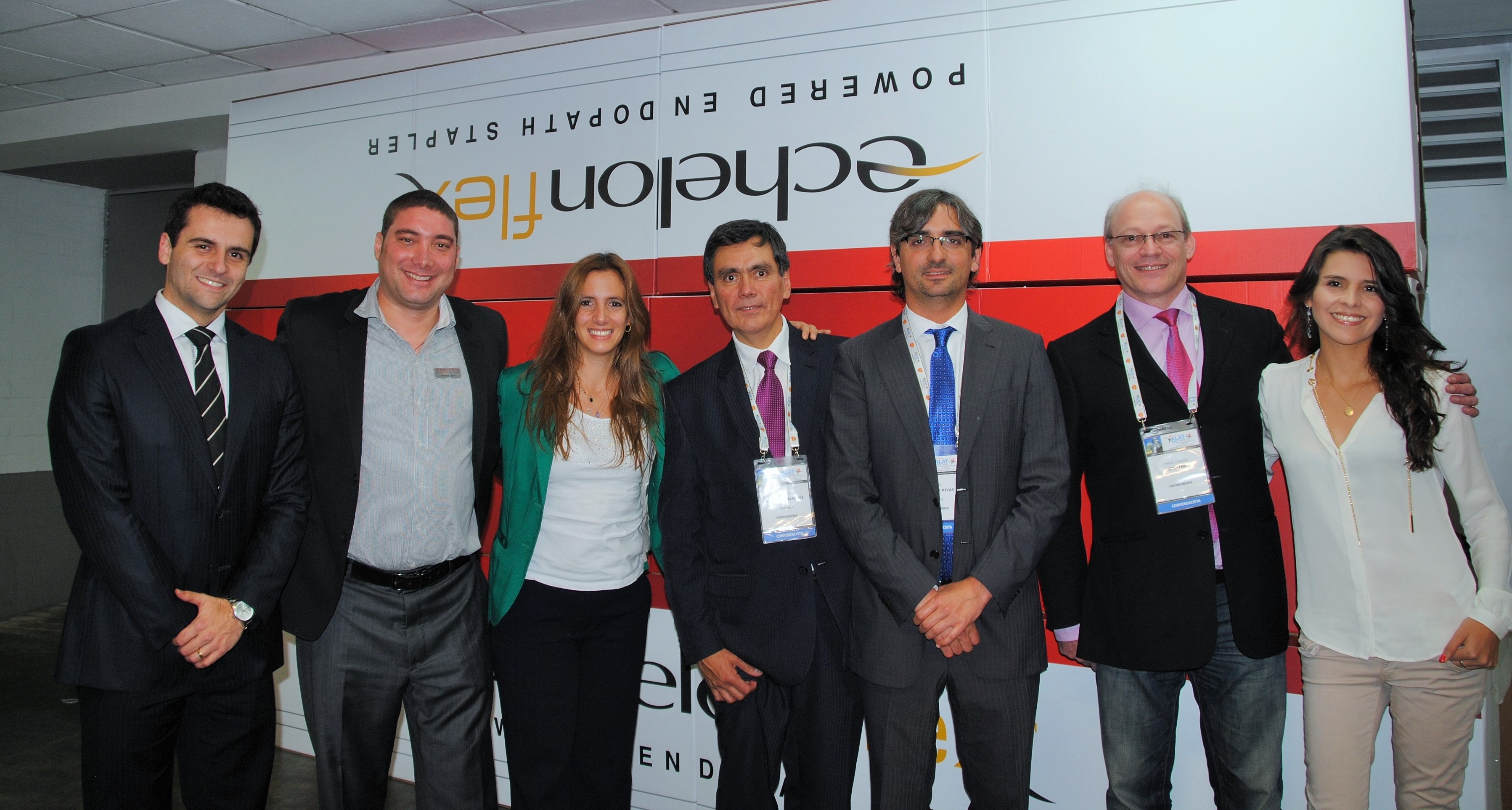
The Ethicon (Johnson & Johnson) sponsored session was by far, the best of the conference – and an excellent overview of modern technologies in thoracic surgery.
starting with Dr. Ricardo Buitrago (purple tie), Dr. Diego Gonzalez Rivas (blue tie) and Dr. Mario Ghefter (pink tie) are changing the future of thoracic surgery
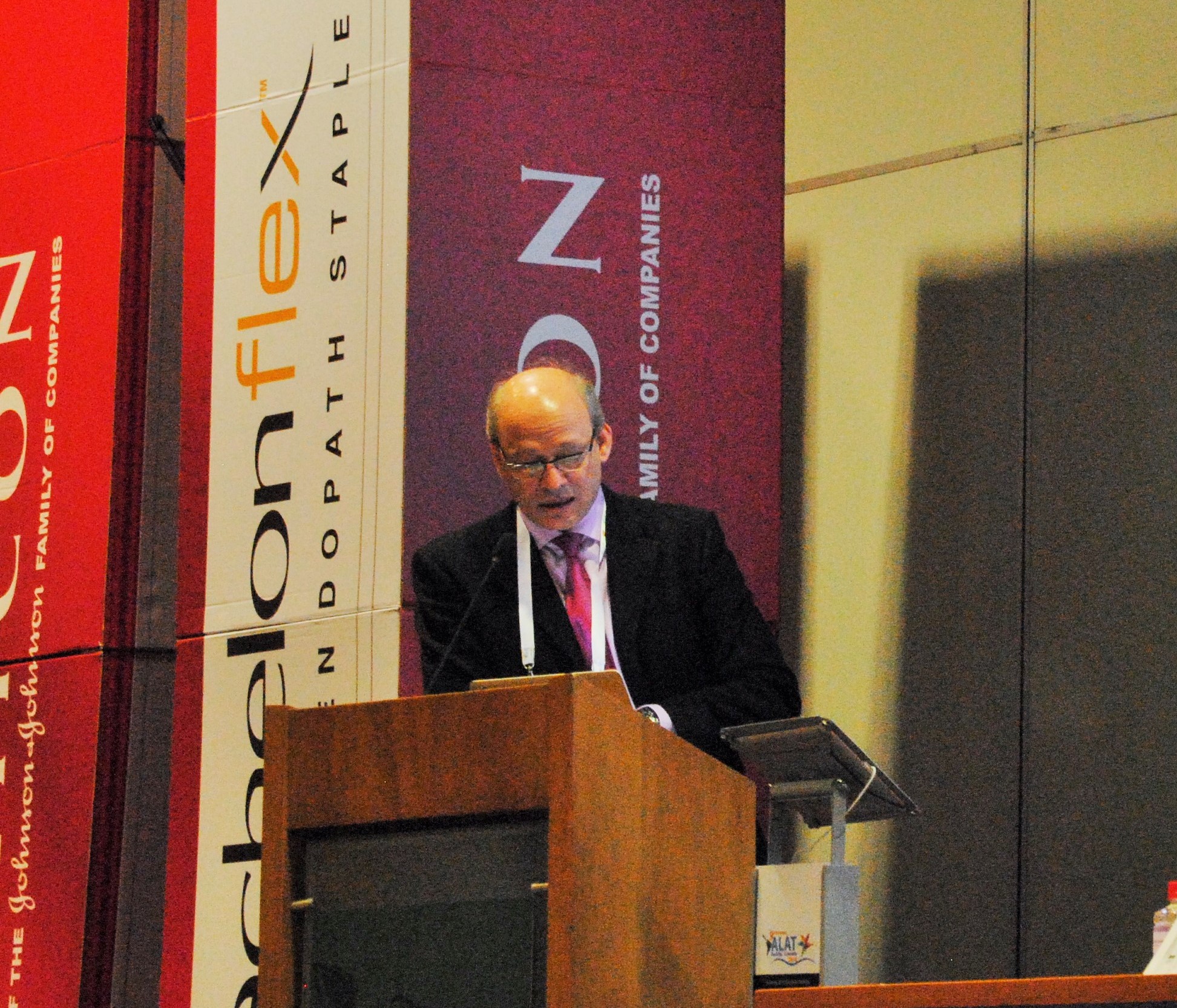
Dr. Diego Gonzalez Rivas
“Is uni-port surgery feasible for advanced cancers?” Short answer: Yes.
The first speaker, was Dr. Diego Gonzalez Rivas of Coruna, Spain. He is a world-renown thoracic surgeon and innovator of uni-port thoracoscopic surgery. He discussed the evolution of single port surgery as well as the most recent developments with this technique, including more advanced and technically challenging cases such as chest wall resections (2013), sleeve resections/ reconstructions (2013), pulmonary artery reconstructions (2013) and surgery on non-intubated, awake patients (2014).
Experience and Management of bleeding
The biggest challenges to surgeons learning this technique is management of bleeding. But as he explained in previous lectures, this can be overcome with a direct approach. (these lectures and YouTube videos, Dr. Gonzalez explains the best ways to manage intra-operative bleeding.) In the vast majority of cases – this did not require deviation or conversion from the uni-port technique.)
As surgeons gain proficiency with this technique which mirrors open surgery, the only contra-indications for surgical resection of cancerous tissue (by single port) are tumors of great size, and surgeon discomfort with the technique.
Dr. Mario Ghefter
My favorite lecture of the series was given by Dr. Mario Ghefter of Sao Paolo, Brazil. While his lecture was ostensibly about video-assisted thoracoscopy (VATS), it was more of a retrospective vision and discussion of the modern history of thoracic surgery as seen through the eyes of a 22 year veteran surgeon.
He talked about the beginnings of VATS surgery and the contributions from such legends as Cefolio and D’Amico, including the 2005 paper – and modern-day thoracic bible, “Troubleshooting video-assisted thoracoscopic lobectomy (Demmy, James, Swanson, McKenna and D’Amico).
Dr. Ghefter also talked about how improved imaging and diagnostic procedures such as PET-CT and EBUS have been able to provide additional diagnostic information pre-operatively that helps surgeons to plan their procedures and treatment strategies more effectively.
Dr. Mario Ghefter
As a counterpoint to both Dr. Gonzalez and Dr. Buitrago, Dr. Ghefter acquitted himself admirably. He reminded audience members that even the newer technologies have some drawbacks – both as procedures and for the surgeons themselves.
He also successfully argued (in my opinion) that while the popularity of procedures such as multiple port VATS and even open thoracotomies have dropped drastically as thoracic surgeons embrace newer technologies, there will always be a place and time for these more traditional procedures.
Dr. Mario Ghefter is the Director of Thoracic Surgery at Hospital do Servidor Público Estadual – Sāo Paulo and on staff at the Hospital Alemão Oswaldo Cruz.
Dr. Ricardo Buitrago
Native Colombian (and my former professor), Dr. Ricardo Buitrago is acknowledged as one of the foremost experts in robotic thoracic surgery in Latin America.
During his presentation, he discussed the principles and basics of use of robotic techniques in thoracic surgery. He reviewed the existing literature surrounding the use of robotic surgery, and comparisons of outcomes between thoracic surgery and traditional lobectomy.
He reviewed several recent robotic surgery cases and the use of robotics as a training tool for residents and fellows.
While he mentioned some of previously discussed limitations of robotic surgery (namely cost of equipment) he cited recent studies demonstrating significant cost savings due to decreased length of stay and a reduced incidence of surgical complications.
He also discussed recent studies (by pioneering surgeons such as Dr. Dylewski) demonstrated short operating times of around 90 minutes.
Discussing the classification and treatment of lung cancer according to the latest revisions (7th edition).
Medical City, Dallas, Texas USA
Sometimes location and timing is everything. Since I can’t attend all of the great thoracic surgery conferences and events, sometimes I just have to wait for something closer to home. But then again, “home” is a relative concept.
As a locum tenens provider, I travel around the country working in various hospital surgical programs on short-term contracts. It’s an interesting and always changing life but one that allows me to pursue my love of thoracic surgery to the fullest.
For the next few weeks, Medical City in Dallas, Texas is my home, as part of the cardiothoracic surgery service. It’s a return trip so it was nice to renew my acquaintance with the surgeons and staff of the CVICU and step-down units.
Today, as part of an ongoing continuing medical education program series, Dr. Mitchell Magee, of Southwest Cardiothoracic Surgeons gave an hour-long lecture entitled, “Lung cancer staging and evolving less invasive surgical treatment alternatives.” The focus of the talk was the changes in lung cancer classification and staging in the 7th edition guidelines. These revisions were proposed to replace previous versions which were based on a very small, select sample of patients at a single site. In comparison, the new revisions are based on over 100,000 patients worldwide.
T, N, M
T – tumor
N – nodes
M – metastasis
This classification system has been in use since the 1940’s and has been revised several times to reflect our growing knowledge. The latest revisions (7th edition) were released in 2012 after several years of research and debate. (For more on this process, see “The science behind the 7th edition Tumour, Node, Metastasis staging system for lung cancer” by Marshall et al, 2012).
Dr. Magee discussed the most recent revisions and how these changes affect both the treatment recommendations and prognoses for our patients. After reviewing these changes, he talked a bit about obtaining sufficient diagnostic information for accurate staging, including the role of EBUS, the new CT scan screening guidelines and the gold standard, mediastinoscopy. He also discussed some of the limitations of PET/CT and other non-invasive diagnostic imaging.
Upstaged/ Downstaged
As part of these changes in the subclassification of tumors, 10 stages have been downstaged (meaning that previously in-operable cases may now be eligible for resection) and seven classifications have been upstaged – meaning that the cancers are now considered more advanced.
For example, patients with two separate tumors in the same lobe of the lung has been upstaged to T3. Two different tumors in the same lung, but a different lobe is now T4 classification.
More specific
Some of the classifications have changed to make findings more specific. For example, T1 staging has now been subdivided into T1a and T1b.
Any invasion of the pleura, including microscopic – is now T2 staging.
He concluded the presentation with a short overview of the history of surgical resection for lung cancer, and the evolution of surgical techniques from open thoracotomies with pneumonectomies to lung sparing procedures utilizing more minimally invasive techniques.
Despite these changes, the hallmarks of a successful cancer operation remain unchanged – the right operation for the individual patient, and the need to respect oncological principles, like surgical margins, and a through lymph node dissection.
Lymph node dissection/ node sampling
Node sampling remains a crucial part of the cancer staging process despite the advent of less invasive imaging studies due to it’s infaliable accuracy. (There is either tumor tissue in the node or there isn’t, where as PET scan results can be false positive or false negative).
For this reason, tissue samples remain the gold standard of treatment and are the most accurate way to predict and prognosticate the extent of disease.
General rules regarding lymph node sampling are:
– More nodes are better. The minimum acceptable number of nodes for accurate staging is at least SIX for at least THREE different stations.
A good way to remember the relationship between node stations and node status is that bode stations are determined by distance from mediastinum; meaning that node station 14 is more peripheral that node 2.
N1 nodes are stations 10 – 14
N2 nodes are the single digit nodes (2, 4, 7 etc.)
Lymph nodes used for diagnosis and staging. Copyright Memorial Sloan-Kettering Cancer Center. Used with permission.
References and additional suggested readingBaltayiannis N, Chandrinos M, Anagnostopoulos D, Zarogoulidis P, Tsakiridis K, Mpakas A, Machairiotis N, Katsikogiannis N, Kougioumtzi I, Courcoutsakis N, Zarogoulidis K. (2013). Lung cancer surgery: an up to date. J Thorac Dis. 2013 Sep;5(Suppl 4):S425-S439. Review. Free pdf. Nice review article discussing the importance of staging for determining optimal treatment for lung cancer, as well as the impact of the latest revisions to the (7th edition) TNM classification system.
IASLC Staging Handbook in Thoracic Oncology – a site-specific guide on the new TNM classification of thoracic malignancies. This publication is published in coordination with the 7th editions of the TNM Classification of Malignant Tumors/UICC and AJCC Cancer Staging Manual.
Goldstraw P, Crowley J, Chansky K et al. (2007). The
IASLC lung cancer project: proposals for the revision of the
TNM stage groupings in the forthcoming (seventh) edition of
the TNM classification of malignant tumours. J Thorac Oncol
2007; 2: 706-714. Figure 1. Powerpoint slides TNM classification revisions for the 7th edition.
Quick and easy summary of the 7th edition classifications for Lung cancer staging – 7th edition Lung cancer staging pdf from the American Joint Commission on Cancer.
Dr. Mitchell Magee, thoracic surgeon at Medical City – Dallas, Texas
Dr. Mitchell Magee is Surgical Director of Thoracic Oncology and the Minimally Invasive Therapy Institute for Lung and Esophagus at Medical City Dallas. While his partner, Dr. Dewey focuses exclusively on cardiac surgeries like cardiac bypass, valve replacement, TAVR, LVADS and cardiac transplantation, Dr. Magee is the thoracic arm of the two surgeon Southwest Cardiothoracic Surgeons practice. This means Dr. Magee is able to devote his time to a sizable portion of all of the esophageal tears, empyemas, mediastinal masses and lung pathology that a city the size of Dallas has to offer.
Dr. Mitchell Magee & Amber Bethea, PA-C
Dr. Magee is also part of the CLEAR Clinic at Medical City – which is the lung cancer screening center at the Medical City Dallas facility.
In Jupiter, Florida talking about robots, lung cancer screening and solitary pulmonary nodules with Dr. K. Adam Lee, thoracic surgeon
Jupiter, Florida
Dr. K. Adam Lee, MD and Dawn Bitgood, FNP
All my prepared questions fly out of my mind as I greet Dr. Lee and his team. It’s been several months since I first contacted Dr. Lee to ask about his new thoracic surgery program at Jupiter Medical Center in coastal Florida, but it has taken this long for me to find a way to Florida. After nine months here, Dr. Lee is well-settled into his new position as medical director of the thoracic surgery and lung center.
Detecting and treating lung cancer
We talk about the regional differences in thoracic surgery, with Dr. Lee confirming that the majority of his practice is surgical oncology; including diagnosed lung cancer and solitary pulmonary nodules. In fact, since coming to Jupiter, Dr. Lee has started a lung cancer screening program based on the newly released CT scan guidelines for the early detection of lung cancer, as well as a lung nodule clinic for the evaluation of lung nodules.
Minimally invasive surgery
With Dr. Lee, “minimally-invasive’ is the theme. “I want patients to ask, ‘do I have to have a thoracotomy?” he states. “I want patients to know that there are minimally invasive options,” he continues as he talks about the advantages of minimally invasive techniques such as robotic-assisted thoracic surgery (RATS) and video-assisted thoracoscopic surgery (VATS). “Why should patients have all the pain [associated with large surgical incisions] if there is no reason not to do minimally invasive surgery?”
Dr. Lee should know; he’s been performing robotic surgery since 2003.
Dr. Lee, performing surgery with the DaVinci robot
Teaching others
As part of his commitment to advancing technologies, he has recently paired with Ethicon Endo-surgery to be able to provide training in minimally invasive surgery to thoracic surgery colleagues all over the world. Twice a month, he travels to other facilities to demonstrate these techniques for other surgeons. The operating rooms at the hospital here have recently been outfitted for web-based broadcasting for the remainder of the time, so that surgeons, regardless of location are able to watch these demonstrations[1].
He reports that learning to perform robotic surgery is easier for surgeons to learn than video-assisted thoracoscopic surgery, explaining that while the instrumentation is different (using robotic arms to perform surgery), the tissue manipulation and techniques are closer to open surgery [compared to VATS], and thus more familiar to conventionally trained surgeons.
I didn’t get to see Dr. Lee in the operating room – but soon, everyone will be able to.
[1] Surgeons interested in learning more can contact either Ethicon Endo-surgery or Dr. Lee directly.
* I was surprised to find out that the lung cancer screening program (CT scan, radiology interpretation/ consultation and a consultation with a thoracic surgeon) is under 300.00 USD. In an age of exorbitant medical fees, this is an affordable option for early detection of lung cancer.
a new article published in Cancer, and summarized at Medscape talks about the importance of Lung Resection for long-term survival in Lung Cancer.
Re-posting an article on the benefits of early surgical intervention on elderly patients with early stage lung cancers from Medscape.com. This is a nice article summarizing the research study conducted by Dr. Nancy Keating at Harvard Medical School in Boston, MA. A link to the original research abstract is here, but no free full-text available.
This article that highlights the importance of surgery – even for patients that primary care physicians and others may not immediately think of as great surgical candidates (frail elderly, COPD, other illnesses.)
Unfortunately, they didn’t address WHO was doing the surgeries – was it thoracic surgeons in high resection geographic areas (on the higher risk patients) as is often the case? Were surgeries in the areas with lower resection rates more likely to be done by general surgeons who are less experienced in operating on more frail thoracic patients? [all thoracic patients are frail to some decrease given the nature of the condition – so specialty trained thoracic surgeons are usually much more experienced in caring for these patients]. It would have been nice to know.
Surgery Rates tied to Lung Cancer Outcomes in the Elderly
David Douglas (Medscape)
NEW YORK (Reuters Health) Aug 24 – People with early non-small cell lung cancer (NSCLC) live longer if they’re in regions of the U.S. where doctors perform more surgeries for that indication, according to a new study.
The link between higher surgery rates and better survival held true even for frailer patients.
“We found that areas with high rates of surgery tended to operate on older and sicker patients, yet still had better outcomes for early-stage lung cancer than areas with lower use of surgery,” said senior investigator Dr. Nancy L. Keating in an email to Reuters Health.
“These data suggest that areas with lower surgery rates may benefit from higher rates of surgery,” she said.
Dr. Keating, from Harvard Medical School in Boston, said, “Resection has by far the highest chance of cure.”
But, she noted, “It may be that fear of harm (surgeons being concerned about causing poor outcomes) may be leading to relative underuse of this effective treatment.”
“While there are some patients for whom the risks certainly outweigh the benefits,” she added, “those patients may be fewer than some physicians recognize.”
Dr. Keating and colleagues studied a population-based cohort of more than 17,000 Medicare beneficiaries at least 66 years old who were diagnosed with stage I or II NSCLC during 2001 to 2005.
Using Surveillance, Epidemiology, and End Results (SEER) data, they compared areas with high and low rates of curative surgery for early stage lung cancer.
Fewer than 63% of patients had operations in low-surgery areas, whereas more than 79% did in high-surgery areas, according to a July 28th online paper in Cancer.
The high-surgery areas saw more operations on older patients and in those with chronic obstructive pulmonary disease (COPD).
The one-year lung-cancer-specific mortality rate was 12% in the high-surgery regions and 17% in low-surgery. The adjusted odds ratio for each 10% increase in the surgery rate was 0.86. There were similar findings for all-cause mortality.
Original article reference information:
Cancer. 2011 Jul 28. doi: 10.1002/cncr.26363. [Epub ahead of print]. Improved outcomes associated with higher surgery rates for older patients with early stage nonsmall cell lung cancer. Gray SW, Landrum MB, Lamont EB, McNeil BJ, Jaklitsch MT, Keating NL.
Creation of a new ‘regional thoracic surgery center’ in Hamilton, Ontario highlights some of the issues we’ve talked about here before: high volume centers, optimizing outcomes, decreasing wait times, and quality/ consistency of care.
Since this center is in Canada – it is also important to note that this change will decrease waiting times (initial presentation to treatment) for patients. For people unfamiliar with socialized medicine – these waiting periods can be significant. The article phrases this differently, stating time of initial presentation to diagnosis – which can have a different meaning – (or if the diagnosis is made from surgical tissue, essentially the same.) However, that time was 95 DAYS (or over three months) and has now been reduced to 35 days.
In other Thoracic Surgery news – I am currently researching articles on high-altitude lung surgery, so there may be a gap in between postings as I continue to review the existing data.
new research results from the University of Pennsylvania on the treatment of pleural mesothelioma.
The University of Pennsylvania reports the latest results of a small study involving 28 patients with pleural mesothelioma.
This limited study compared combination treatment using photodynamic therapy along with a lesser lung surgery (14 patients) in comparison to extrapleural pneumonectomy alone (14 patients). 22 of the 28 patients also received chemotherapy.
Patient population: 28 patients – 12 /14 patients in either group with advanced (stage III/IV ) disease
Results: Extrapleural pneumonectomy group had a median survival of 8.9 months. The combination photodynamic/ surgery group median survival exceeded two years (when the study ended).
Take away message for readers: It’s too early, and the study groups are far too small for us to generalize these findings. However, these preliminary results are encouraging and should prompt more, larger scale studies / trials looking at photodynamic therapy as adjuvant therapy along with thoracic surgery for pleural mesothelioma.
Update: 06/15/2011:
the mesothelioma study from PA just got picked up by a major wire service, so expect to read and hear a lot more about it.
Update: 08/15/2014: Mesothelioma.net has asked that I link with their site. They offer some informational services for people facing mesothelioma. Please let me know if this site is spam-plagued or otherwise dubious and I will remove the link (the site is a bit ‘shiny’ and circular for my taste.)
A look at the literature, including a recent systemic review: exercise is not only safe for lung cancer patients – but improves quality of life, and may (according to Jones) improve post-operative outcomes..
There’s an article ( Jones et. al 2010) on the benefits of pre-operative and post-operative exercise regimens for patients with lung cancer, that was conducted down at Duke. They use a lot of abbreviations in the article, but the gist is that a patient’s post-operative risk can be determined by their peak oxygen consumption (which is measurable), and that this can be modified (improved) prior to surgery with a targeted fitness program. This program notably included weight training in addition to aerobic exercise.
A systematic review was recently published (Feb 2011) looking at a compilation of these types of studies to give an overview of the preponderance of evidence by Granger et. al. This isn’t an open access article so I can’t post the link here – but I can link the abstract which is a short summary of his findings..
So basically Granger looked at the above mentioned study, and fifteen others – and drew limited conclusions.. He limited his conclusions to saying exercise was safe, and helpful in lung cancer patients and improved the ‘health related quality of life’. It’s a bit different from what Jones had to say – but the message is the same; exercise is not only safe for lung cancer patients – but improves quality of life, and may (according to Jones) improve post-operative outcomes..
Nagarajan et al. (2011) reviewed several published papers on the topic of pulmonary rehabilitation in an effort to answer the questions of whether pre-operative pulmonary rehabilitation reduced post-operative complications & overall length of stay. While they were unable to conclusively answer these questions – they did find that pre-operative pulmonary rehabilitation increased exercise capacity, and preserved pulmonary function in patients with COPD. This may not sound important – until you realize that these are critical measures of quality of life. (for example – what if “increased exercise capacity” means a person can now walk to around their home, and perform daily activities of living such as showering and getting dressed without becoming short of breath?)
How to maximize your chances before lung surgery to speed healing, post-operative recovery and reduce the incidence of complications.
As most of my patients from my native Virginia can attest; pre and post-operative surgical optimization is a critical component to a successful lung surgery. In most cases, lung surgery is performed on the very patients who are more likely to encounter pulmonary (lung) problems; either from underlying chronic diseases such as emphysema, or asthma or from the nature of the surgery itself.
Plainly speaking: the people who need lung surgery the most, are the people with bad lungs which makes surgery itself more risky.
During surgery, the surgeon has to operate using something called ‘unilung ventilation’. This means that while the surgeon is trying to get the tumor out – you, the patient, have to be able to tolerate using only one lung (so he can operate on the other.)
Pre-surgical optimization is akin to training for a marathon; it’s the process of enhancing a patient’s wellness prior to undergoing a surgical procedure. For diabetics, this means controlling blood sugars prior to surgery to prevent and reduce the risk of infection, and obtaining current vaccinations (flu and pneumonia) six weeks prior to surgery. For smokers, ideally it means stopping smoking 4 to 6 weeks prior to surgery.(1) It also means Pulmonary Rehabilitation.
Pulmonary Rehabilitation is a training program, available at most hospitals and rehabilitation centers that maximizes and builds lung capacity. Numerous studies have show the benefits of pre-surgical pulmonary rehabilitation programs for lung patients. Not only does pulmonary rehabilitation speed recovery, reduce the incidence of post-operative pneumonia,(2) and reduce the need for supplemental oxygen, it also may determine the aggressiveness of your treatment altogether.
In very simple terms, when talking about lung cancer; remember: “Better out than in.” This means patients that are able to have surgical resection (surgical removal) of their lung cancers do better, and live longer than patients who receive other forms of treatment such as chemotherapy or radiation.
If you are fortunate enough to have your lung cancer discovered at a point where it is possible to consider surgical excision – then we need you to take the next step, so you are eligible for the best surgery possible.
We need you to enhance your lung function through a supervised walking and lung exercise program so the surgeon can take as much lung as needed. In patients with marginal lung function,(3) the only option is for wedge resection of the tumor itself. This is a little pie slice taken out of the lung, with the tumor in it. This is better than chemotherapy or radiation, and is sometimes used with both – but it’s not the best cancer operation because there are often little, tiny, microscopic tumor cells left behind in the remaining lung tissue.
The best cancer operation is called a lobectomy, where the entire lobe containing the tumor is removed. (People have five lobes, so your lung function needs to be good enough for you to survive with only four.(4) This is the best chance to prevent a recurrence, because all of the surrounding tissue where tumors spread by direct extension is removed as well. Doctors also take all the surrounding lymph nodes, where cancer usually spreads to first. This is the best chance for five year survival, and by definition, cure. But since doctors are taking more lung, patients need to have better lung function , and this is where Pulmonary Rehab. comes in. In six weeks of dedicated pulmonary rehab – many patients who initially would not qualify for lobectomy, or for surgery at all – can improve their lung function to the point that surgery is possible.
Post-operatively, it is important to continue the principles of Pulmonary rehab with rapid extubation (from the ventilator), early ambulation (walking the hallways of the hospitals (5) and frequent ‘pulmonary toileting’ ie. coughing, deep breathing and incentive spirometry.
All of these things are important, where ever you have your surgery, but it’s particularly important here in Bogota due to the increased altitude.
One last thing for today:
a. Make sure to have post-pulmonary rehab Pulmonary Function Testing (PFTs, or spirometry) to measure your improvement to bring to your surgeon,
b. walk daily before surgery (training for a marathon, remember)
c. bring home (and use religiously!) the incentive spirometer provided by rehab.
ALL of the things mentioned here today, are things YOU can do to help yourself.
Footnotes:
1. Even after a diagnosis of lung cancer, stopping smoking 4 to 6 weeks before surgery will promote healing and speed recovery. Long term, it reduces the risk of developing new cancers.
2. Which can be fatal.
3. Lung function that permits only a small portion (or wedge section) to be removed
4. A gross measure of lung function is stair climbing; if you can climb three flights of stairs without stopping, you can probably tolerate a lobectomy.
5. This is why chest tube drainage systems have handles. (so get up and walk!)
and the snowball effect of atrial fibrillation after surgery. Discussion includes beta blockers and vitamin C as methods to reduce the incidence of post-operative atrial fibrillation with discussion of the literature supporting its use.
In previous posts, we’ve talked about prevention and management of respiratory complications of lung surgery. However, one of the more common complications of lung surgery, is atrial fibrillation, or an abnormal heart rate and rhythm. Most of the time, atrial fibrillation after surgery is temporary – but that does not make it a benign problem.
Developing atrial fibrillation is problematic for patients because increases length of stay (while we attempt to treat it) and increases the risk of other problems (such as stroke – particularly if we can’t get the heart rhythm to return to normal).
‘The Cootie Factor’ Length of stay is important for more than cost and convenience. One of the things I try to explain to my patients – is that hospitals are full of sick people, and in general, my surgery patients are not sick– they’ve had surgery..
But surgery increases their chance and susceptibility to contracting infections from other patients, and visitors. I call this ‘the cootie factor’. (Everyone laughs when you say cooties – but everyone knows exactly what you mean.) So the reason I am rushing my patients out the door is more than just for patient convenience and the comforts of home – it’s to prevent infection, and other serious complications that come from being hospitalized, in close quarters, with people who have may have some very bad cooties indeed (MRSA, resistant Klebsiella, VRE, Tuberculosis and other nasties.)
But besides, length of stay – atrial fibrillation, or a very rapid quivering of the atrial of the heart (250+ times per minute) increases the chance of clots forming within the atrial of the heart, and then being ejected by the ventricles straight up into central circulation – towards the brain – causing an embolic stroke.. Now that’s pretty nasty too..
Atrial fibrillation risk reduction
But there are some easy things we can do to reduce the chance of this happening..
One of the easiest ways to prevent / reduce the incidence of post-operative atrial fibrillation – to slow down the heart rate. We KNOW that just by slowing down the heart by 10 – 15 beats per minute, we can often prevent abnormal heart rhythms.
Most of the time we do this by pre-operative beta blockade, which is a fancy term for using a certain class of drugs, beta blockers (such as metoprolol, carvedilol, atenolol) to slow the heart rate, just a little bit before, during and after surgery.
In fact, this is so important – national/ and international criteria uses heart rate (and whether patients received these medications prior to surgery) as part of the ‘grading’ criteria for rating surgery/ surgeons/ and surgery programs. It’s part of both NSQIPs and the Surgical Apgar Scale – both of which are important tools for preventing intra-operative and post-operative problems..
The good thing is, most of these drugs are cheap (on the $4 plan), very safe, and easily tolerated by patients. Also, most patients only need to be on these medications for a few days before and after surgery – not forever.
Now, if you do develop atrial fibrillation (a. fib) after surgery – we will have to give you stronger (more expensive, more side effects) drugs such as amiodarone, or even digoxin (old, but effective) to try to control or convert your heart rhythm back to normal.
If you heart rhythm does not go back to normal in a day or two – we will have to start you on a blood thinner like warfarin to prevent the blood clots we talked about previously. (Then you may have to have another procedure – cardioversion, and more medicines, if it continues, so you can start to see why it’s so important to try to prevent it in the first place).
Research has also looked at statin drugs to prevent atrial fibrillation after surgery – results haven’t been encouraging, but if you are already on cholesterol medications prior to surgery, there are plenty of other reasons for us to continue statins during and after surgery.. (Now, since the literature is mixed on whether statins help prevent a. fib – I wouldn’t start them on patients having lung surgery, but that’s a different matter.)
Now Dr. Shu S. Lin, and some of the other cardiac surgeons did some studies down at Duke looking at pre-operative vitamin C (along with quite a few others) and the results have been interesting.. That doesn’t mean patients should go crazy with the supplements.. anything, even Vitamin C can harm you, if taken willy-nilly (though the risk with vitamin C is usually minimal).
In fact, the evidence was strong enough (and risk of adverse effects was low enough) that we always prescribed it to our pre-operative patients for both heart and lung surgery. (Heart patients are at high risk of atrial fibrillation too.) We prescribed 500mg twice a day for a week before surgery, until discharge – which is similar to several studies. I’ve included some of these studies before – please note most of them focus on atrial fibrillation after heart surgery.
Contrary to popular belief, performing a VATS procedure (versus open surgery) does not eliminate the risk of post-operative atrial fibrillation.
Now Dr. Onaitis, D’Amico and Harpole published some interesting results last year (and of course, as Duke Thoracic surgeons, I am partial) – but I can’t repost here since it’s limited access articles..
Discussion of treatment goals, and patient centered care for Malignant pleural effusions. This is the first in a series of articles on lung cancer, and lung surgery topics. Originally posted at our sister site.
Not all conditions are curable, and not all treatments are curative. Some treatments are based on improving quality of life, and alleviating symptoms. This is a hallmark of patient centered care – doing what we can to make the patient feel better even when we can’t ‘fix’ or cure the underlying disease. No where is this more evident than in the treatment of malignant effusions.
By definition, a Malignant Effusion is the development of fluid in the fluids related to an underlying (and sometimes previously undiagnosed) malignancy. Malignant effusions can be seen with several different kinds of cancers, most commonly lung and breast cancers. The development of a malignant effusion is a poor prognostic sign as it is an indicator of metastasis to the pleural tissue/ space.
The development of a malignant effusion usually presents with symptoms of shortness of breath, and difficulty breathing. While the treatment of the underlying cancer may vary, the primary goal of treatment of an effusion is palliative (or symptom relief). The best way to relieve symptoms is by removing the fluid.
This can be done several ways – but each has its own drawbacks.
Thoracentesis:
The fluid can be drawn out with a needle (thoracentesis) either bedside or under fluroscopy. This procedure is quick, and can be performed on an out-patient basis, in a doctor’s office, or in radiology.
The potential drawbacks with this treatment strategy are two-fold:
1. There is a chance that during the procedure, the needle will ‘poke’ or ‘pop’ the lung, causing a pneumothorax (or collapse of the lung). This then requires a chest tube to be placed so the lung can re-expand while it heals. However, if the procedure is performed uneventfully, (like it usually does) the patient can go home the same day.
2. The other complication – is rapid re – accumulation – since you haven’t treated the underlying cause, but have only removed the fluid. This also happens when the cause of the effusion (nonmalignant) is from congestive heart failure. This means the fluid (and symptoms of shortness of breath) may return quickly, requiring the patient to return to the hospital – which is hard of the patient and their family.
Video- Assisted Thoracoscopy: (VATs)
Malignant effusions can also be treated by VATS – this is a good option if we are uncertain of the etiology (or the reason) for the effusion. While all fluid removed is routinely sent for cytopathology (when removed during surgery, thoracentesis or chest tube placement) – but cytopathology can be notoriously inaccurate with false negative reports, because the diagnosis is dependent on the pathologist actually seeing cancer cells in the fluid. However, during the VATs procedure – the surgeon can take tissue samples, and photos along with fluid for diagnostic testing. This is important because I have had cases in the operating room (VATS) where the surgeon actually sees the tumor(s)** with the camera but the fluid comes back as negative.
** in these cases, we send biopsies of the tumor tissue – which is much more accurate and definitive.
But a VATS procedure requires an operation, chest tube placement and several days in the hospital.
Chest tube placement:
Another option is chest tube placement – which also requires several days in the hospital..
During both chest tube placement and VATS, a procedure called pleurodesis can be performed to try to prevent the fluid from re-accumulating.
But what if we know it’s a malignant effusion? What are the other options for treatment?
Catheter based treatments: (aka PleurX style catheter, or Heimlich valve)
(note: catheter means a small tube – a foley catheter is the type used to drain urine, but other types are used for many things – even an IV is a catheter.)
One of the options used in our practice was pleur X (brand) catheter placement. This catheter was a small flexible tube that could be placed under local anesthesia – either in the office or the operating room – as an ambulatory procedure. After some patient teaching, including a short video, most family members felt comfortable emptying the catheter every two or three days at home, to prevent fluid re -accumulation (and allowing the patient to continue normal activities, at home.)
PleurX catheter placement is preferred in many cases due to ease of use, and patient convenience. The Heimlich valve is messier – as it tends to leak, and harder for patients to hide under clothing.
Sometimes a visiting nurse would go out and empty the catheter, and in several cases, patients would come to the office, where I would do the same thing – it was a nice way to relieve the patient’s symptoms without requiring hospitalization, and several studies have shown that repeated drainage often caused spontaneous pleurodesis (fluid no longer accumulated.) We would then take the catheter out in the office.. Now, like any procedure, there is a chance for problems with this therapy as well, infection, catheter can clog, etc..
But here’s another study, showing that even frail patients benefit from home-based therapy – which is important when we go back and consider our original treatment goals:
-Improving quality of life
-Relieving symptoms
In the article, the authors used talc with the catheters and then applied a Heimlich valve, which is another technique very similar to pleurX catheter placement. (Sterile talc is used for the pleurodesis procedure – which we will talk about in more detail in the future.)