Guest post: Report from the 3rd Mediterranean Symposium in Thoracic Surgical Oncology – VATS

An invited report from Dr. Marcello Migliore on the recent Italian conference on VATS and lung cancer


 Report from the 3rd Mediterranean Symposium in Thoracic Surgical Oncology on VATS RESECTIONS FOR LUNG CANCER: moving toward standard of care.

Speakers and moderators at the 3rd Mediterranean Symposium on Thoracic Surgical Oncology

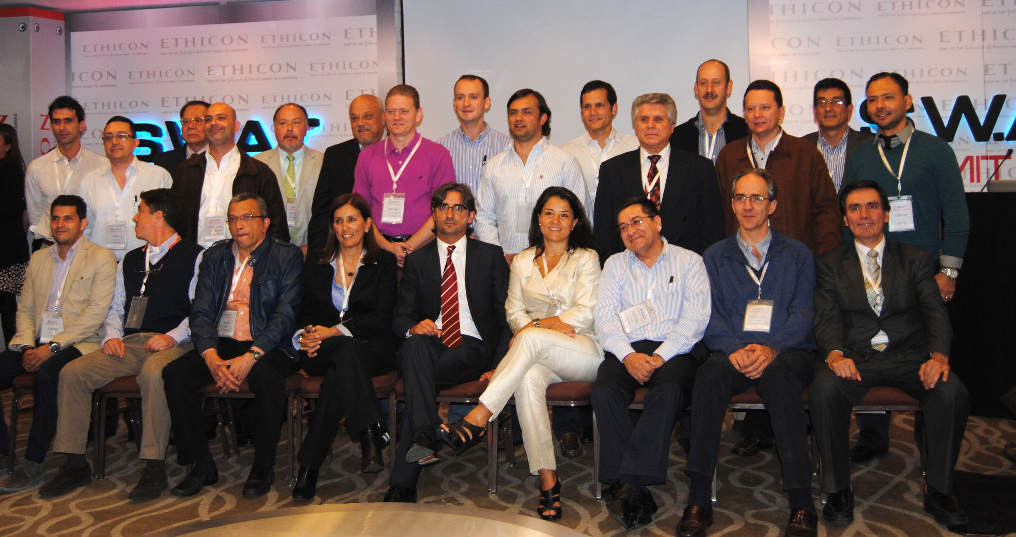
 The third mediterranean symposium on thoracic surgical oncology was  successful.  The symposium was held the 21st – 22nd april 2016  at the Aula Magna of the Faculty of Medicine at the University of Catania. More than 150 people attended, and among them there were thoracic surgeons, general surgeons, oncologists, chest physicians, residents and medical students.  This year, we had speakers from Europe and the USA.  The main topic was VATS resections for lung cancer (Photo 1). During the opening ceremony, the Rector Giacomo Pignataro awarded a medal to Professor Tom Treasure for enhancing our outstanding education and research experience (Photo 2).

Photo 2: The Rector of the University of Catania  is giving the medal to Professor Tom Treasure

Although the concept of operating thru a small port was born and developed in Europe (1- 7) it has been noted that 90% of papers on uniportal VATS lobectomy come from East Asian countries (8-11).  Throughout the symposium  different speakers agreed that a  proper definition of uniportal VATS is mandatory to speak the same language worldwide.

Awake thoracic surgery was discussed together with the need of accurate  preoperative staging procedures such as endobronchial ultrasound, videomediastinoscopy or Video-assisted mediastinal lympadenectomy.   It was concluded that a wide spectrum of factors must be considered when determining the appropriate tests to assess the lymph nodes in  NSCLC, which includes not only the sensitivity and specificity of the test,  but also the ability to perform  the procedure on an individual patient.

Data from New York showed very clearly that there have been no large-scale randomized control trials to compare open and VATS lobectomy. Although most may agree with the short-term superiority of VATs lobectomy over its open counterpart, many argue that is an in adequate oncologic procedure. Hence whether the approach is equivalent in overall and cancer specific survival to its open counterpart is not known. He also reported an important recent analysis of SEER-Medicare which confirmed that VATS lobectomy appears to have similar survival to its open counterparts (12).

A magnificent video was presented to explain every step of the lobectomies performed through a small skin incision.  A long discussion followed and all auditorium proposed that ‘single incision’ VATS probably define better than uniportal VATS what surgeons are doing worldwide. Certainly the length of skin incision is important and should be taken in serious consideration.  We felt that a consensus conference is probably necessary  consensus conference is probably necessary.  The indication for a Wedge resection rather than lobectomy in initial stage lung cancer is still weak.

The Italian VATS group was formed in 2013 , and nowadays there are 65 participating centres and that 2800 VATS lobectomy have already been included. In Catania we joined the group few months ago (13)

A very interesting session for juniors and medical students from UK and Italy was carried out,  and 12 abstracts have been presented as interactive posters.  Two of them have been chosen for possible publication in Future Oncology.

Finally, the first data survival seems to benefit little from the various even growing “personal” modifications of the standard VATS technique. Since there is a limited variation between VATS and uniportal VATS, the likelihood is that either VATS and uniportal VATS will be operative in the near future. Its success will depend on survival advantages and decrease chest pain  and not just on new technical instrumentation. To protect patient’s safety, the length  of the skin incision should  be chosen on the basis of several clinical factors and not in relation of modern “demand”.  Although the trial VIOLET is ongoing in UK to demonstrate if VATS resection for lung cancer is better than open thoracotomy, doubts arises  as standard postero-lateral thoracotomy for lung cancer seems to be an incision which is performed rarely today.  A skin incision of 6-8 cm (mini-thoracotomy) with video assistance is enough for most of lung resections. The question which arises is if a mini-thoracotomy of 6 cm should be called “uniportal” or not.

Marcello Migliore, MD

Thoracic surgeon and invited commentator

Dr. Marcello Migliore
  1. Migliore M Initial History of Uniportal Video-Assisted Thoracoscopic Surgery. Ann Thorac Surg 2016;101 (1), 412-3.
  2. Migliore M, Calvo D, Criscione A, Borrata F. Uniportal video assisted thoracic surgery: summary of experience, mini-review and perspectives. Journal of Thoracic Disease 2015; 7 (9), E378-E380
  3. Migliore, M., Giuliano, R., & Deodato, G. (2000). Video assisted thoracic surgery through a single port. In Thoracic Surgery and Interdisciplinary Symposium on the threshold of the Third Millennium. An International Continuing Medical Education Programme. Naples, Italy (pp. 29-30).
  4. Migliore, M., Deodato, G. (2001). A single-trocar technique for minimally invasive surgery of the chest. Surgical Endoscopy, 8(15), 899-901.
  5. Migliore M. Efficacy and safety of single-trocar technique for minimally invasive surgery of the chest in the treatment of noncomplex pleural disease. J Thorac Cardiovasc Surg 2003;126:1618-23.
  6. Rocco, G., Martin-Ucar, A., & Passera, E. (2004). Uniportal VATS wedge pulmonary resections. The Annals of Thoracic Surgery, 77(2), 726-728.
  7. Gonzalez D, Paradela M, Garcia J, et al. Single-port video-assisted thoracoscopic lobectomy. Interact Cardiovasc Thorac Surg 2011;12:514-5.
  8. Yang HC, Noh D. Single incision thoracoscopic lobectomy through a 2.5 cm skin incision. J Thorac Dis  2015;7:E122-5.
  9. Ocakcioglu I, Sayir F, Dinc M. A 3-cm Single-port Video-assisted Thoracoscopic Lobectomy for Lung Cancer. Surg Laparosc Endosc Percutan Tech 2015;25:351-3.
  10. Kamiyoshihara M, Igai H, Ibe T, et al. A 3.5-cm Single-Incision VATS Anatomical Segmentectomy for Lung Ann Thorac Cardiovasc Surg 2015;21:178-82.
  11. Zhu Y, Xu G, Zheng B, et al. Single-port video-assisted thoracoscopic surgery lung resection: experiences in Fujian Medical University Union Hospital. J Thorac Dis 2015;7:1241-51.
  12. Paul S, Isaacs AJ, Treasure T, Altorki NK, Sedrakyan A. Long term survival with thoracoscopic versus open lobectomy: propensity matched comparative analysis using SEER-Medicare database. BMJ 2014;349:g5575
  13. Migliore, M., Criscione, A., Calvo, D., Borrata, F., Gangemi, M., & Attinà, G. (2015). Preliminary experience with video-assisted thoracic surgery lobectomy for lung malignancies: general considerations moving toward standard practice. Future Oncology, 11(24s), 43-46.
  14. Migliore M. Will the widespread use of uniportal surgery influence the need of surgeons ? Postgrad Med J 2016 (in press).


Upcoming course on Minimally Invasive thoracoscopy – Latin America

upcoming minimally invasive thoracoscopy course for my Spanish speaking readers at the Clinica Alemana in Santiago, Chile

Cirugia de Torax won’t be there this year – but Clinica Alemana, one of the highest ranked hospitals in Latin America is holding another course on Minimally invasive thoracic surgery this October.

INFO WEB_Cirugia Toracica_23.09.14 (1)-page-001

The facility, which is in Santiago, Chile is also home to Dr. Raimundo Santolaya, who was one of our first interviews here at Cirugia de Torax.

Information about the course:

INFO WEB_Cirugia Toracica_23.09.14

 II Curso de Cirugía Torácica Mínimamente Invasiva

20 y 21 de octubre de 2014

 Aula Magna de Clínica Alemana de Santiago

Avda. Manquehue Norte 1410, Vitacura

Featured speakers include Dr. Miguel Congregado (Seville, Spain), Dr. Pablo Moreno de Santa Barajas (Vigo, Spain), Dr. David Smith (Buenos Aires, Argentina) and Dr. Patricio Varela of the University of Chile.

Course content is sponsored by the Chilean Society of Respiratory Diseases, The Chilean Society of Surgeons, the American College of Surgeons and the Faculty of Medicine at Clinica Alemana – University of Desarrollo.

Interested surgeons should contact the clinic at this address: dmedico@alemana.cl or cursosclinicos1@alemana.cl or enroll on-line at www.alemana.cl

The event is jointly sponsored by M. Kaplan, Johnson & Johnson, IMP, Solumed and Stryker.

Course schedule:

INFO WEB_Cirugia Toracica_23.09.14 (1)-page-002INFO WEB_Cirugia Toracica_23.09.14 (1)-page-003

VGTT: video-guided tube thoracostomy

Dr. Chin Hao Chen revisits one of the basic procedures in thoracic surgery: Chest tube placement

Even Hippocrates placed chest tubes or the history of tube thoracostomy

Chest tube placement has been performed since ancient Greek times.  Early physicians, including Hippocrates himself, performed (and wrote about) the use of tube thoracostomy for the treatment of lung abscesses and empyema.  Often this procedure is performed using a ‘blind approach’ based entirely on external anatomic features (intercostal spaces) and a fundamental knowledge of internal and chest wall anatomy.  Over the years, surgeons have developed guidelines to this technique using palpation/ and other tactile information but none of these techniques challenged initial insertion technique.

With any blind procedure, there is a risk of inadvertent injury due to the lack of visualization, particularly in patients with previous thoracic procedures or infections (adhesions), or when performed by less experienced staff.

Direct visualization during this procedure (akin to VATS) may lessen this risk.  However, little has been published on alternatives to the traditional technique.

VGTT: video-guided tube thoracostomy

Our latest post comes directly from Dr. Chih-Hao Chen at Mackay Memorial Hospital in Taiwan.

Dr. Chen presents a video clip demonstrating video-guided tube thoracostomy (VGTT), a technique used to avoid tube-related injury during the course of tube thoracostomy (versus blind insertion).  This visualization technique is helpful particularly when performed by inexperienced staff, such as residents or in emergent situations.

A complete description of this technique was recently published in the Annals of Thoracic Surgery.

Chen, et. al (2013). Video-guided tube thoracostomy with use of a nonfiberoptic endoscope. Ann Thorac Surg, 2013;96: 1450-5.  Articles commentary also available.

This paper describes the technique as well as discussing the clinical experience of Dr. Chen and his team in applying this technique to several patients.

Dr. Chin-Hao Chen is a thoracic surgeon at Mackay Memorial Hospital in Taiwan.  Dr. Chen is a frequent and valued contributor here at Cirugia de Torax.  He has provided several case studies as well as articles and videos on surgical techniques.

Dr. Gonzalez Rivas, Johnson & Johnson and Single-port thoracic surgery

the 2013 S.W.A.T conference, presented by Johnson & Johnson. Featured presenters Dr. Diego Gonzalez Rivas and Dr. Paula Ugalde discuss single port thoracoscopy and topics in minimally invasive surgery

Very pleased that despite the initial difficulties, we are able to provide information regarding the recent conference.

Talking about Single-port surgery in Bogotá, Colombia – 2013 S.W.A.T. Summit

Dr. Diego Gonzalez Rivas and Dr. Paula Ugalde were the headliners at the recent Johnson and Johnson thoracic surgery summit on minimally invasive surgery.  Both surgeons gave multiple presentations on several topics.  They were joined at the lectern by several local Colombian surgeons including Dr. Stella Martinez Jaramillo (Bogotá), Dr. Luis Fernando Rueda (Barranquilla), Dr. Jose Maineri (Venezuela) Dr. Mario Lopez (Bogotá) and Dr. Pardo (Cartagena).

Thoracic surgeons at the 2013 S.W.A.T Summit in Bogota, Colombia. Drs. Gonzalez-Rivas and Dr. Paula Ugalde are center, front-row

Target audience missing from conference

The audience was made up of thirty Latin American surgeons from Colombia, Costa Rica and Venezuela.  This surgeons were hand-picked for this invitation-only event.  Unfortunately, while Johnson and Johnson organized and presented a lovely event; their apparent lack of knowledge about the local (Colombian) thoracic surgery community resulted in the exclusion of several key surgeons including Dr. Mauricio Velasquez, one of Colombia’s earliest adopters of single-port thoracoscopy.  Also excluded were the junior members of the community, including Dr. Castano, Dr. Carlos Carvajal, and current thoracic surgery fellows.  It was an otherwise outstandingand informative event.

However, this oversight represents a lost-opportunity for the parent company of Scanlon surgical instruments, the makers of specialized single port thoracoscopic instrumentation endorsed and designed by Dr. Gonzalez-Rivas himself, including the Gonzalez-Rivas dissector.

The Gonzalez - Rivas dissector, photo courtesy of Scanlon International
The Gonzalez – Rivas dissector, photo courtesy of Scanlon International

As discussed in multiple publications, previous posts as well as during the conference itself, it is these younger members who are more likely to adopt newer surgical techniques versus older, more experienced surgeons.  More seasoned surgeons may be hesitant to change their practices since they are more comfortable and accustomed to open surgical procedures.

Despite their absence, it was an engaging and interesting conference which engendered lively discussion among the surgeons present.

Of course, the highlight of the conference actually occurred the day before, when Dr. Gonzalez- Rivas demonstrated his technique during two separate cases at the National Cancer Institute in Bogotá, Colombia. (Case report).

Dr. Gonzalez-Rivas and Dr. Ricardo Buitrago performing single port thoracoscopy at the National Cancer Institute

Featured presenters:

Dr. Diego Gonzalez – Rivas is a world-renown thoracic surgeon jointly credited (along with Dr. Gaetano Rocco) with the development of single-port thoracoscopic (uni-port) surgery.  He and his colleagues at the Minimally Invasive Surgery Unit in La Coruna, Spain give classes and lectures on this technique internationally.  Recent publications include three papers in July alone detailing the application of this surgical approach, as well as several YouTube videos demonstrating use of this technique for a wide variety of cases.

Dr. Gonzalez Rivas
Dr. Gonzalez Rivas

Dr. Paula Ugalde, a Chilean-borne thoracic surgeon (from Brazil) who gave several presentations on minimally-invasive surgery topics. She is currently affiliated with a facility in Quebec, Canada.

Dr. Paula Ugalde
Dr. Paula Ugalde

Refuting the folklore

Part of the conference focused on refuting the ‘folklore’ of minimally-invasive procedures.   Some of these falsehoods have plagued minimally-invasive surgery since the beginning of VATS (in 1991), such as the belief that VATS should not be applied in oncology cases. The presenters also discussed how uniportal VATS actually provides improved visibility and spatial perception over traditional VATS (Bertolaccini et al. 2013).

However, Gonzalez-Rivas, Ugalde and the other surgeons in attendance presented a wealth of data, and publications to demonstrate:

–          VATS is safe and feasible for surgical resection in patients with cancer. (Like all surgeries, oncological principles like obtaining clear margins, and performing a thorough lymph node dissection need to be maintained).

–          Thorough and complete lymph node dissection is possible using minimally invasive techniques like single-port surgery.  Multiple studies have demonstrated that on average, surgeons using this technique obtain more nodes than surgeons using more traditional methods.

–          Large surgeries like pneumonectomies and sleeve resections are reasonable and feasible to perform with single-port thoracoscopy.  Using these techniques may reduce morbidity, pain and length of stay in these patients.

–          Rates of conversion to open surgery are very low (rare occurrence).  In single-port surgery, “conversion” usually means adding another port – not making a larger incision.

–          Learning curve fallacies:  the learning curve varies with each individual surgeon – but in general, surgeons proficient in traditional VATS and younger surgeons (the “X box generation”) will readily adapt to single-port surgery.

–          Bleeding, even significant bleeding can be managed using single-port thoracoscopy.  Dr. Gonzalez Rivas gave a separate presentation using several operative videos to demonstrate methods of controlling bleeding during single-port surgery – since this is a common concern among surgeons hesitant to apply these advanced surgical techniques.

Additional References / Readings about Single-Port Thoracoscopy

 Scanlon single-port thoracoscopy kits  – informational brochure about specially designed instruments endorsed by Dr. Gonzalez Rivas.

Dr. Diego Gonzalez Rivas – YouTube channel : Dr. Gonzalez Rivas maintains an active YouTube channel with multiple videos demonstrating his surgical technique during a variety of cases.  Below is a full-length video demonstrating the uniportal technique.

Additional posts at Cirugia de Torax about Dr. Diego Gonzalez- Rivas

2012 interview in Santiago, Chile

Dr. Gonzalez-Rivas “TedTalk” –

SITS lobectomy – discussion on previous publication/ case report.

Dr. Gonzalez Rivas and the future of thoracic surgery

Upcoming conference in Florida – information about registering for September conference for hands-on course in single-port thoracoscopic surgery with Dr. Gonzalez-Rivas

Youtube video for web conference on Single-port thoracoscopic surgery

Bertolaccini, L., Rocco, G., Viti, A. & Terzi, A. (2013).  Surgical technique: Geometrical characteristics of uniportal VATSJ. Thorac Dis. 2013, Apr 07.    Article from thoracic surgeons at the National Cancer Institute in Naples, Italy explains how the geometric advantages of uniportal VATS improves visibility and spatial perception over traditional VATS, and mimics open surgery.

Calvin, S. H. Ng (2013). Uniportal VATS in Asia.  J Thorac Dis 2013 Jun 20.  Article discussing the spread of uniportal techniques in Taiwan, China and other parts of Asia.

Gonzalez Rivas, D., Fieira, E., Delgado, M., Mendez, L., Fernandez, R. & De la Torre, M. (2013). Surgical technique: Uniportal video-assisted thoracoscopic lobectomyJ. Thorac Dis. 2013 July 4.

Gonzalez Rivas, D., Delgado, M., Fieira, E., Mendez, L. Fernandez, R. & De la Torre, M. (2013).  Surgical technique: Uniportal video-assisted thoracoscopic pneumonectomy.  J. Thorac Dis. 2013 July 4.

Rocco, G. (2013). VATS and uniportal VATS: a glimpse into the future.  J. Thorac Dis. 2013 July 04.  After coming across several articles by Dr. Gaetano Rocco, and actively pursuing several other publications by this Italian thoracic surgeon, I have become increasingly convinced that Gaetano Rocco, along with Dr. Gonzalez Rivas is one of the world’s leading innovators in thoracic surgery.  Hopefully, cirugia de torax will be able to catch up to Dr. Rocco at some point for an in-depth discussion.

While I advance criticism of this event – it was a fantastic conference.  My only reservations were to the exclusivity of the event.  While this was certainly related to the costs of providing facilities and services for this event – hopefully, the next J & J thoracic event will be open to more interested individuals including young surgeons and nurses.

Living legends and Cirugia de Torax

writing about Dr. Diego Gonzalez Rivas and the other living legends in thoracic surgery and connecting people to the world of thoracic surgery

Readers at Cirugia de Torax have certainly noticed that there are numerous articles regarding the work of Dr. Diego Gonzalez Rivas.  This week in particular, after a recent thoracic surgery conference and an afternoon in the operating room – there is a lot to say about the Spanish surgeon.

It’s also hard to escape that fact that I regard him in considerable awe and esteem for his numerous contributions to thoracic surgery and prolific publications.  I imagine that this is similar to how many people felt about Drs. Cooley, Pearson or Debakey during their prime.

Making thoracic surgery accessible

But the difference is Dr. Diego Gonzalez Rivas himself.  Despite the international fame and critical surgical acclaim, he remains friendly and approachable. He has also been extremely supportive of my work, at a time when not many people in thoracic surgery see the necessity or utility of a nurse-run website.

After all, the internet is filled with other options for readers; CTSnet.org, multiple societies like the Society of Thoracic Surgeons (STS), and massive compilations like journal-based sites (Annals of Thoracic Surgery, Journal of Thoracic Disease, Interactive Journal of Cardiothoracic Surgery).

But the difference between Cirugia de Torax and those sites is like the difference between Dr. Gonzalez Rivas and many of the original masters of surgery: Approach-ability and accessibility.

This site is specifically designed for a wider range of appeal, for both professionals in thoracic surgery, and for our consumers – the patients and their families.  Research, innovation, news and development matters to all of us, not just the professionals in the hallowed halls of academia.  But sometimes it doesn’t feel that way.

Serving practicing surgeons

For practice-based clinicians, and international surgeons publication in an academia-based journal requires a significant effort.  These surgeons usually don’t have research assistants, residents and government grants to support their efforts, collect their data and clean up their grammar.   Often English is a second or third language.  But that doesn’t mean that they don’t make valuable contributions to their patients and the practice of thoracic surgery.   This is their platform, to bring their efforts to their peers and the world.

Heady aspirations

That may sound like a lofty goal, but we have readers from over a 110 countries, with hundreds of subscribers along with over 6,000 people with Cirugia de Torax directly on their smart phone.  Each month, we attract more hits and more readers.

Patient-focused information

That’s important for the other half of our mission – connecting our patients with the world of thoracic surgery. Discussing research findings, describing procedures and presenting information to the people who are actually undergoing the procedures we are writing about.  Letting them know what’s new, what’s changed – and what to expect.  

Every day, at least 200 people read “Blebs, Bullae and Spontanous Pneumothorax”.  Why?  Because it’s a concise article that explains what blebs are, how a pneumothorax occurs and how it’s treated.  Another hundred people usually go on to read the accompanying case report about blebectomy, for similar reasons.  There are links for more information, CT scans and intra-operative photos included, so that people can find exactly what they need with a minimum of effort.

Avoiding ‘Google overload’

With the massive volume of information available on the internet, high-quality, easily understood, applicable information has actually become even more difficult for patients to find than ever before.  Patients spend hours upon hours browsing through academic jargon, commercial websites and biased materials while attempting to sift through the reams of information for pertinent and easily understandable information.  There is also a lot of great material out there – so we provide links to reputable sites, recommend well-written articles and discuss related research.

Connecting patients to surgeons

We also provide patients with more information about the people they are entrusting their bodies, their hopes and their lives to.  It’s important that they know about the Dr. Benny Wekslers, the Dr. Hanao Chens, and the Dr. Diego Gonzalez Rivas out there.

Update:  June 2019

After multiple reader requests from this site, we have launched a service to assist readers in pursuiting minimally invasive thoracic surgery, uniportal surgery, HITHOC and other state-of-the-art thoracic surgery procedures with the modern masters of thoracic surgery.  We won’t talk a lot about this on the site, but we do want readers to know that we are here to help you.  If you are wondering what surgery costs like with one of the world’s experts – it’s often surprisingly affordable.

If you are interested in knowing more, please head to our sister site, www.americanphysiciansnetwork.org or send an email to kristin@americanphysiciansnetwork.org.

Keeping it ‘real’

Looking over the shoulder of Dr. Gonzalez Rivas in the operating room

As much as I may admire the work and the accomplishments of Dr. Gonzalez-Rivas – it’s important not to place him on a pedestal.  He and his colleagues are real, practicing surgeons who operating on regular people, not just heads of state and celebrities.  So when we interview these surgeons and head to the OR, it’s time to forget about the accolades, the published papers and the fancy titles. It’s time to focus on the operations, the techniques, the patients and the outcomes because ‘master of thoracic surgery’ or rural surgeon – the operation and patient are all that really matters.

K. Eckland

Rocco et al. “Ten year experience on 644 patients undergoing single-port (uniportal) video-assisted surgery

Reviewing “Ten years experience on 644 patients undergoing single-port (uniportal) video-assisted” by Gaetano Rocco et al. at the National Cancer Institute in Naples, Italy

In this month’s issue of the Annals of Thoracic Surgery, Dr. Gaetano Rocco and his colleagues at the National Cancer Institute, Pascale Foundation in Naples, Italy reported their findings on ten year’s worth of single-port surgery in their institution.

Who:  644 patients; (334 males, 310 females)

Indications:

Annals of thoracic surgery - Rocco et. al (2013)
Annals of thoracic surgery – Rocco et. al (2013)


What:  Outcomes and experiences in single port thoracic surgery over a ten-year period.  All procedures performed by a single surgeon at this institution, and single-port VATS accounted for 27.7% of all surgeries performed during this time period.

When: data collected on thoracic surgery patients from January 2000 – December 2010.

Technical Notes:

Pre-operative CT scan was used for incision placement planning.  Incision was up to 2.5 cm (1 inch) in length depending on indications for surgery.

Since manual palpation of non-visible nodules is not possible using this technique, an ultrasound probe was used to identify these lesions.

Mean operating time was 18 minutes (diagnostic VATS) and 22 minutes for wedge resections.

Outcomes:

30 day Mortality: 0.6% (4 patients – all who presented with malignant effusions).

Major Morbidity: 2.8%

Persistent drainage requiring re-do talc pleurodesis: 13 patients

Prolonged airleak (more than 5 days): 13 patients

Atrial fibrillation: 4 patients

Pancreatitis: 1 patient

Conversion rate:  3.7% (overall)

Conversion rate to 2 or 3 port VATS: 2.2% (14 patients)

Conversion to mini-thoracotomy: 1.5% (10 patients)

Patients underwent conversion due to incomplete lung collapse (22 patients) and bleeding (2 patients).

There were no re-operations or “take backs”.  The four patients with malignant effusions who died within the 30 day post-op period were re-admitted to the ICU.

Post-operatively:

Otherwise, all patients were admitted to either the floor or the step-down unit following surgery.

Pain management: post-operative pain was managed with a non-narcotic regimen consisting of a 24 hour IV infusion pump of ketorolac (20mg) and tramadol (100mg*).  After the first 24 hours, patients were managed with oral analgesics such as paracetamol (acetaminophen).

Limitations:  in this study, uni-port VATS was not used for major resections, as seen in the work of Dr. Diego Gonzalez and others.  This may be due to the fact that uni-port VATS was an emerging technique at the initiation of this study.

Strengths:  This is one of the largest studies examining the use of single-port thoracic surgery – and showed low morbidity and mortality.  (Arguably, the 30 day mortality in this study was related to the patients’ underlying cancers, rather than the surgical procedure itself.)

*Intravenous tramadol is not available in the United States.

Reference article

Rocco, G., Martucci, N., La Manna, C., Jones, D. R., De Luca, G., La Rocca, A., Cuomo, A. & Accardo, R. (2013).  Ten years experience on 644 patients undergoing single-port (uniportal) video-assisted surgery.  Annals of Thoracic Surgery, 2013, Aug, 96(2): 434-438.

Additional work by these authors on uni-port VATS: (many of these articles require subscription).

Rocco G, Martin-Ucar A, Passera E. Uniportal VATS wedge pulmonary resections. Ann Thorac Surg. 2004;77:726–728.

Rocco G. Single port video-assisted thoracic surgery (uniportal) in the routine general thoracic surgical practiceOp Tech (Society of Thoracic and Cardiovascular Surgeons). 2009;14:326–335.

Rocco G, Khalil M, Jutley R. Uniportal video-assisted thoracoscopic surgery wedge lung biopsy in the diagnosis of interstitial lung diseasesJ Thorac Cardiovasc Surg. 2005;129:947–948.

Rocco G, Brunelli A, Jutley R, et al. Uniportal VATS for mediastinal nodal diagnosis and stagingInteract Cardiovasc Thorac Surg. 2006;5:430–432

Rocco G, La Rocca A, La Manna C, et al. Uniportal video-assisted thoracoscopic surgery pericardial window. J Thorac Cardiovasc Surg. 2006;131:921–922.

Jutley RS, Khalil MW, Rocco G Uniportal vs standard three-port VATS technique for spontaneous pneumothorax: comparison of post-operative pain and residual paraesthesiaEur J Cardiothorac Surg 2005;28:43-46.

Salati M, Brunelli A, Rocco G. Uniportal video-assisted thoracic surgery for diagnosis and treatment of intrathoracic conditions. Thorac Surg Clin. 2008;18:305–310.

Rocco G, Cicalese M, La Manna C, La Rocca A, Martucci N, Salvi R. Ultrasonographic identification of peripheral pulmonary nodules through uniportal video-assisted thoracic surgeryAnn Thorac Surg. 2011;92:1099–1101.

Rocco G, La Rocca A, Martucci N, Accardo R. Awake single-access (uniportal) video-assisted thoracoscopic surgery for spontaneous pneumothorax. J Thorac Cardiovasc Surg. 2011;142:944–945.

Rocco G, Romano V, Accardo R, et al. Awake single-access (uniportal) video-assisted thoracoscopic surgery for peripheral pulmonary nodules in a complete ambulatory setting. Ann Thorac Surg. 2010;89:1625–1627.

Recommended reading: Rocco G. (2012). One-port (uniportal) video assisted thoracic surgical resections – a clear advance. J Thorac Cardiovasc Surg.2012;144:S27–S31.

Additional articles on single-port surgery can be found in the new single-port surgery section, under “Surgical Procedures

Awake Epidural Anesthesia for thoracoscopic pleurodesis

Awake epidural anesthesia for thoracoscopic pleurodesis: A prospective cohort study. a new publication from Dr. Mauricio Velasquez and his surgical team reviewing results from their 36 month study

On the heels of a recent announcement on CTSnet.org soliciting surgeon input on their experiences with non-general anesthesia for thoracic surgery procedures, Cirugia de torax is revisiting one of the surgeons we interviewed last year, Dr. Mauricio Velasquez at Fundacion Valle de Lili in Cali, Colombia.

Dr. Velasquez in the operating room with Lina Caicedo Quintero (nurse) Valle de Lili, Cali, Colombia
Dr. Velasquez in the operating room with Lina Caicedo Quintero (nurse) Valle de Lili, Cali, Colombia

The trip to Cali was primarily to discuss Dr. Velasquez’s Thoracic Surgery Registry, and to observe him performing several single port surgery cases.  However, during the trip, Dr. Velasquez also spoke about several other aspects of his current practice including some of his recent cases, and the thoracic surgery program at Fundacion Valle de Lili.

Dr. Mauricio Velasquez after another successful case

We also talked with his wife, (and lead author), the talented Dra. Cujiño, an anesthesiologist who subspecializes in thoracic anesthesia.   Together, they have successfully performed several thoracic cases using thoracic epidural anesthesia on awake patients.

By chance, they published articles in both  Revista Colombianas de anesthesia and Neumologia y cirugía de torax in the last few weeks.

Revista Colombianas de anesthesia

Patients receiving epidural anesthesia received a small dose of midazolam prior to insertion of epidural needle at the T3 – T4 intervertebral space.  During the case, patients received bolus administration via epidural of 0.5% bupivacaine on a prn basis.

Short surgeries, single port approach

All patients, regardless of anesthesia type underwent single port thoracoscopic surgery for the talc pleurodesis procedure.  Surgery times were brief, averaging 30 to 35 minutes  for all cases (range 25 – 45 minutes) with the epidural patient cases being slightly shorter.

Dr. Mauricio Velasquez performing single port thorascopic surgery

Dramatic reduction in length of stay

In their study, patients receiving awake anesthesia had an average length of stay of four days compared with ten days for the general anesthesia group.

Decreased incidence of post-operative complications

There was a marked reduction in the incidence of post-operative respiratory complications (19 in general anesthesia group) versus 3 patients in the awake anesthesia group.  Post-operative mortality was also decreased (six in general anesthesia) versus two deaths in the awake anesthesia group.  However, the mortality statistics may also be impacted by the overall poor prognosis and median survival time of patients presenting with malignant effusions.

Post-operative pain

Study patients also self-reported less post-operative pain in the awake anesthesia group – with only one patient reporting severe pain versus seven patients in the general anesthesia group.

Conclusions

Cujiño, Velasquez and their team found awake thoracic epidural anesthesia (ATEA) was a safe and effective method for intra-operative anesthesia and was associated with a decreased post-operative pain, decreased length of stay (LOS) and decreased incidence of post-operative complications.

Notes

This study was funded by the authors with no relevant disclosures or outside financial support.

References

Indira F. Cujiño,  Mauricio Velásquez,  Fredy Ariza,  Jhon Harry Loaiza (2013).    Awake epidural anesthesia for thoracoscopic pleurodesis: A prospective cohort studyRev Colomb Anestesiol. 2013;41:10-5.  A 36 month study involving 47 cancer patients comparing (standard) general anesthesia outcomes with awake epidural anesthesia.

en Espanol: Anestesia epidural para pleurodesis por toracoscopia: un estudio prospectivo de cohort.

The second article has not been posted online yet.  Look for updates in the coming weeks.

Case Report: Repair of diaphragm defect in Hepatic Hydrothorax

Case report with video of SITS (single incision thoracoscopic surgery) repair of diaphragm defect in a case of hepatic hydrothorax resulting from liver cirrhosis with Dr. Chih-Hao Chen, MAckay Memorial Hospital, Taiwan

Case Report: Single incision thoracoscopic repair of diaphragmatic defect in a patient with hepatic hydrothorax

Dr. Chih-Hao Chen, Thoracic Surgeon
MAckay Memorial Hospital, Taiwan

Dr. Chen, Thoracic Surgeon

Clinical History:

Patient is an elderly woman who was admitted after a motor-vehicle accident  with a traumatic fracture of the humerus and femoral neck. She was brought to our ED immediately and was intubated due to acute respiratory failure.

Subsequent Chest radiograph showed diffuse opacity in right hemithorax and concomitant fracture in left side humerus and femoral neck. Attempt for tapping of the pleural effusion showed clear in nature.

According to previous medical records, she had no relevant history. She was admitted to ICU for further evaluation and management.

Fluid analysis in emergency department showed transudate.

Relevant Diagnostics: 

The initial effusion analysis :

Pleural Fluid Analysis

Color                    Yellow

Appearance               clear

Specific Gravity         1.009

Rivalta                  Negative

RBC                      274       /cmm

WBC                      27        /cmm

L:N:OTC

L                    2

N                    18

Other cells          7

【Pleural】

P-glucose                134       mg/dL

P-protein                0.872     g/dL

P-LDH                    46        IU/L

Additional Labs:

Coags:  14.1 sec.  INR 1.41 APTT:  38.9 sec.

CBC: Hb 10.0 g/dL HCT  30.1 %  RBC 3.00  MCV  100.3 fL  WBC 5.80 10^3/uL  Platelet 57

LFTS: Total Bilirubin  2.7 mg/dL  AST 116 ALT 68 Albumin 2.3 g/dL Direct Bilirubin H 1.1 mg/dL

Chem panel: BUN 83 mg/dL Creatinine 1.6 mg/dL  K 3.2 mEq/L Na 144 mEq/L

Chest radiograph on admission showed a massive right-sided pleural effusion.

Chest radiograph on admission

For symptomatic control, the physician performed intermittent thoracentesis. Because the traumatic site is left aspect of the trunk ( fracture in left side humerus and left side femoral neck ) and right side effusion was very clear.

Hepatic hydrothorax was suspected. Later peritoneal scan confirmed the diagnosis.

Peritoneal scan
Peritoneal scan

The scan showed left side pleural space was sparring from radioisotope. Direct communication between right side pleural cavity and the abdomen. The diagnosis is confirmed with such findings.

CT scans are not diagnostic for this condition, and were not indicated for her other injuries. Therefore, we did not arrange CT scan of the chest / abdomen.

Abdominal ultrasound showed moderate to massive ascites. Along with hepatic encephalopathy, moderate to massive ascites, prolonged PT/PTT, low albumin, higher bilirubin, the extent of cirrhosis is Child’s class C.

Operative Procedure:  Single incision thoracoscopic repair of a diaphragmatic defect.  Theoretically, with SITS, the wound can be very tiny. However, in our experience (fifteen total cases to date), diaphragm surgery through single port may be a bit difficult because we did not know where the defect is. We have to inspect very carefully and to search for the defect where the fluid came out. In this case, we made one small wound around 2 cm in length at the 6th ICS along the anterior axillary line.

Repair of the diaphragmatic defect was performed using silk suture similar to that used to repair inguinal hernias. Intra-operatively, the defect was 2 -3 mm in diameter.

At the conclusion of the procedure, using the original incision, we placed one Fr.24 chest tube to monitor the drainage and may consider chemical pleurodesis if the drainage persists.  The operative procedure was accomplished within 30 minutes.

chesttube

Post-operative Chest Radiograph

post-op

Post-operative condition of the chest film showed near complete resolution of the effusion and lung re-expansion was complete.

Pathology/ Fluid Cytology: fluid analysis and peritoneal scan showed communication between peritoneal space and right side pleural space confirming pre-operative diagnosis.  No tissue specimens were taken during this procedure.

Discussion:

Hepatic hydrothorax is the development of a pleural effusion in a patient with liver disease in the absence of cardiopulmonary pathology, making it a diagnosis of exclusion in many cases.  It can occur in patients with and without ascites and may be the first presenting symptom in patients with undiagnosed liver disease.  Similar to catamenial pneumothorax; hepatic hydrothorax is predominantly a right-sided disease.  This is due to an anatomic gutter or diaphragmatic defect that occurs, and allows the passage of material or fluid from the abdominal cavity into the pleural space.  This can be seen and identified on peritoneal studies(Peritoneal scan) like the study showed in our case study above.  (Similar pathologies can occur in related conditions such as renal failure related hydrothorax due to this defect). Such defect is usually identified in the tendon part of the diaphragm. Peritoneal scan can confirm there is communication between the abdominal cavity and the pleural space. However, the definite location, size and number of defects can not been identified by the scan alone. Thoracoscopic inspection is the only method to search for such defect(s).

Video-assisted thoracoscopic surgery (VATS) has been shown to be a safe and effective method of treating this condition, by allowing surgeons to correct the defect, and thus prevent recurrence (Saito et al. 2012). The cure rate varied greatly in the literature. The key is whether the defect can be repaired. For one to two obvious defects, direct suture repair usually cured the disease. (the cure rate more than 80%) However, for some undetectable defects or defects with fenestration type, the cure rate is very low, ( around 30-50% ).  Alternative strategies have to be considered in such condition, such as tissue glue, abrasion pleurodesis, mesh interposition and  using sclerosing agents(OK432, bleomycin, Minocin, talc, etc).  This is in distinct contrast to the numerous non-surgical drainage procedures such as thoracentesis, which removes accumulated fluid but does not correct the underlying pathology.  However, the hallmark of this condition, liver failure predisposes patients to complications such as bleeding, infection and poor wound healing.  These risks are one of the primary reasons treatment was often limited to drainage procedures prior to the popularization of lower risk VATS procedures. In the past, patients with Child’s class C liver cirrhosis are basically not proper surgical candidates because of extremely high mortality/morbidity rate. In recent experience of single-port approach, some patients with Child B and C are still safe with minimal postoperative complications. The advance of these minimally invasive technologies such as uni-port thoracoscopy permits fewer and more limited incisions which is believed to further reduce these risks while providing patients with definitive treatment options.  More case studies such as this one, along with larger studies are needed to demonstrate the benefits of this technique for hepatic hydrothorax.

References

Doraiswamy V, Riar S, Shrestha P, Pi J, Alsumrain M, Bennet-Venner A, Kam J, Klukowicz A, Miller R. (2011).  Hepatic hydrothorax without any evidence of ascites.  ScientificWorldJournal. 2011 Mar 7;11:587-91. doi: 10.1100/tsw.2011.68  Case study.

Gurung P, Goldblatt M, Huggins JT, Doelken P, Nietert PJ, Sahn SA (2011).   Pleural fluid analysis and radiographic, sonographic, and echocardiographic characteristics of hepatic hydrothorax. Chest. 2011 Aug;140(2):448-53. doi: 10.1378/chest.10-2134. Epub 2011 Jan 27.

Kim YS, Susanto I, Lazar CA, Zarrinpar A, Eshaghian P, Smith MI, Busuttil R, Wang TS. (2012). Ex-vacuo or “trapped lung” in the setting of hepatic hydrothorax.. BMC Pulm Med. 2012 Dec 17;12(1):78.

Lee WJ, Kim HJ, Park JH, Park DI, Cho YK, Sohn CI, Jeon WK, Kim BI. (2011).  Chemical pleurodesis for the management of refractory hepatic hydrothorax in patients with decompensated liver cirrhosis.  Korean J Hepatol. 2011 Dec;17(4):292-8. doi: 0.3350/kjhep.2011.17.4.292.  Eleven patient Korean study looking at the effectiveness of pleurodesis in patients with hepatic hydrothorax.  While the procedure was successful in 8 patients, the authors noted a high rate of procedural-associated complications. (Notably, the researchers used several different agents for chemical pleurodesis.)

Luh SP, Chen CY. (2009).  Video-assisted thoracoscopic surgery (VATS) for the treatment of hepatic hydrothorax: report of twelve cases. J Zhejiang Univ Sci B. 2009 Jul;10(7):547-51. doi: 10.1631/jzus.B0820374

Nishina M, Iwazaki M, Koizumi M, Masuda R, Kakuta T, Endoh M, Fukagawa M, Takagi A. (2012). Case of peritoneal dialysis-related acute hydrothorax, which was successfully treated by thoracoscopic surgery, using collagen fleece.  Tokai J Exp Clin Med. 2011 Dec 20;36(4):91-4.

Saito M, Nakagawa T, Tokunaga Y, Kondo T. (2012).  Thoracoscopic surgical treatment for pleuroperitoneal communication.  Interact Cardiovasc Thorac Surg. 2012 Oct;15(4):788-9. Epub 2012 Jun 29

Sawant P, Vashishtha C, Nasa M. (2011).  Management of cardiopulmonary complications of cirrhosis.  Int J Hepatol. 2011;2011:280569. doi: 10.4061/2011/280569. Epub 2011 Jul 19.  Article discussing complications of cirrhosis including hydrothorax.

Sen S, Senturk E. (2010).  Diaphragmoplasty with patch on the hepatic hydrothorax due to pleuroperitoneal fistula. Arch Bronconeumol. 2010 Dec;46(12):662-3. doi: 10.1016/j.arbres.2010.06.016. Epub 2010 Aug 7.  Letter with case report, photos and diagnostic imaging.

Sherman KE. (2011).  Advanced liver disease: what every hepatitis C virus treater should know.  Top Antivir Med. 2011 Aug-Sep;19(3):121-5. Review

Wojcikiewicz TG, Gupta S. (2009).  Primary biliary cirrhosis presenting with ascites and a hepatic hydrothorax: a case report.  A case report on patient with unilateral pleural effusion as part of initial presentation of hepatic malignancy.  J Med Case Rep. 2009 Jul 14;3:7371. doi: 10.4076/1752-1947-3-7371

Case Report: Dual port thoracoscopy for decortication, part II

case report on dual port thoracoscopy

This case study was prepared with assistance from Dr. Carlos Ochoa. Since we have been discussing the relevance of case reports and providing tips on case report writing for new academic writers – we have written the following case report in the style advocated by McCarthy & Reilley (2000) using their case report worksheet to demonstrate the ease of doing so in this style.

Since the previous presentation of dual-port thoracoscopy for decortication was missing essential materials, we are presenting a second case report.

Authors: K. Eckland, ACNP-BC, MSN, RN & Carlos Ochoa, MD

Case Report: Dual port thoracoscopy for decortication of a parapneumonic effusion

Abstract:  The use of increasingly minimally invasive techniques for the treatment of thoracic disease is becoming more widespread. Dual and even single port thoracoscopy is becoming more frequent in the treatment of parapneumonic effusions and empyema.

Clinical question/problem: the effectiveness and utility of dual port thoracoscopy for parapneumonic effusions.

Analysis of literature review: Despite the increasing frequency of dual and single port thoracoscopic techniques, there remains a dearth of literature or case reports on this topic.  Pubmed and related searches reveal only a scattering of reports.

Summary: As the case report suggests, dual port thoracoscopy is a feasible and reasonable option for the treatment of parapneumonic effusion.

Case history:  50-year-old patient with a three-week history of pneumonia, with complaints of right-sided chest pain, cough and increased phlegm production.  Additional past medical history is significant for poorly controlled diabetes, hypertension, and obesity with central adiposity.  Medications included glyburide and lisinopril.

After being seen and evaluated by an internal medicine physician, the patient was started on oral antibiotics.  After three weeks, when his symptoms failed to improve, he was referred by internal medicine to thoracic surgery for out-patient evaluation.

On exam: middle-aged obese diabetic gentleman in no immediate distress, resting comfortable in the exam room.  Face appeared moderately flushed, but skin cool and dry to the touch, no evidence of fever.

On auscultation, he had diminished breath sounds over the right lower lobe with egophony over the same area.  The remainder of the exam was essentially normal.

Lab studies showed a mildly elevated WBC of 11.6, decreased Hgb of 10.4 / HCT 32.5.   Hemoglobin A1c 10.6, Fasting glucose 228, HDL mildly low at 40.

EKG showed slight axis deviation, with slightly prolonged QRS complex (.16) with no evidence of loss of R, St elevation or other abnormalities.  He was cleared by internal medicine for surgery.

Radiographic data:

Chest x-ray showing right-sided loculated effusion
CT slices, tissue window

After risks, benefits and alternatives to VATS decortication were explained to the patient – the patient consented to proceed with surgical decortication.  After scheduling surgery, the patient was seen by anesthesia in preparation for the procedure.

Surgical procedure:  Dual-port thoracoscopy with decortication of parapneumonic effusion.

Dual port thoracoscopy

After being prepped and drapped in sterile fashion and confirmation of dual lumen endotracheal tube placement, a small 2 cm incision was made for insertion of a 10mm port.  Following entry into the chest with the thoracoscope, the right lung was deflated for optimal inspection and decortication of loculations.  After completing the majority of the procedure, a second access port was created for better visualization and to ensure that a thorough decortication was completed.  The lung and pleural were separated from the chest wall, and diaphragm, and demonstrated good re-expansion with lung re-inflation prior to completion of the procedure.

chest tubes at conclusion of case

At the conclusion of the procedure, two 28 french chest tubes were placed in the existing incisions.  These were sutured into place, and connected to a pleurovac drainage system before applying a sterile gauze dressing.  The patient remained hemodynamically stable throughout the case, with no episodes of hypoxia or desaturation.  Following surgery, the patient was transferred to the PACU in stable condition.

Post-operative course was uncomplicated.  Chest tubes were water-sealed on POD#3 and chest tubes were removed POD#4, with the patient being subsequently discharged after chest x-ray.

close up view of dual port thoracoscopy

Literature Review

A literature review was performed on PubMed using “dual port thoracoscopy”, “dual port VATS”, “2 port” as well as minimally invasive thoracoscopic surgery “

Results of search:  A limited number of case studies (3) described thoracoscopic surgery with a single port.  There was one case found describing cases conducted with two ports, and the majority of reports involved three or more access ports.

Discussion/ Conclusion

While convention medical wisdom dictates a trial and error treatment approach with initial trial of antibiotic therapy followed by chest tube placement (Light, 1995), surgeons have long argued that this delay in definitive treatment places the patient at increased risk of significant morbidity and mortality (Richardson, 1891). Multiple recent reviews of the literature and research comparisons continue to demonstrate optimal outcomes with surgery based approaches versus antibiotics alone, TPA and tube thoracostomy.  The ability to perform these procedures in the least invasive fashion (VATS versus thoracotomy approaches) defies the arguments against surgical intervention as advanced by interventionalists (radiologists and pulmonologists.)  Successful decortication with the use of dual port thoracoscopy is another example of how technology is advancing to better serve the patient and provide optimal outcomes, and offers a minimally invasive option when single port surgery may not be feasible.

During the case above, visibility and access to the thoracic cavity was excellent.  However, in cases requiring additional access, reversion to the standard VATS configuration can be done easily enough with significant delays or additional risks to the patient.

References/ Resources

Foroulis CN, Anastasiadis K, Charokopos N, Antonitsis P, Halvatzoulis HV, Karapanagiotidis GT, Grosomanidis V, Papakonstantinou C. (2012). A modified two-port thoracoscopic technique versus axillary minithoracotomy for the treatment of recurrent spontaneous pneumothorax: a prospective randomized study.  Surg Endosc. 2012 Mar;26(3):607-14. [free full text not available.]

Gonzalez – Rivas, D., Fernandez, R., De la Torre, M., & Martin – Ucar, A. E. (2012).  Thoracoscopic lobectomy through a single incision.  Multimedia manual cardio-thoracic surgery, Volume 2012This is an excellent article which gives a detailed description, and overview of the techniques used in single incision surgery.  Contains illustrations, full color photos and videos of the procedure.

Gonzalez-Rivas D, Paradela M, Fieira E, Velasco C. (2012).  Single-incision video-assisted thoracoscopic lobectomy: initial results.  J Thorac Cardiovasc Surg. 2012 Mar;143(3):745-7.

Gonzalez-Rivas D, de la Torre M, Fernandez R, Mosquera VX. (2011).  Single-port video-assisted thoracoscopic left upper lobectomyInteract Cardiovasc Thorac Surg. 2011 Nov;13(5):539-41.

Intrathoracic Hyperthermic Chemotherapy (Hithoc) in advanced non-small lung cancer: the Nara Experience

As part of a continuing discussion of HITHOC (Hyperthermic IntraThoracic intraOperative Chemotherapy), today we are talking about the results of a small study conducted at the Nara Medical University, School of Medicine in Nara, Japan.

As part of a continuing discussion of HITHOC (Hyperthermic IntraThoracic intraOperative Chemotherapy), today we are talking about the results of a small study conducted at the Nara Medical University, School of Medicine in Nara, Japan.

While the study is small (just 19 patients in three groups), it’s important because the patients involved all had advanced lung cancer, with malignant pleural effusions or disseminated disease discovered at the time of surgery. This is important, as readers know, because lung cancers are often diagnosed late, (after patients develop malignant effusions), and that the prognosis for patients with malignant effusions is grim.

Population: 19 patients.

Notably, the treatment group C consisting of seven patients (which received no intrathoracic thermic treatment) who were treated during an earlier period (2001 – 2003). Group C had an average age of 64. Essentially a control group.

The remaining patients were treated during 2006 – 2008 and are divided into two groups;

Group A which received hyperthermic (hot) saline infusion with a 30 minute dwell time – consisted of seven patients.  This group was also older (average age 72).

Group B, consisting of five patients who received hyperthermic chemotherapy (cisplatin) infusion into the chest cavity with a 30 minute dwell time.

Note: Infusion in this post refers to instillation of fluid into the chest cavity, not an intravenous treatment.    All patients received post-operative adjuvant chemotherapy.

The grouping of A and B serves to distinguish whether the mechanism of treatment is related to the application of heat alone, or the application of heated chemotherapeutic agents.  Current theories about the effectiveness of HITHOC suggest that the heat of the chemotherapy allows the drugs to penetrate more deeply into the tissues compared to application of chemotherapy alone, but requires studies such as this to support this theory.

Interestingly, the pre-operative staging of these patients differed significantly from intra-operative findings with 8 patients diagnosed with early disease (stage I), five patients with stage II and only six patients as stage IIIA pre-operatively.  (Presence of a pleural effusion denotes stage IV).  Malignant effusions were not seen during pre-operative workup. (It is not uncommon to find more advanced cancer at the time of surgery.)

Surgery: All of the patients underwent a VATS procedure (video-assisted thoracoscopy).  The majority of patients of patients (16) underwent surgery to remove the primary lesion (cytoreductive surgery) with ten patients undergoing lobectomy and six patients undergoing wedge resection.

Intra-operative findings:  16 patients found to have malignant effusions, 10 patients with disseminated disease.

Results: No intra-operative/ post-operative deaths.

Group A (hot saline group): no deaths during follow-up period, with a median follow-up period of almost 20 months.  No recurrence of pleural effusions.

Group B (heated chemotherapy group): 4 deaths in follow-up period; median survival time was 41 months, one patient with recurrent pleural effusion 26 months after treatment.

Group C: (VATs alone): 5 deaths (during follow-up period) median survival 25 months, 4 patients with recurrent pleural effusions (average time to recurrent effusion: 3 months).

While this study is too small (with only five patients receiving intrathoracic chemotherapy) to generalize the results – it should prompt researchers into conducting more studies and trials into the use of hyperthermic intrathoracic chemotherapy in patients with late stage lung cancers.

The decreased incidence of pleural effusion in the treatment groups (A and B) is important also for quality of life issues.  However, these findings are also limited by the small study size.

I have written to Dr. Naito (corresponding author on this article) for further comment and information.

Reference:

1. Kimura, M., Tojo, T., Naito, H., Nagata, Y., Kawai, N., & Taniquichi, S. (2010). Effects of a simple intraoperative intrathoracic hyperthermotherapy for lung cancer with malignant pleural effusion or dissemination. Interactive Cardiovascular & Thoracic Surgery 2010, April, 10 (4); 568 -71.  (linked to pdf).