the “rapper whose lungs collapse” raps about his life with bleb disease
When I started Cirugia de Torax in April 2011, I wanted bridge the gap between medical specialty societies (and professionals only) websites and the message boards frequented by people suffering from lung disease and thoracic conditions.
I wanted the people with these conditions to be able to read what we are reading; scholarly articles, informative case reports and ground-breaking research.
I also wanted to introduce readers to the ‘heros and cowboys‘ of thoracic surgery – the researchers and surgeons out there who spend long hours (and often sacrifice much of their personal lives) in pursuit of medical and surgical advances for our patients.
The response I have received has been overwhelmingly positive. From the very first post, surgeons were encouraging and generous with their time. They invited me into their offices and their operating rooms.
Clinica Shaio, Bogotá, Colombia
Researchers from around the world took the time to answer questions about their articles and explain their work.
Other doctors and health care providers have contacted with their own case reports, questions and experiences.
The patients and their families have been friendly and welcoming. They have invited me to share their stories with others.
Readers respond:
Almost immediately, readers began contacting me; with questions, comments and stories about their own experiences. Their questions prompted me to do more reading, more writing and more interviews.
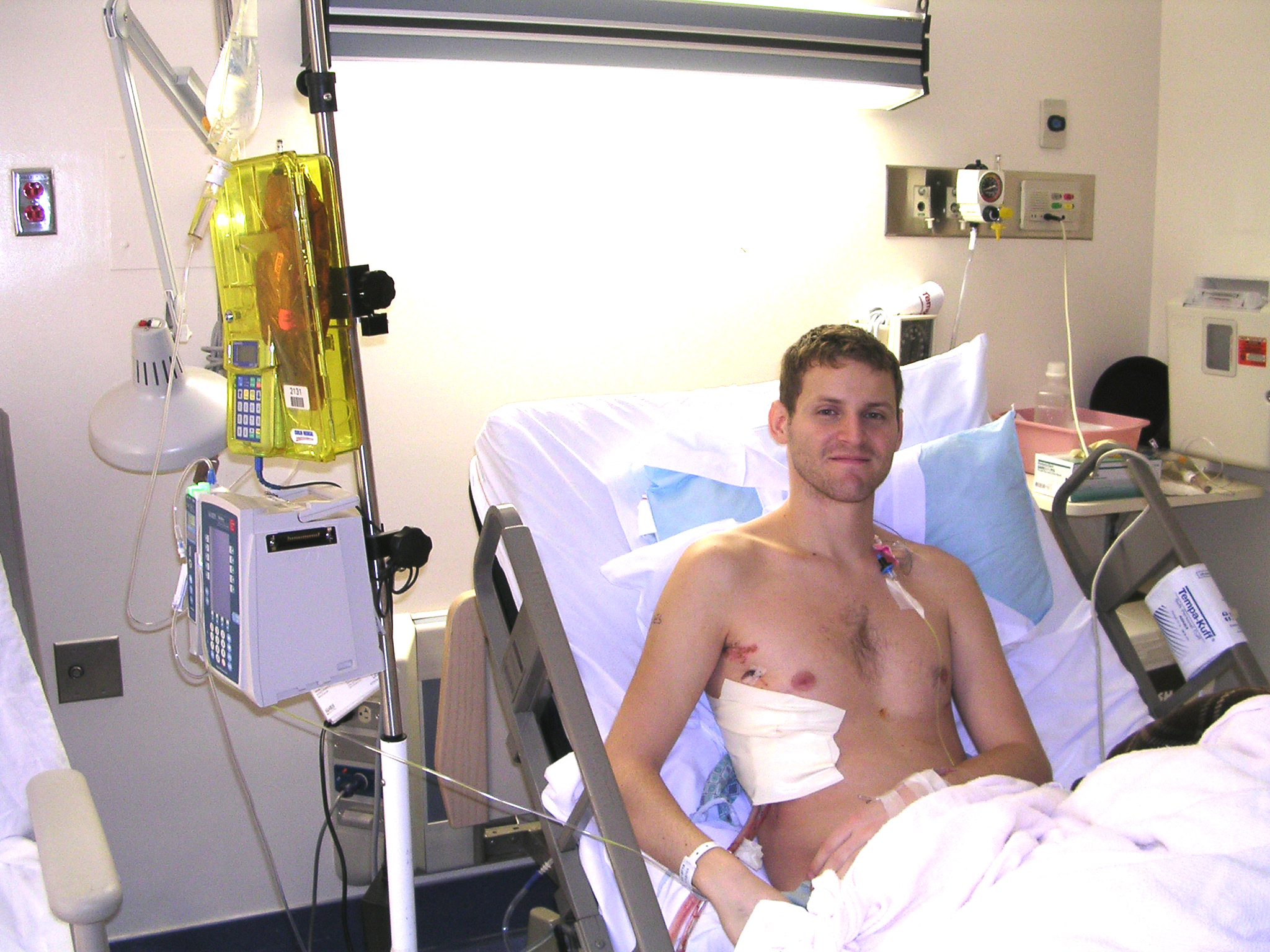
But their stories often touched my heart.. Dann Furia is one of these people.
Meet Dann Furia
A Pennsylvania resident in his mid-30’s, Mr. Furia has bleb disease. Over the years, these blebs have ruptured many times, requiring multiple chest tube placements and thoracic surgery.
Overview of spontaneus pneumothorax and treatment modalities.
There are multiple classifications of pneumothoraces – primary, secondary, iatrogenic, traumatic, tension etc. This article is a limited overview of the most common type(s) of pneumothorax, and methods of treatment.
What are blebs?
The lung is made up of lung tissue itself (consisting of alveoli, bronchi and bronchioles) and a thin, membranous covering called the pleura. This covering serves to prevent inhaled air from travelling from the lung to the area inside the thoracic cavity. ‘Blebs’ are blister-like air pockets that form on the surface of the lung. Bulla (or Bullae for pleural) is the term used for air-filled cavities within the lung tissue.
Who gets/ who has blebs and/or bullae?
Blebs and bullae may be related to an underlying disease process such as emphysema / chronic obstructive pulmonary disease, but they (blebs in particular) may also be found in young, healthy people with no other medical issues. Indeed, the ‘classic’ scenario for a primary spontaneous pneumothorax is a young adult male (18 – 20’s), tall and thin in appearance and no other known medical history who presents with complaints of shortness of breath or dyspnea.
Smoking, and smoking cannabis have been implicated in the development of spontaneous pneumothorax in young (otherwise healthy) patients.
Bullae, or air pockets within the lung tissue are more commonly associated with chronic disease processes such as chronic obstructive pulmonary disease (emphysema). It can be also part of the clinical picture in cystic fibrosis and other lung diseases.
When these blebs rupture or ‘pop’ inhaled air is able to travel from the airways to the thoracic cavity, creating a pneumothorax or lung collapse.
The symptoms of a pneumothorax depend on the amount of lung collapse and the baseline respiratory status of the patient. In young, otherwise healthy patients, the symptoms may be more subtle even with a large pneumothorax. In patients with limited reserve (chronic smokers, COPD, pulmonary fibrosis, sarcoidosis) patients may experience shortness of breath, dyspnea/ difficulty breathing, chest and chest wall pain. With large pneumothoraces or complete collapse of a lung, patients may become cyanotic, or develop respiratory distress.
In cases of pneumothorax caused by external puncture of the lung, or other traumatic circumstances, a patient may develop a life-threatening condition from a tension pneumothorax. This can happen with a simple, primary lung collapse from bleb rupture, but it is uncommon.
How is this treated?
Simple (or first-time) pneumothorax
Oxygen therapy – traditional treatment for small pneumothorax in asymptomatic or minimally symptomatic patients was oxygen via a face mask or non-rebreather. Much of the more recent literature has discredited this as an effective treatment.
Tube thoracostomy (aka chest tube placement) – a chest tube is placed to evacuate air from the thoracic cavity, to allow the lung to re-expand. The chest tube is initially placed to suction until the lung surface heals, and the lung is fully expanded. After a waterseal trial, the chest tube is removed.
Recurrent pneumothorax / other circumstances;
Blebectomy via:
VATS (video-assisted thoracoscopy)
Open thoracotomy or mini-thoracotomy
As we have discussed previously, the VATS procedure / open thoracotomy and mini-thoracotomy are not really stand alone procedures but are the surgical approaches or techniques used to gain entry into the chest. Using a VATS technique involves the creation of one or more ‘ports’ or opening for the use of thoracoscopic surgical tools, and a thoracoscope (or camera.) There are rigid and flexible scopes available; but most thoracic surgeons prefer the rigid scopes for better visibility and control of tissue during the operation[1].
blebs seen during VATS procedure
Open thoracotomy or mini-thoracotomy incisions may be used to gain access to the lung, particularly for resection of bullae (lung volume reduction) surgeries for the treatment of chronic disease.
During this procedure, fibrin sealants may be used. Investigational use of both radio-frequency and other ablative therapies have also been used (Linchevskyy, Makarov & Getman, 2010, Funai, Suzuki, Shimizu & Shiiya 2011**).
Pleurodesis may also be used – in combination with either tube thoracostomy or surgical resection. Pleurodesis can be performed either mechanically, chemically or both. Mechanical pleurodesis is accomplished by irritated the pleura by physical means (such as scratching or rubbing the pleura with the bovie scratch pad or surgical brushes. A chest tube also produces a small amount of mechanical pleurodesis as the tube rubs on the chest wall during patient movement.
Chemical pleurodesis is the instillation of either sterile talc or erythromycin to produce irritation or inflammation of the pleura. With bedside pleurodesis or tube thoracostomy pleurodesis, sterile talc is mixed with lidocaine and sterile water to create a talc slurry. (If you like your patient, carry it in your pocket for 10 – 20 minutes to allow the solution to warm to at least room temperature. This will help reduce the discomfort during instillation.) The mixture should be in a 60cc syringe or similar delivery device – shake briskly before use. The mixture is then instilled via the existing thoracostomy tube. The chest tube is clamped for 30 – 60 minutes (dwell time) and the patient is re-positioned every 10 to 20 minutes. Despite the lidocaine, the talc will produce a burning sensation, so pre-medication is desirable. This procedure has largely fallen out of fashion in many facilities. Post-pleurodesis, pleural inflammation may cause a brief temperature elevation. This is best treated with incentive spirometry, and pulmonary toileting.
Chemical pleurodesis can also be performed in the operating room. Loose sterile talc can be insufflated, or instilled using multiple delivery devices including aerosolized talc. As discussed in previous articles, pleurodesis can also be used for the treatment of pleural effusions.
Special conditions and circumstances related to Pneumothorax:
Catamenial pneumothorax – this a pneumothorax that occurs in menstruating women. It usually occurs on the right-side and is associated with endometriosis, and defects in the diaphragm. A related case study can be viewed here. Several recent studies suggest catamenial pneumothorax may be more common that previously believed and should be suspected in all women presenting with right-sided pneumothorax, particularly if pneumothorax occurs within 48 – 72 hours of menstrual cycle. This may be the first indication of underlying endometrial disease.
[1] Flexible scopes are usually preferred for GI procedures such as colonoscopy, where the camera is inserted into a soft tissue orifice. By comparison, the thoracic cavity with the bony rib cage is more easily navigated with the use of a firm instrument.
** I have contacted the primary authors on both of these papers for more information.
Like all materials presented on this site, this paper is presented for information only. It should not be considered medical advice or treatment. Also, all information provided is generalized information and (outside of clinical case presentations) is not intended to treat of diagnose any disease or condition. If you have questions about the content, please contact us. If you have medical questions, please consult your thoracic surgeon or pulmonologist.