Talking about Lung Cancer staging with Dr. Mitchell Magee

Discussing the classification and treatment of lung cancer according to the latest revisions (7th edition).

Medical City, Dallas, Texas USA

Sometimes location and timing is everything.  Since I can’t attend all of the great thoracic surgery conferences and events, sometimes I just have to wait for something closer to home.  But then again, “home” is a relative concept.

As a locum tenens provider, I travel around the country working in various hospital surgical programs on short-term contracts.  It’s an interesting and always changing life but one that allows me to pursue my love of thoracic surgery to the fullest.

For the next few weeks, Medical City in Dallas, Texas is my home, as part of the cardiothoracic surgery service.  It’s a return trip so it was nice to renew my acquaintance with the surgeons and staff of the CVICU and step-down units.

Today, as part of an ongoing continuing medical education program series, Dr. Mitchell Magee, of Southwest Cardiothoracic Surgeons gave an hour-long lecture entitled, “Lung cancer staging and evolving less invasive surgical treatment alternatives.”  The focus of the talk was the changes in lung cancer classification and staging in the 7th edition guidelines.  These revisions were proposed to replace previous versions which were based on a very small, select sample of patients at a single site.  In comparison, the new revisions are based on over 100,000 patients worldwide.

T, N, M

T – tumor

N – nodes

M – metastasis

This classification system has been in use since the 1940’s and has been revised several times to reflect our growing knowledge.  The latest revisions (7th edition) were released in 2012 after several years of research and debate.  (For more on this process, see “The science behind the 7th edition Tumour, Node, Metastasis staging system for lung cancer” by Marshall et al, 2012).

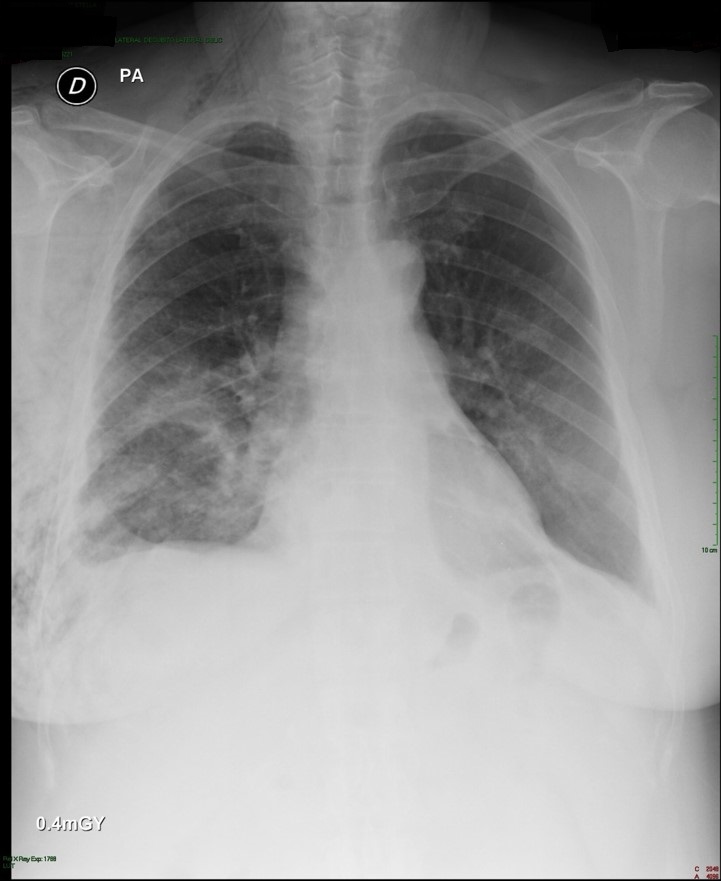
Dr. Magee discussed the most recent revisions and how these changes affect both the treatment recommendations and prognoses for our patients.  After reviewing these changes, he talked a bit about obtaining sufficient diagnostic information for accurate staging, including the role of EBUS, the new CT scan screening guidelines and the gold standard, mediastinoscopy.  He also discussed some of the limitations of PET/CT and other non-invasive diagnostic imaging.

Upstaged/ Downstaged

As part of these changes in the subclassification of tumors, 10 stages have been downstaged (meaning that previously in-operable cases may now be eligible for resection) and seven classifications have been upstaged – meaning that the cancers are now considered more advanced.

For example, patients with two separate tumors in the same lobe of the lung has been upstaged to T3.  Two different tumors in the same lung, but a different lobe is now T4 classification.

More specific

Some of the classifications have changed to make findings more specific.  For example, T1 staging has now been subdivided into T1a and T1b.

Any invasion of the pleura, including microscopic – is now T2 staging.

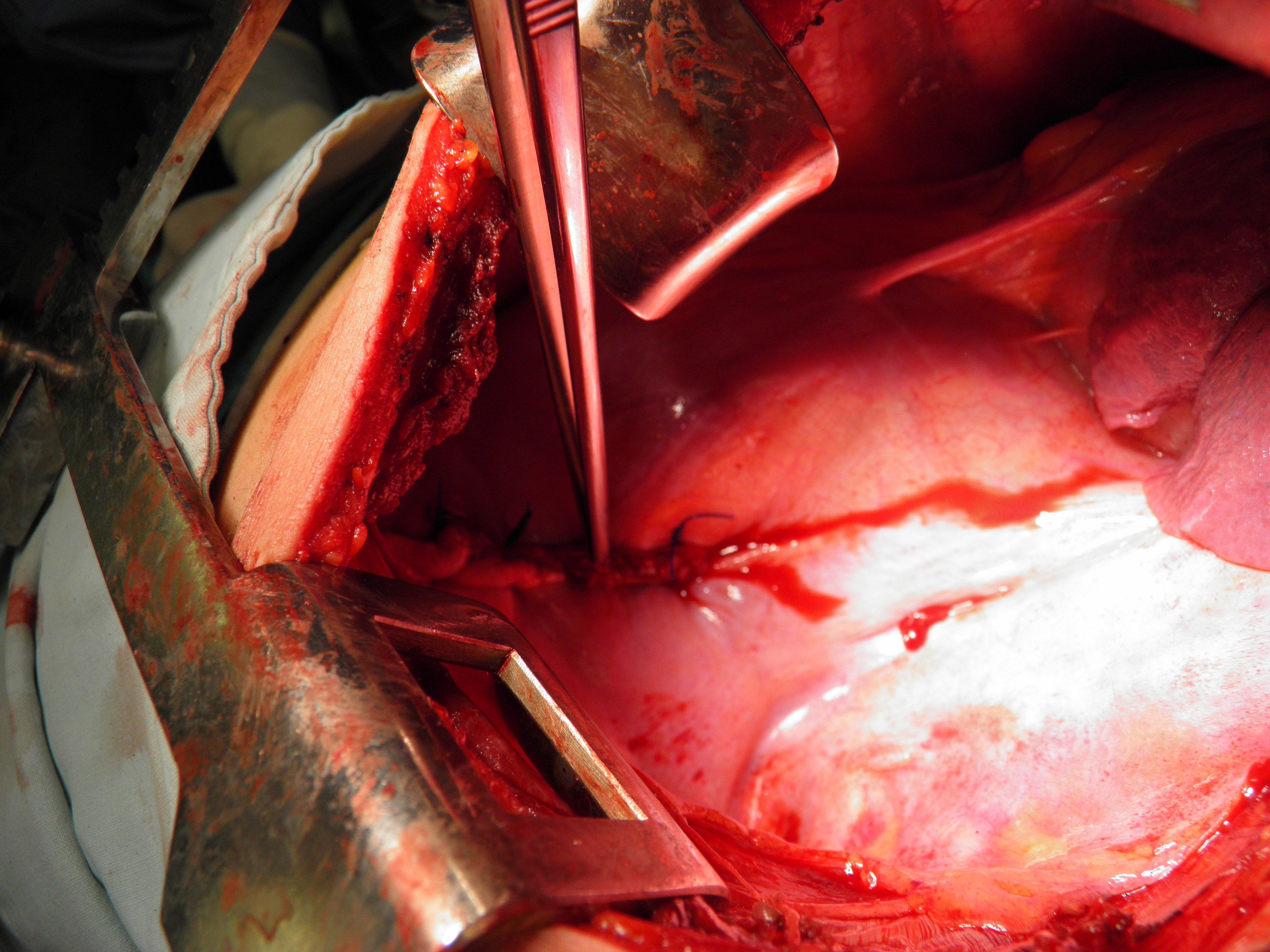
He concluded the presentation with a short overview of the history of surgical resection for lung cancer, and the evolution of surgical techniques from open thoracotomies with pneumonectomies to lung sparing procedures utilizing more minimally invasive techniques.

Despite these changes, the hallmarks of a successful cancer operation remain unchanged – the right operation for the individual patient, and the need to respect oncological principles, like surgical margins, and a through lymph node dissection.

Lymph node dissection/ node sampling

Node sampling remains a crucial part of the cancer staging process despite the advent of less invasive imaging studies due to it’s infaliable accuracy.  (There is either tumor tissue in the node or there isn’t, where as PET scan results can be false positive or false negative).

For this reason, tissue samples remain the gold standard of treatment and are the most accurate way to predict and prognosticate the extent of disease.

General rules regarding lymph node sampling are:

– More nodes are better.  The minimum acceptable number of nodes for accurate staging is at least SIX for at least THREE different stations.

A good way to remember the relationship between node stations and node status is that bode stations are determined by distance from mediastinum; meaning that node station 14  is more peripheral that node 2.

N1 nodes are stations 10 – 14

N2 nodes are the single digit nodes (2, 4, 7 etc.)

Lymph nodes used for diagnosis and staging.  Copyright Memorial Sloan-Kettering Cancer Center. Used with permission.
Lymph nodes used for diagnosis and staging. Copyright Memorial Sloan-Kettering Cancer Center. Used with permission.

References and additional suggested readingBaltayiannis N, Chandrinos M, Anagnostopoulos D, Zarogoulidis P, Tsakiridis K, Mpakas A, Machairiotis N, Katsikogiannis N, Kougioumtzi I, Courcoutsakis N, Zarogoulidis K. (2013).  Lung cancer surgery: an up to date. J Thorac Dis. 2013 Sep;5(Suppl 4):S425-S439. Review.  Free pdf.   Nice review article discussing the importance of staging for determining optimal  treatment for lung cancer, as well as the impact of the latest revisions to the (7th edition) TNM classification system.

IASLC Staging Handbook in Thoracic Oncology – a site-specific guide on the new TNM classification of thoracic malignancies. This publication is published in coordination with the 7th editions of the TNM Classification of Malignant Tumors/UICC and AJCC Cancer Staging Manual.

Goldstraw P, Crowley J, Chansky K et al. (2007). The
IASLC lung cancer project: proposals for the revision of the
TNM stage groupings in the forthcoming (seventh) edition of
the TNM classification of malignant tumours. J Thorac Oncol
2007; 2: 706-714. Figure 1.  Powerpoint slides TNM classification revisions for the 7th edition.

Quick and easy summary of the 7th edition classifications for Lung cancer staging – 7th edition Lung cancer staging pdf from the American Joint Commission on Cancer.

Lung cancer screening guidelines – screening questions for patients to determine if they need lung cancer screening.

Li S, Zheng Q, Ma Y, Wang Y, Feng Y, Zhao B, Yang Y. (2013).  Implications of False Negative and False Positive Diagnosis in Lymph Node Staging of NSCLC by Means of (18)F-FDG PET/CT.  PLoS One. 2013 Oct 25;8(10):e78552. doi: 10.1371/journal.pone.0078552.  Incidence of false negatives/ positives and who is most at risk for false findings.
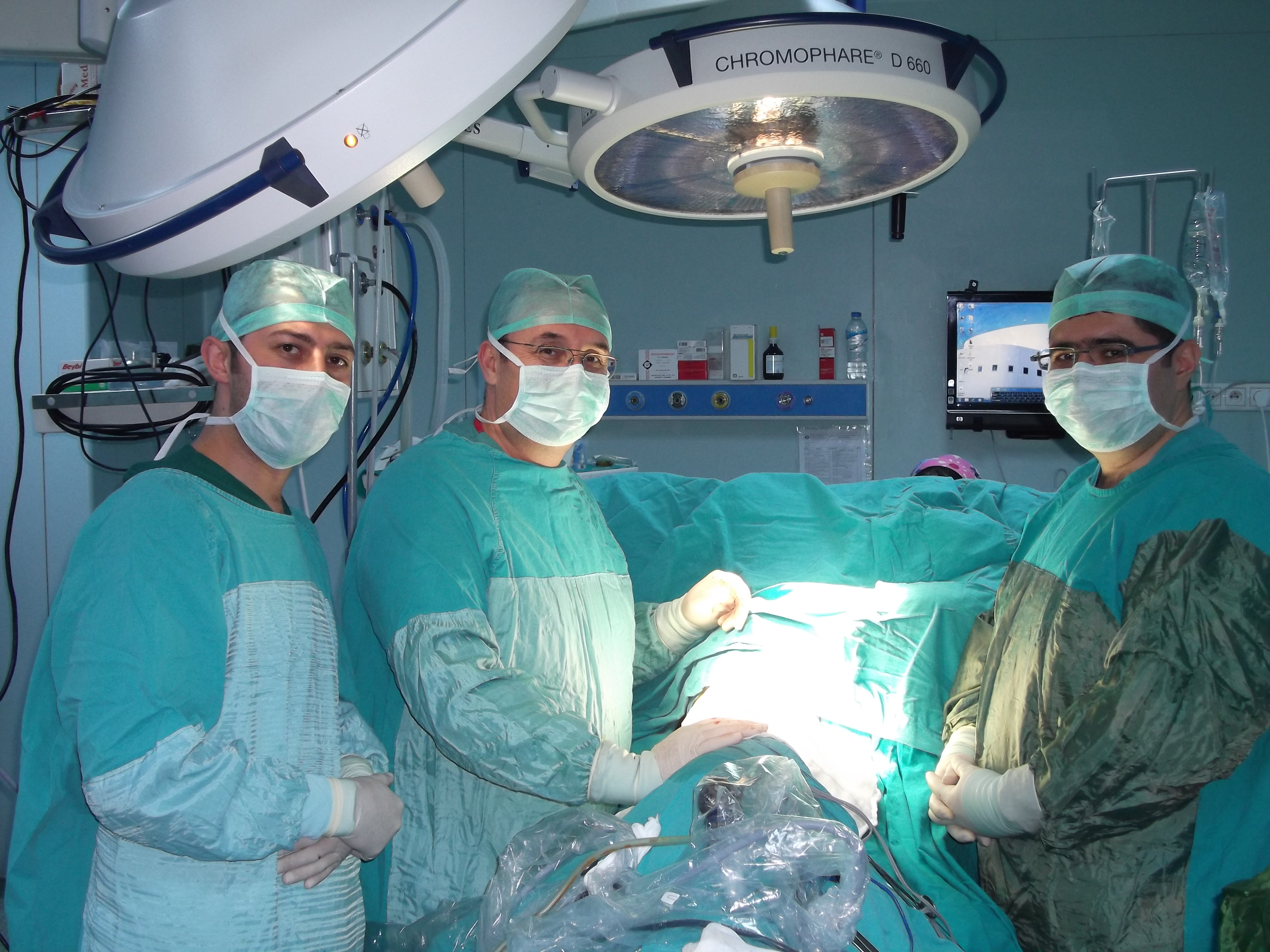
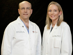
About Dr. Magee

Dr. Mitchell Magee, thoracic surgeon at Medical City - Dallas, Texas
Dr. Mitchell Magee, thoracic surgeon at Medical City – Dallas, Texas

Dr. Mitchell Magee is Surgical Director of Thoracic Oncology and the Minimally Invasive Therapy Institute for Lung and Esophagus at Medical City Dallas.  While his partner, Dr. Dewey focuses exclusively on cardiac surgeries like cardiac bypass, valve replacement, TAVR, LVADS and cardiac transplantation, Dr. Magee is the thoracic arm of the two surgeon Southwest Cardiothoracic Surgeons practice.  This means Dr. Magee is able to devote his time to a sizable portion of all of the esophageal tears, empyemas, mediastinal masses and lung pathology that a city the size of Dallas has to offer.

Dr. Mitchell Magee with Amber Bethea, PA-C
Dr. Mitchell Magee & Amber Bethea, PA-C

Dr. Magee is also part of the CLEAR Clinic at Medical City – which is the lung cancer screening center at the Medical City Dallas facility.

Southwest Cardiothoracic Surgeons

7777 Forest Lane, A307

Dallas, TX 75230

(972) 566-4866

Dr. Joseph Skoda

Dr. Joseph Skoda, Nihilist, Skeptic, Dermatologist.

stethoscope

 Why we auscultate and percuss: Dr. Joseph Skoda (1805 – 1881)

Not all of thoracic surgery’s founding fathers were surgeons.  In fact, one of the most important contributors to thoracic medicine, anatomy and physical examination, Dr. Joseph Skoda, was actually a dermatologist.

Walked to Vienna

The Czech born physician was also determined.  Stricken with tuberculosis (consumption) as a child, yet determined to follow in the footsteps of his older brother, Franz, he defied his parents’ wishes that he enter the priesthood. While he initially studied theology, his affinity for mathematics, physics and natural sciences led him to pursue medicine.  Of limited means, his education was financed by the beneficence of Madame Bischoff, the wife of a  wealthy local industrialist.

Thus, instead of entering the church, in 1925, he spent six days traveling by foot; walking from his native Pilsen (Bohemia) to Vienna to attend medical school.

After obtaining his doctorate in medicine from the University of Vienna in 1831, Dr. Skoda spent a year in his native Bohemia studying Cholera before returning to the famed General Hospital of Vienna as an unpaid assistant physician.  Over the next few years he worked in several different wards, including the Tuberculosis ward.

Multiple publications during the 1830’s*

During the early portion of his career, Dr. Skoda was a prolific author of medical publications such as “About percussion,” “About the Percussion of the Heart and the Sounds Originated by Heart Movements, and Its Application to the Investigation of Organs of the Abdomen”, “About the Diagnosis of Defects of Heart Valves” but it was his original research publication,  “A treatise on auscultation and percussion” for which he is best known. He revived previously  published but little known (or used) techniques of percussion and auscultation.  He promoted the use of the stethoscope for physical examination and developed much of the terminology used for diagnosing and describing cardiopulmonary conditions.

However, these publications did little to earn the respect or admiration of his colleagues.  Instead, he was demoted to ward physician for the insane ward as a punitive measure for disturbing patients with his methods of physical examination.  But this animosity was not unilateral in nature.

Despite his enthusiasm for anatomy and physical examination – Dr. Skoda was not generally well-liked by his colleagues.  In fact, he was appointed to several of his professional positions based on the recommendations of his close friend, Dr. Carl Freiherr von Rokitansky over the objections of his peers; including his appointment as a professor of the newly established “Modern” Medical School of Vienna. (This was a change in the traditional school of thought regarding medical education).

Some of their distaste may have stemmed from the fact that Dr. Skoda deviated from traditional dictates of the time and became the first professor to lecture in German instead of Latin.  He more likely earned their enmity due to his failure to be duly impressed by their therapeutic marvels, medicinal treatments and patent medicines.  Dr. Skoda was skeptical in regards to the actual therapeutic benefits to many of the medical treatments of the era, and frequently attributed the restoration of health to the fundamentals of healthy food, clean air and basic hygiene.

Therapeutic Nihilism

In an age where tinctures of mercury, laudanum and turpentine were popular remedies alongside bleeding, cupping and leeches, Dr. Skoda’s adage of “To do nothing is best in internal medicine” was probably more correct than his peers. This skepticism earned him the label of “therapeutic nihilist”  who disdained modern medical interventions but this is far from the truth.

He was more like the fictional television physician, “Dr. House” of his generation.

First pericardial aspiration

Despite his mistrust of traditional medical and pharmaceutical quackery, Dr. Skoda ventured to experiment with specific medical interventions such as pleural aspiration, and pericardiocentesis as well as use of newer investigational medications such as salicylates (which later became modern-day aspirin).  Dr. Skoda along with Dr. Franz Schul performed the first known pericardial aspiration in 1840.

Advancing physical examination and stethoscope use

Through the use of diagnostic tools like the stethoscope (which he promoted and refined after re-discovering and advancing the work of Viennese physician, Leopold Auenbrugger, as well as French physicians; Rene Laennec and Pierre Piorry), Dr. Skoda was instrumental in advancing physical examination as a tool for diagnosis.

Cardiac Murmurs and Adventitious Breath sounds

He first described and diagnosed the ‘drum-like’ sound of pneumonia, the Skodaic resonance of pleural effusions and supported the earlier work of Boullaud & Rouanet on cardiac sounds including the grades and distinguishing sounds of different murmurs. He gave us much of the terminology we use today, to describe rales, friction rubs, crepitus, bronchophony and voice conduction.

The next time you hear the tympanic sound during percussion [indicating a pleural effusion], remember this “Skodaic resonance” finding and Dr. Joseph Skoda.

* His major work in skin diseases and contributions to the specialty of dermatology begin in 1841.  He continued to make various contributions to the field of medicine until his death at the age of 76 due to cardiac disease.

 References

Davies MK, Hollman A.  (1997).  Joseph Skoda (1805-1881).  Heart. 1997 Jun;77(6):492.

Sakula, A. (1981).  Joseph Skoda 1805-81: a centenary tribute to a pioneer of thoracic medicine.  Thorax. 1981 Jun;36(6):404-11

JAMA editiorial, “Joseph Skoda, Physican Diagnostician” October 19, 1964.

Additional Resources

A Practical Guide to Clinical Examination: Lungs – University of California, San Diego.

Basics of lung percussion – Loyola University Medical Education Network

A Travelers Guide to the History of Biology and Medicine: Austria

The Auscultation Assistant – UCLA site with examples of heart and lung sounds

Single port thoracoscopy for diaphragmatic disorders

a report from Dr. Chin Hao Chen and his colleagues at Mackay Memorial Hospital on 21 cases of diaphragmatic plication via single and dual port thoracoscopy.

Dr. Chen and his colleagues at Mackay Memorial Hospital in Taiwan published a new article on their experiences using single and dual port thoracoscopy for diaphragm plication.

The report follows 21 cases from July of 2008 to December of 2011.   All 21 cases with left-sided eventrations.  11 were plicated using dual port thoracoscopy in the time period prior to January 2010.  In January of 2010, single port thoracoscopy became routine practice at Mackay Memorial.  The 10 subsequent cases were all performed by single-port thoracoscopy.

Surgical procedure:  The average surgical time between dual port and single port varied by ten minutes with dual port surgery taking longer, averaging 92 minutes. ( see Table 1 of original article).  2.0 silk suture was used for plication of the diaphragm.

Port placement: 

In cases using dual port thoracoscopy, the surgeons made the first port at the 7th ICS near the MCL with a second port at the 4th or 5th ICS along the anterior axillary line.

For single port cases, the sole port was 1.5 to 2.0 cm in length and was placed at the 6th ICS along the anterior axillary line.

Example of sutured diaphragm - (view  from thoracotomy)  Photo courtesy of Dr. Ochoa, 2011.
Example of sutured diaphragm – (view from thoracotomy) Photo courtesy of Dr. Ochoa, 2012.

At the conclusion of the VATS procedure for all patients, a single 24fr or 28fr chest tube was placed, and marcaine was administered as a intercostal block.  Patients were extubated prior to leaving the operating room.

The chest tube was removed on the first or second post-operative day.  Patients were discharged home following chest tube removal.  Post-operative pain scores were minimal, and there was no operative mortality.

The authors discuss surgical technique, and port location for a significant portion of the article.  Interested readers are advised to read the original for more details.

Discussion:

Interestingly, while much of the literature on diaphragmatic eventration focuses on early repairs of this condition (neonates and pediatric cases), all of the patients in this series were adults, with an average age of 54 – 55 years of age.  Both genders were represented; 15 women and 6 men, with an almost equal distribution among single and dual port cases.  (3 men in each group, 7 women in single port, 8 in dual port.)

Unlike traumatic diaphragmatic tear or rupture, diaphragmatic eventration is usually a congenital condition and may be asymptomatic.  It is often discovered incidentally after patients undergo radiographic studies for other conditions.  However, this condition may predispose patients to other conditions such as respiratory distress or dyspnea by compromising respiratory function on the affected side. In fact, the affected lung may appear tiny, and underdeveloped at the time of repair.

In Dr. Wu and Dr. Chen’s study, patients who underwent dual or single port thoracoscopy reported pain scores of four or less at 24 and 36 hours post-operatively.  Post-operative hospitalization was short, with patients being discharged on the first or second post-operative day, with no recurrences or mortality.

Reference Article: 

Hsin-Hung Wu, Chih-Hao Chen, Ho Chang, Hung-Chang Liu, Tzu-Ti Hung and Shih-Yi Lee (2013).  A preliminary report on the feasibility of single-port thoracoscopic surgery for diaphragm plication in the treatment of diaphragm eventration.  Journal of Cardiothoracic Surgery 2013, 8:224.  Provisional pdf of free full text article, with radiographs, color photographs.

Resources for Additional Information

Eventration of the diaphragm at Learning Radiology

A. P. Kansal, Vishal Chopra, A. S. Chahal, Charanpreet S. Grover, Harpreet Singh, and Saurabh Kansal (2009).  Right-sided diaphragmatic eventration: A rare entityLung India. 2009 Apr-Jun; 26(2): 48–50.

Radhiana M Y H, Mubarak MY. (2011). A case of focal eventration of left hemidiaphragm with transthoracic left kidney confused with a traumatic diaphragmatic hernia.  Med J Malaysia. 2011 Mar;66(1):60-1.  Case report.

Visouli AN, Mpakas A, Zarogoulidis P, Machairiotis N, Stylianaki A, Katsikogiannis N, Tsakiridis K, Courcoutsakis N, Zarogoulidis K. (2012).  Video assisted thoracoscopic plication of the left hemidiaphragm in symptomatic eventration in adulthoodJ Thorac Dis. 2012 Nov;4(Suppl 1):6-16.  Three port VATS in an adult.

CTSnet recognizes Dr. Diego Gonzalez Rivas

Dr. Diego Gonzalez Rivas receives recognition from the global network of cardiothoracic surgeons, CTSnet.

CTSnet.org, the largest global network of cardiothoracic surgery professionals has recently recognized Dr. Diego Gonzalez Rivas for his pioneering efforts in thoracic surgery.

a TEDtalk favorite

This comes on the heels of a recent TEDtalk on Dr. Gonzalez and the process of innovation in surgery. During this 18 minute talk, Dr. Gonzalez talks about his own experiences in surgery.

Dr. Diego Gonzalez Rivas, a “fan” favorite here at Cirugia de Torax, is at the forefront of the field due to his contributions to minimally invasive surgery in the area of single-port thoracoscopy.

The dynamic young Spaniard has been making headlines over the last decade as he introduced and then refined the single port surgical technique.  He and his colleagues, Dr. Maria Mercedes
de la Torre Bravos and Dr. Ricardo Fernandez Prado at the Minimally Invasive Thoracic Surgery Unit (UCTMI) in Coruna, Spain have successfully used this technique on thousands of patients, for a wide variety of procedures including sleeve lobectomies, pneumonectomies, bilobectomies and other complex procedures.

Dr. Gonzalez-Rivas demonstrates single port thoracoscopy at the National Cancer Institute in Bogota, Colombia
Dr. Gonzalez-Rivas demonstrates single port thoracoscopy at the National Cancer Institute in Bogotá, Colombia

Despite this widespread fame, Dr. Gonzalez Rivas remains unaffected and approachable.  He spends much of his time in operating rooms around the world, teaching his technique to his peers.  Next week, he heads to Guangzhou, China.

Pleural fluid cytopathology

How to prepare a proper specimen for pleural fluid cytology & cytopathological analysis.

Pleural Fluid Cytopathology

Pleural fluid analysis is more than a typical ‘rounds’ question for interns and students.  This fluid contains important indicators of disease status.  Who among us hasn’t memorized pH levels, glucose and protein values?  (For a discussion on transudate versus exudate effusions, see the Medscape article by Jeffrey Rubins below.)

While pleural fluid analysis can be used to assist in the differential diagnosis of multiple conditions; pleural fluid cytopathology is often ordered when a more sinister condition like metastatic cancer is suspected.  Therefore, it is especially important for clinicians to ensure that pleural fluid cytopathology samples are collected, and sent in the most efficient and effective manner possible.  While there are few written guidelines regarding this process, here are some helpful tips based on interviews with several pathologists and the available literature.

Biopsy is best but fluid analysis is still helpful

While the gold standard for diagnosis is always tissue biopsy (in this case pleural tissue biopsy), this does not mean that cytopathological analysis is completely unnecessary.  In many cases, this fluid analysis gives a first look that aids in the diagnosis and staging of disease.  It is particularly useful for patients undergoing thoracentesis procedures, particularly when thoracentesis is performed in lieu of a more invasive procedure such as VATS (which allows for direct tissue biopsy.)

But do I still need to do a biopsy if the fluid analysis is negative?

However, there is often a catch-22 in the use of pleural fluid pathology which can lead to some confusion among patients and providers.  This catch-22 is related to the sometimes variable reliability of pleural fluid cytopathology for diagnosis of malignancy.  This means that the results aren’t always accurate.  As anyone in thoracic surgery can tell you, there have been numerous times when the fluid analysis results are reported as negative (for malignancy) even when the surgeon is (literally) staring at a pleural tumor in the operating room.  This means that a negative pleural fluid cytopathology result can not be used to rule out malignancy.

However, when the fluid is positive, it may save the patient from an additional procedure*.

Cytology versus cytopathology

“Cytology” is the generic term for the study of cells.  Cytopathology is the actual pathological investigation of free cells and tissue fragments, often for the diagnosis or treatment of cancer.

When used clinically, cytopathology is often used to distinguish between other more basic studies of bodily fluids or tissues.  This in-depth cellular examination is more critical in many cases than basic pleural fluid analysis.  This examination may include identification of immunological factors and tumor markers.  This is one of the tests that clinicians use to try and answer the question,“Is it cancer?”.  However, the answer is not always as straight forward.

Reliability and Predictive Value

This question is difficult to answer due to sometimes variable prognostic value of the fluid itself.  Even under the best of circumstances, reliability of this test (like most diagnostics) is less than 100%.  Different studies calculate the accuracy of pleural fluid analysis at detecting cancer vary wildly;anywhere from 10 to 80% has been reported in the literature with false negatives as the most frequent error (when discussing sensitivity and specificity). However, poorly prepared specimens may contribute to false results as well.

Ensuring optimal results by obtaining proper specimens

 Over the years, during different discussions with multiple pathologists as well as laboratory technicians, a common theme has emerged regarding the use of pleural fluid for pathology analysis.  Several of these individuals remarked that obtaining an accurate diagnosis was often difficult due to improper or suboptimal preparation of the pleural fluid, in addition to characteristics of the fluid itself.  What constitutes a ‘proper’ or ‘optimal’ pleural fluid is still (among lab technicians and pathologists) up to debate, but here are some general guidelines:

1. Send it all.

Due to the nature of pathology analysis which replies of the presence and identification of malignant cells within the fluid itself, a larger fluid specimen provides for a better sample.  When thoracentesis/ VATS or other drainage is being performed, and this yields 2 liters of fluid – send all two liters.  Don’t select out the first 25ml in a urine specimen cup, send it all.

There are no set guidelines for the amount of fluid necessary for cytopathology analysis. While malignancies have been successfully detected in amounts as small as 4ml, the rationale behind providing larger samples has been explained as ‘increasingly the likelihood of detecting the presence of cells indicative of malignancy’.

While the amount of fluid needed is currently up for debate among pathologists, sending too little fluid may result in a missed diagnosis, whereas an overabundance of fluid is more of an inconvenience to lab technicians.

Be sure to include the last frothy bits, which often contain more sediment/ cellular material than fluid recovered at the beginning of the sample.  (The content of this fluid may even vary due to the patient’s position – which is another reason to take a larger sample.) In a conscious patient, this may mean several minutes of discomfort, but encourage patients to take deep breaths, and cough so that as much fluid as possible can be removed.  (In patients with very large effusions, this may be a lengthy process as ‘short breaks’ are taken during the procedure to accommodate for fluid shifts.  This brings us to # 2.

2.  Keep it fresh: Talk to the lab about whether you should consider adding an additive like heparin or EDTA to your sample at the time of collection to prevent the degradation of cells.  Depending on when / where your sample is collected and sent – there may be significant delays in the processing of the collected sample.  Many pathologists report that after 4 hours there are significant changes in untreated pleural fluid kept at room temperature.

Consider this as you gather your sample;

–          Did you leave it in the patient’s room for the nursing staff to deliver?

–          Is it possible it may sit for several hours before arriving to the lab?

–          Is the lab well-staffed or will the fluid sit waiting for analysis by overworked, and stressed employees at a lab that may be inundated with many more urgent requests?

Guzman et al. (1992) and other researchers found that with the addition of EDTA to pleural fluid specimens, tumor cells were easily identified even after four days of storage.

Even if your facility doesn’t provide EDTA for your specimens, it’s a good bet that sending a syringe full of fluid from the bottom of a week-old pleurovac is probably not your best bet.

3.  Eliminate errors: Don’t make them guess!

Always personally label fluid and tissue samples completely with the patient’s name, reference number (as used by your institution), body site (ie. Right pleural space) as well as the ordering clinician’s name.  Include your phone number if you want to be called with the results or questions.

On the actual order, or lab requisition, provide additional information including patient symptoms, and pertinent history (ie. 63 year old with 40+ pk years of smoking, and history of asbestos exposure in Navy shipyards, now presents with pleural effusion, chest pain and 25 pound weight loss.)  Provide any special instructions as needed.   This allows the pathologist examining the patient’s specimens to correlate clinical history, symptoms and other available diagnostics with cellular findings and stains.

4.  Now do it again.  If the patient develops a second pleural effusion, go ahead and send that fluid too – particularly if the first sample was non-diagnostic.

*Depending on the patient’s clinical status/ symptomology.  As mentioned in a previous post, many patients with malignant pleural effusions may undergo additional procedures at some point in time for palliation of symptoms.

References

 American Society of Cytopathology – a great resource for interested readers.  The website also contains a ‘virtual slide atlas’ which includes case studies and several slides showing pleural fluid cytopathology. Click here for the case study of a 60 year old with pleural effusion.

Antonangelo L, Capelozzi VL. (2006). Collection and preservation of the pleural fluid and pleural biopsy. J Bras Pneumol. 2006;32 Suppl 4:S163-9. Portuguese.  These Brazilian authors from the University of Sao Paulo discuss the proper collection of pleural fluid specimens.  In this article, the authors make recommendations for the collection, storage and examination of pleural fluid for a variety of laboratory and microscopic tests.

Brandstetter RD, Velazquez V, Viejo C, Karetzky M. (1994). Postural changes in pleural fluid constituents. Chest. 1994 May;105(5):1458-61.

Guzman J, Arbogast S, Bross KJ, Finke R, Costabel U (1992).  Effect of storage time of pleural effusions on immunocytochemical cell surface analysis of tumor cells. Anal Quant Cytol Histol. 1992 Jun;14(3):203-9. No free full text available.

Porcel JM.  (2011).  Pearls and myths in pleural fluid analysis. Respirology. 2011 Jan;16(1):44-52.  Porcel advocates for smaller volumes, but an ‘inadequate sample’ should never be a reason for a missed diagnosis.  He also advises the addition of an additive if there are any anticipated delays (4+hours) in specimen processing.

Salyer WR, Eggleston JC, Erozan YS. (1975).  Efficacy of pleural needle biopsy and pleural fluid cytopathology in the diagnosis of malignant neoplasm involving the pleura. Chest 1975 May, (5) 536-9.  Classic article on the predictive value of pleural fluid cytopathology. A  pdf of Salyer et al is available here.

Rubins, J. (2013).  Pleural effusion workup.  From Medscape/ Emedicine.com.  Pleural Effusion Workup pdf version.

Additional Resources

Shidham, V. B. & Falzon, M. (2010). Serous cavities.  Chapter 3 in  Diagnostic Cytopathology: Expert Consult: Online and Print (2010).  Grey & Kocjan (Eds).   Elsevier Health Sciences.

Chung et. al. Optimal timing of thoracoscopic drainage and decortication for empyema

A new Korean study looks at the best time to perform surgical interventions on patients with empyema thoracis.

In a recent issue of the Annals of Thoracic Surgery, Chung et al. attempt to answer the long-standing question over the optimal timing of surgical intervention for empyema.  This has been a long-standing debate among surgeons and other medical specialists.  Despite advances in thoracic surgery (such as video-assisted thoracoscopy) as well as wealth of surgical commentary suggesting earlier intervention, surgery is usually considered a last resort; often after weeks or months of antibiotics, tube thoracoscopy or fibrinolytic therapy.

Chung et. al raise the stakes for this discussion with their research into this issue.  In “Optimal timing of thoracoscopic drainage and decortication for empyema,” over the course of 8 years, the authors compared outcomes such as chest tube duration, number of persistent air leaks and overall length of stay by the time interval of symptomology and surgical treatment.

Why this is important

As discussed in previous posts, empyema is a serious infection with a mortality rate of approximately 1 in 5 patients.  Empyema is frequently found in the chronically ill, debilitated or malnourished.  Delays in definitive treatment (surgical decortication) plays a role in the high rate of mortality with this condition; with increased hospital stays, and increased patient debility as the patient continues to sicken, and consume their health reserves while less effective treatments are initiated.

What is early?  What is optimal?

Early surgical intervention has been theorized since the turn of the 20th century to lessen morbidity and mortality, however there have been very few actual studies to address the question of timing.  This study, while small, directly compares outcomes in patients receiving VATS at different points in the infectious process/ illness spectrum.

Using a retrospective study design, patients from April 2004 to March 2012 were subdivided into three different time intervals; symptoms for less than 2 weeks, 2 to 4 weeks and patients with symptoms persisting longer than 4 weeks prior to surgery.  Of the 128 empyema patients, the vast majority (93.7%) were treated with VATS, with only 8 patients undergoing open procedures like thoracotomy.

Who:

Patients included in the study met criteria set forth in 2000 by the American College of Chest Physicians for diagnoses of Empyema and Loculated pleural effusions with thickened parietal pleura.  Patient symptomology including symptoms such as dyspnea, persistent fever and sepsis were also taken into account when determining eligibility for surgical management.

Patients by intervals:

Less than 2 weeks (group 1) : 73 patients

2 to 4 weeks (group 2) : 43 patients

More than 4 weeks (group 3) : 14 patients

The vast majority of patients were male, with only 14 female patients in this study – spread throughout the groups.  Additional variables such as age and pre-existing and co-morbid conditions (diabetes, liver disease, TB or other lung disease, etc.) were also collected*.   The percentage of patients in each group who had undergone additional treatments for empyema pre-operatively (thoracentesis, antibiotics, etc.) was similar in all groups for antibiotics (ranging from 93% in group 1 to 100% of group 3) with around a quarter of both group 1 (24.6%) and group 3 (28.6%) requiring immediate surgery due to deteriorating status.

The 8 thoracotomy patients were used as a comparison group to evaluate the effectiveness of VATS for chronic empyema.  All eight open surgical had pre-operative empyemas of 4 weeks duration or more.

What was compared/ measured?

While patient pre-operative characteristics were collected and analyzed as part of the evaluation of the empyema groups, the main surgical outcome measures were:

–          Duration of procedure: Shorter in group 1 (average 100 minutes) versus group 2 (125 minutes) and group 3 (138 minutes).

–          Chest tube duration – shorter in groups 1 (6.92 days) and 2 (7.45 days) than group 3 (10 days).

–          Length of overall post-operative hospitalization: shorter in group 1 (9.49 days) and 2 (9.73 days) compared to group 3 (13.5 days).

–          Length of post-operative ICU stay: no significant difference

Other surgical outcomes

Overall post-operative mortality was zero.  There was no incidence of post-operative respiratory failure.

Re-operations/ Additional procedures:

Group 1 (73 patients) 2 patients with treatment failure/ empyema reoccurrence requiring re-operation with VATS (2.7%).

1 patient without complete resolution with VATS, required conversion to open decortication.

1 patient that developed a contralateral pleural effusion that required chest tube placement.

Total re-operations: 3 (4.0%)

Group 2: No re-operations.

Group 3 (14 patients)

1 patient required re-operation with VATS for treatment failure.

Prolonged air leak

Group 1: 2 patients (2.7%)

Group 2: 1 patient (3%)

Group 3: 4 patients (28.6%)

 Study Weaknesses

The biggest weaknesses in this study are the small number of participants in group 3, and the lack of a control group.

Small numbers = reduced strength of findings

How much more powerful would similar results be, had the numbers of participants in all the groups been equivalent?  For example, if Chung et al. presented data showing that air leaks occurred in over 28% of patients with older empyemas (group 3) in comparison with groups 1 (2.7% and group 2: (3%) if these groups had been equally populated, would be a much stronger argument for earlier intervention.

As it is, with just 14 participants in group 3, 28.6% is less of a dramatic finding than false precision from small numbers.  However, it serves as a credit to Dr. Chung and his medicine colleagues, as clinicians, that the majority of these patients received definitive treatment at earlier stages.

Lack of control group

Had researchers included patients who were treated only by non-surgical methods (up to 2 weeks duration) and compared the overall length of stay, incidence of respiratory failure, ICU days and mortality in these patients, the data would have had an increased impact.  However, questions remain regarding the use of VATS versus the current standards of treatment in this ‘early’ group.  While we can partially speculate that treatment failures of patients in this group represent later patients (i.e. Patient fails treatment and is referred for surgery and becomes part of group 1, 2 or 3), the missing information may have represented a crucial factor to drive the decision-making process.

For example, how many patients in that same time period, presented with early empyemas and:

–          Received antibiotics and recovered?

–          Were admitted to the hospital and died of respiratory failure/ sepsis etc. without ever making it to surgery?

But, presumably, the difficulties of collecting this data retrospectively were prohibitive.  However, does the high rate of immediate surgery in group 1 due to patient deterioration serve as a partial stand-in for this data?

Conclusions

 While further study is warranted to determine the optimal time for surgical intervention in empyema thoracis, this study does an adequate job at demonstrating the benefits of earlier surgical intervention.  While there was no mortality in any of the groups, patients who were operated on 4 weeks or more after being diagnosed/ demonstrating symptoms of empyema, required longer operations, developed more persistent air leaks post-operatively, with longer chest tube duration and longer overall hospital stays.

Further research in this area could include the use of experimental algorithms and protocols to ‘fast-track’ patients with loculated effusions/ empyemas to surgical decortication in an attempt to replicate or further demonstrate improved patient outcomes with earlier surgical intervention.  These algorithms would explore the use of surgery as a first-line treatment with adjuvant antibiotics independent of thoracentesis, tube thoracostomy.

*Full information is provided within several tables in the original article.

Chung, Jae Ho et al. (2013).  Optimal timing of thoracoscopic drainage and decortication for empyema.  Annals of Thoracic Surgery, 2013.

Early ambulation after lung surgery: How early?

Dr. Khandhar of Inova Fairfax Hospital in Falls Church, Virginia and early ambulation after lung surgery

One of the critical benchmarks of recovery from thoracic surgery is early ambulation (walking) after surgery – but “How early?” is a frequently encountered question.

Now, Dr. Sandeep Khandhar, thoracic surgeon of Inova Fairfax Hospital in Falls Church, Virginia aims to answer this question.

The answer, Dr. Khandhar reports is : Within 1 hour of extubation.

In a recent article by Zosia Chustecka for Medscape, she summarizes Dr. Khandhar’s recent study on post-operative ambulation in thoracic patients.  Dr. Khandhar presented these findings at the 2013 World Conference on Lung Cancer this month.

In this study, involving 750 patients who were given a goal of walking 250 feet within an hour after extubation.  In this 3 year project, only 10% of patients were unable to  walk within one hour after extubation.  60% of patients were able to walk the full distance of 250 feet within an hour of extubation.

In these patients, early mobilization was associated with a significant reduction in length of stay: from 3 to 5 days down to just 1.6 days, as well as a decreased need for intravenous narcotics post-operatively.

We have contacted Dr. Khandhar for additional information about this study.

Chustecka, Z. (2013). Lung Cancer Patients Up and Walking Within Hour of Surgery.  Medscape, 28 October 2013.    [Medscape requires a free subscription to review articles and news].

16th National Continuing Education Forum in General Thoracic Surgery

Report from the recent 16th National Forum on Thoracic Surgery in Shanghai, China.

Shanghai Pulmonary Hospital, Tongji University

Shanghai, China

October 18th – 19th, 2013

Dr. Gonzalez Rivas demonstrates the single port technique during a live surgery presentation
Dr. Gonzalez Rivas (second from the left) demonstrates the single port technique during a live surgery presentation

As readers know, we strive to feature information about thoracic surgery from around the world.  This report on the 16th National Continuing Education Forum in General Thoracic Surgery comes from featured speaker, Dr. Diego Gonzalez Rivas of Coruna, Spain.

Some of the biggest names in thoracic surgery were in attendance, to present their research and surgical techniques to a crowd of over 600 Chinese thoracic surgeons. The lectures (and live surgery) were also broadcast across China.

World-renown thoracic surgeons at the 16th National Forum in Shanghai, China

Invited International Speakers included:

Dr. G. Alexander Patterson, thoracic surgeon/ lung transplant from the Washington School of Medicine in St. Louis, Mo. (USA).  Dr. Patterson gave a lecture on clinical experiences and advances in Lung Transplantation.  He also lectured on pancoast tumors.

Dr. Claude Deschamps, French Canadian thoracic surgeon and Chair of Surgery at the Mayo Clinic, Rochester, MN (USA). Dr. Deschamps talked about the use of anti-reflux surgery.

Dr. Gaetano Rocco, of the National Cancer Institute in Naples, Italy.  Dr. Rocco talked about advances in chest wall reconstruction.  He gave another lecture on uniport surgery.

Dr. Alan Sihoe from the University of Hong Kong discussed management of air leaks.

Surgeons from Taiwan and mainland China presented on a variety of topics including tracheal surgery, management of empyema, sympathectomy for hyperhidrosis and surgical treatment of tuberculosis.  (The full list of speakers and topics presented is available here*.)

Conference Spotlight: Single port surgery 

But the focal point of the forum was single port (uniportal) surgery.  Saturday (the 19th) was devoted to lectures and demonstrations of the single port thoracoscopic technique, including live surgical demonstrations performed by Dr. Diego Gonzalez Rivas.  His live surgery presentation was viewed by 500 surgeons at the conference as well as hundreds of other surgeons via a live feed.

Dr. Gonzalez Rivas demonstrates the uniport technique in Shanghai, China
Dr. Gonzalez Rivas demonstrates the single port (uniport) technique in Shanghai, China

Thank you to Dr. Gonzalez Rivas for his submission.  We welcome reports, photographs and discussions on recent and upcoming thoracic surgery conferences.  If you have a meeting, paper or presentation to share, please contact us at k.eckland@gmail.com

*Information is translated from Mandarin using google software with some obvious translational errors, particularly names of several of the Chinese surgeons.

VGTT: video-guided tube thoracostomy

Dr. Chin Hao Chen revisits one of the basic procedures in thoracic surgery: Chest tube placement

Even Hippocrates placed chest tubes or the history of tube thoracostomy

Chest tube placement has been performed since ancient Greek times.  Early physicians, including Hippocrates himself, performed (and wrote about) the use of tube thoracostomy for the treatment of lung abscesses and empyema.  Often this procedure is performed using a ‘blind approach’ based entirely on external anatomic features (intercostal spaces) and a fundamental knowledge of internal and chest wall anatomy.  Over the years, surgeons have developed guidelines to this technique using palpation/ and other tactile information but none of these techniques challenged initial insertion technique.

With any blind procedure, there is a risk of inadvertent injury due to the lack of visualization, particularly in patients with previous thoracic procedures or infections (adhesions), or when performed by less experienced staff.

Direct visualization during this procedure (akin to VATS) may lessen this risk.  However, little has been published on alternatives to the traditional technique.

VGTT: video-guided tube thoracostomy

Our latest post comes directly from Dr. Chih-Hao Chen at Mackay Memorial Hospital in Taiwan.

Dr. Chen presents a video clip demonstrating video-guided tube thoracostomy (VGTT), a technique used to avoid tube-related injury during the course of tube thoracostomy (versus blind insertion).  This visualization technique is helpful particularly when performed by inexperienced staff, such as residents or in emergent situations.

A complete description of this technique was recently published in the Annals of Thoracic Surgery.

Chen, et. al (2013). Video-guided tube thoracostomy with use of a nonfiberoptic endoscope. Ann Thorac Surg, 2013;96: 1450-5.  Articles commentary also available.

This paper describes the technique as well as discussing the clinical experience of Dr. Chen and his team in applying this technique to several patients.

Dr. Chin-Hao Chen is a thoracic surgeon at Mackay Memorial Hospital in Taiwan.  Dr. Chen is a frequent and valued contributor here at Cirugia de Torax.  He has provided several case studies as well as articles and videos on surgical techniques.

Case Report: Hilar mass resection using single port thoracoscopy with Dr. Diego Gonzalez – Rivas

in the operating room with Dr. Diego Gonzalez Rivas for single port thoracoscopic (uniportal) surgery.

Hilar mass resection using single port thoracoscopy with Dr. Diego Gonzalez – Rivas

K. Eckland & Andres M. Neira, MD

Instituto Nacional de Cancerlogia

Bogota, Colombia

Surgeon(s): Dr. Diego Gonzalez Rivas and Dr. Ricardo Buitrago

Dr. Diego Gonzalez Rivas demonstrates single port thoracoscopy

Case History:

59-year-old female with past medical history significant for recurrent mediastinal mass previously resectioned via right VATS.  Additional past medical history included prior right-sided nephrectomy.

Pre-operative labs:

CBC:  WBC 7230   Neu 73%  Hgb:14.1  Hct 37  platelets 365000

Pt 12.1  / INR1.1  PTT: 28.3

Diagnostics:

Pre-operative CT scan: chest

edited to preserve patient privacy

Procedure:  Single port thoracoscopy with resection of mediastinal mass and lymph node sampling

After review of relevant patient history including radiographs, patient was positioned for a right-sided procedure. After being prepped, and draped, surgery procedure in sterile fashion.  A linear incision was made in the anterior chest – mid clavicular line at approximately the fifth intercostal space.  A 10mm port was briefly inserted and the chest cavity inspected.  The port was then removed, and the incision was expanded by an additional centimeter to allow for the passage of multiple instruments; including camera, grasper and suction catheter.

Dr. Gonzalez Rivas and Dr. Ricardo Buitrago at National Cancer Institute

The chest cavity, pleura and lung were inspected.  The medial mediastinal mass was then identified.

instruments

As previously indicated on pre-operative CT scan, the mass was located adjacent and adherent to the vessels of the hilum.  This area was carefully dissected free, in a painstaking fashion.  After freeing the mediastinal mass from the hilum, the remaining surfaces of the mass were resected.  The mass was fixed to the artery pulmonary and infiltrating it) .  The mass was removed en-bloc.  Care was then taken to identify, and sample the adjacent lymph nodes which were located at stations (4, 7 and 10).

GonzalezRivas 051

Following removal of the tumor and lymph nodes, the area was re-inspected, and the lung was re-inflated.  A 28 french chest tube was inserted in the original incision, with suturing of the fascia, subcutaneous and skin layers.

closing the single port incision

Hemostasis was maintained during the procedure with minimal blood loss.

Patient was hemodynamically stable throughout the case, and maintained appropriate oxygen saturations.  Following surgery, the patient was awakened, extubated and transferred to the surgical intensive care unit.

Post-operative:  Post-operative chest x-ray confirmed appropriate chest tube placement and no significant bleeding or pneumothorax.

Immediate post-operative film (chest tube visible)

Patient did well post-operatively.  Chest tube was discontinued on POD#2 and discharged home.

PA & LAT films on post-operative day 2

pod2

Discussion: Since the initial published reports of single-port thoracoscopy, this procedure has been applied to an increasing range of cases.  Dr. Gonzalez and his team have published reports demonstrating the safety and utility of the single-port technique for multiple procedures including lobectomies, sleeve resections, segmentectomies, pneumonectomies and mediastinal mass resections. Dr. Hanao Chen (Taiwan) has reported several successful esophagectomies using this technical, as well as bilateral pleural drainage using a unilateral single-port approach.

Contrary to popular perception, the use of a single-port versus traditional VATS procedures (three or more) is actually associated with better visibility and accessibility for surgeons.  Surgeons using this technical have also reported better ergonomics with less operating fatigue related to awkward body positioning while operating.

The learn curve for this surgical approach is less than anticipated due to the reasons cited above, and has been cited at 5 to 20 cases by Dr. Gonzalez, the creator of this approach.

The main limitations for surgeons using this technique is often related to anticipated (but potentially unrealized) fears regarding the need for urgent conversion to open thoracotomy.  In reality, many of the complications that may lead to urgent conversion, such as major bleeding, are manageable thoracoscopically once surgeons are experienced and comfortable with this approach.

Dr. Gonzalez and his colleagues have reported a conversion rate of less than 1% in their practice.  Subsequent reports by Dr. Gonzalez and his colleagues have documented these findings.

Other barriers to adoption of this technique are surgeon-based, and may be related to the individual surgeon’s willingness or reluctance to adopt new techniques and technology.   Many of these surgeons would be surprised by how this technique mimics open surgery.

The successful adoption of this technique by numerous thoracic surgery fellows shows the feasibility and ease of learning single-port thoracoscopy by surgeons interested in adopting and advancing their surgical proficiency in minimally invasive surgery.

The benefits for utilizing this technique include decreased length of stay, decreased patient discomfort and greater patient satisfaction.

References/ Additional Readings

Bertolaccini, L., Rocco, G., Viti, A. & Terzi, A. (2013).  Surgical technique: Geometrical characteristics of uniportal VATS.  J. Thorac Dis. 2013, Apr 07.  Article from thoracic surgeons at the National Cancer Institute in Naples, Italy explains how the geometric advantages of uniportal VATS improves visibility and spatial perception over traditional VATS, and mimics open surgery.

Calvin, S. H. Ng (2013). Uniportal VATS in Asia.  J Thorac Dis 2013 Jun 20.  Article discussing the spread of uniportal techniques in Taiwan, China and other parts of Asia.

Chen, Chin-Hao, Lin, Wei-Sha, Chang, Ho, Lee, Shih-Yi, Tzu-Ti, Hung & Tai, Chih-Yin (2013).  Treatment of bilateral empyema thoracis using unilateral single-port thoracoscopic approach.  Ann Thorac Cardiovasc Surg 2013.

Gonzalez Rivas, D., Fieira, E., Delgado, M., Mendez, L., Fernandez, R. & De la Torre, M. (2013). Surgical technique: Uniportal video-assisted thoracoscopic lobectomy.  J. Thorac Dis. 2013 July 4.

Gonzalez Rivas, D., Delgado, M., Fieira, E., Mendez, L. Fernandez, R. & De la Torre, M. (2013).  Surgical technique: Uniportal video-assisted thoracoscopic pneumonectomy.  J. Thorac Dis. 2013 July 4.

Rocco, G. (2013). VATS and uniportal VATS: a glimpse into the future.  J. Thorac Dis. 2013 July 04.  After coming across several articles by Dr. Gaetano Rocco, and actively pursuing several other publications by this Italian thoracic surgeon, I have become increasingly convinced that Gaetano Rocco, along with Dr. Gonzalez Rivas is one of the world’s leading innovators in thoracic surgery.  Hopefully, cirugia de torax will be able to catch up to Dr. Rocco at some point for an in-depth discussion.

Rocco, G., Martucci, N., La Manna, C., Jones, D. R., De Luca, G., La Rocca, A., Cuomo, A. & Accardo, R. (2013).  Ten years experience on 644 patients undergoing single-port (uniportal) video-assisted surgery.  Annals of Thoracic Surgery, 2013, Aug, 96(2): 434-438.

Rocco G, Martin-Ucar A, Passera E. Uniportal VATS wedge pulmonary resections. Ann Thorac Surg. 2004;77:726–728.

Rocco G. Single port video-assisted thoracic surgery (uniportal) in the routine general thoracic surgical practiceOp Tech (Society of Thoracic and Cardiovascular Surgeons). 2009;14:326–335.

Rocco G, Khalil M, Jutley R. Uniportal video-assisted thoracoscopic surgery wedge lung biopsy in the diagnosis of interstitial lung diseasesJ Thorac Cardiovasc Surg. 2005;129:947–948.

Rocco G, Brunelli A, Jutley R, et al. Uniportal VATS for mediastinal nodal diagnosis and stagingInteract Cardiovasc Thorac Surg. 2006;5:430–432

Rocco G, La Rocca A, La Manna C, et al. Uniportal video-assisted thoracoscopic surgery pericardial window. J Thorac Cardiovasc Surg. 2006;131:921–922.

Jutley RS, Khalil MW, Rocco G Uniportal vs standard three-port VATS technique for spontaneous pneumothorax: comparison of post-operative pain and residual paraesthesiaEur J Cardiothorac Surg 2005;28:43-46.

Salati M, Brunelli A, Rocco G. Uniportal video-assisted thoracic surgery for diagnosis and treatment of intrathoracic conditions. Thorac Surg Clin. 2008;18:305–310.

Rocco G, Cicalese M, La Manna C, La Rocca A, Martucci N, Salvi R. Ultrasonographic identification of peripheral pulmonary nodules through uniportal video-assisted thoracic surgeryAnn Thorac Surg. 2011;92:1099–1101.

Rocco G, La Rocca A, Martucci N, Accardo R. Awake single-access (uniportal) video-assisted thoracoscopic surgery for spontaneous pneumothorax. J Thorac Cardiovasc Surg. 2011;142:944–945.

Rocco G, Romano V, Accardo R, et al. Awake single-access (uniportal) video-assisted thoracoscopic surgery for peripheral pulmonary nodules in a complete ambulatory setting. Ann Thorac Surg. 2010;89:1625–1627.

Rocco G. (2012). One-port (uniportal) video assisted thoracic surgical resections – a clear advance. J Thorac Cardiovasc Surg.2012;144:S27–S31.

Additional publications on single-port thoracoscopy (Dr. Gonzalez Rivas)

1 / Single-port video-assisted thoracoscopic anatomic segmentectomy and right upper lobectomy.  Gonzalez-Rivas D, Fieira E, Mendez L, Garcia J. Eur J Cardiothorac Surg. 2012 Aug 24

2 / Single-incision video-assisted thoracoscopic lobectomy: Initial results. Gonzalez-Rivas D, Paradela M, Fieira E, Velasco C.J Thorac Cardiovasc Surg. 2012;143(3):745-7

3 / Single-incision video-assisted thoracoscopic right pneumonectomy.  Gonzalez Rivas D, De la Torre M, Fernandez R, Garcia J. Surgical Endoscopy. Jan 11. 2012 (Epub ahead of print)

4 / Single-port video-assisted thoracoscopic left upper lobectomy.  Gonzalez-Rivas D, de la Torre M, Fernandez R, Mosquera VX. Interact Cardiovasc Thorac Surg. 2011 Nov;13(5):539-41

5 / Video-assisted thoracic surgery lobectomy: 3-year initial experience with 200 cases.  Gonzalez D, De la Torre M, Paradela M, Fernandez R, Delgado M, Garcia J,Fieira E, Mendez L. Eur J Cardiothorac Surg. 2011 40(1):e21-8.

6 / Single-port Video-Assisted Thoracoscopic Anatomical Resection: Initial Experience.  Diego Gonzalez , Ricardo Fernandez, Mercedes De La Torre, Maria Delgado, Marina Paradela, Lucia Mendez. Innovations.Vol 6.Number 3. May/jun 2011. Page 165.

Dr. Gonzalez Rivas, Johnson & Johnson and Single-port thoracic surgery

the 2013 S.W.A.T conference, presented by Johnson & Johnson. Featured presenters Dr. Diego Gonzalez Rivas and Dr. Paula Ugalde discuss single port thoracoscopy and topics in minimally invasive surgery

Very pleased that despite the initial difficulties, we are able to provide information regarding the recent conference.

Talking about Single-port surgery in Bogotá, Colombia – 2013 S.W.A.T. Summit

Dr. Diego Gonzalez Rivas and Dr. Paula Ugalde were the headliners at the recent Johnson and Johnson thoracic surgery summit on minimally invasive surgery.  Both surgeons gave multiple presentations on several topics.  They were joined at the lectern by several local Colombian surgeons including Dr. Stella Martinez Jaramillo (Bogotá), Dr. Luis Fernando Rueda (Barranquilla), Dr. Jose Maineri (Venezuela) Dr. Mario Lopez (Bogotá) and Dr. Pardo (Cartagena).

Thoracic surgeons at the 2013 S.W.A.T Summit in Bogota, Colombia. Drs. Gonzalez-Rivas and Dr. Paula Ugalde are center, front-row

Target audience missing from conference

The audience was made up of thirty Latin American surgeons from Colombia, Costa Rica and Venezuela.  This surgeons were hand-picked for this invitation-only event.  Unfortunately, while Johnson and Johnson organized and presented a lovely event; their apparent lack of knowledge about the local (Colombian) thoracic surgery community resulted in the exclusion of several key surgeons including Dr. Mauricio Velasquez, one of Colombia’s earliest adopters of single-port thoracoscopy.  Also excluded were the junior members of the community, including Dr. Castano, Dr. Carlos Carvajal, and current thoracic surgery fellows.  It was an otherwise outstandingand informative event.

However, this oversight represents a lost-opportunity for the parent company of Scanlon surgical instruments, the makers of specialized single port thoracoscopic instrumentation endorsed and designed by Dr. Gonzalez-Rivas himself, including the Gonzalez-Rivas dissector.

The Gonzalez - Rivas dissector, photo courtesy of Scanlon International
The Gonzalez – Rivas dissector, photo courtesy of Scanlon International

As discussed in multiple publications, previous posts as well as during the conference itself, it is these younger members who are more likely to adopt newer surgical techniques versus older, more experienced surgeons.  More seasoned surgeons may be hesitant to change their practices since they are more comfortable and accustomed to open surgical procedures.

Despite their absence, it was an engaging and interesting conference which engendered lively discussion among the surgeons present.

Of course, the highlight of the conference actually occurred the day before, when Dr. Gonzalez- Rivas demonstrated his technique during two separate cases at the National Cancer Institute in Bogotá, Colombia. (Case report).

Dr. Gonzalez-Rivas and Dr. Ricardo Buitrago performing single port thoracoscopy at the National Cancer Institute

Featured presenters:

Dr. Diego Gonzalez – Rivas is a world-renown thoracic surgeon jointly credited (along with Dr. Gaetano Rocco) with the development of single-port thoracoscopic (uni-port) surgery.  He and his colleagues at the Minimally Invasive Surgery Unit in La Coruna, Spain give classes and lectures on this technique internationally.  Recent publications include three papers in July alone detailing the application of this surgical approach, as well as several YouTube videos demonstrating use of this technique for a wide variety of cases.

Dr. Gonzalez Rivas
Dr. Gonzalez Rivas

Dr. Paula Ugalde, a Chilean-borne thoracic surgeon (from Brazil) who gave several presentations on minimally-invasive surgery topics. She is currently affiliated with a facility in Quebec, Canada.

Dr. Paula Ugalde
Dr. Paula Ugalde

Refuting the folklore

Part of the conference focused on refuting the ‘folklore’ of minimally-invasive procedures.   Some of these falsehoods have plagued minimally-invasive surgery since the beginning of VATS (in 1991), such as the belief that VATS should not be applied in oncology cases. The presenters also discussed how uniportal VATS actually provides improved visibility and spatial perception over traditional VATS (Bertolaccini et al. 2013).

However, Gonzalez-Rivas, Ugalde and the other surgeons in attendance presented a wealth of data, and publications to demonstrate:

–          VATS is safe and feasible for surgical resection in patients with cancer. (Like all surgeries, oncological principles like obtaining clear margins, and performing a thorough lymph node dissection need to be maintained).

–          Thorough and complete lymph node dissection is possible using minimally invasive techniques like single-port surgery.  Multiple studies have demonstrated that on average, surgeons using this technique obtain more nodes than surgeons using more traditional methods.

–          Large surgeries like pneumonectomies and sleeve resections are reasonable and feasible to perform with single-port thoracoscopy.  Using these techniques may reduce morbidity, pain and length of stay in these patients.

–          Rates of conversion to open surgery are very low (rare occurrence).  In single-port surgery, “conversion” usually means adding another port – not making a larger incision.

–          Learning curve fallacies:  the learning curve varies with each individual surgeon – but in general, surgeons proficient in traditional VATS and younger surgeons (the “X box generation”) will readily adapt to single-port surgery.

–          Bleeding, even significant bleeding can be managed using single-port thoracoscopy.  Dr. Gonzalez Rivas gave a separate presentation using several operative videos to demonstrate methods of controlling bleeding during single-port surgery – since this is a common concern among surgeons hesitant to apply these advanced surgical techniques.

Additional References / Readings about Single-Port Thoracoscopy

 Scanlon single-port thoracoscopy kits  – informational brochure about specially designed instruments endorsed by Dr. Gonzalez Rivas.

Dr. Diego Gonzalez Rivas – YouTube channel : Dr. Gonzalez Rivas maintains an active YouTube channel with multiple videos demonstrating his surgical technique during a variety of cases.  Below is a full-length video demonstrating the uniportal technique.

Additional posts at Cirugia de Torax about Dr. Diego Gonzalez- Rivas

2012 interview in Santiago, Chile

Dr. Gonzalez-Rivas “TedTalk” –

SITS lobectomy – discussion on previous publication/ case report.

Dr. Gonzalez Rivas and the future of thoracic surgery

Upcoming conference in Florida – information about registering for September conference for hands-on course in single-port thoracoscopic surgery with Dr. Gonzalez-Rivas

Youtube video for web conference on Single-port thoracoscopic surgery

Bertolaccini, L., Rocco, G., Viti, A. & Terzi, A. (2013).  Surgical technique: Geometrical characteristics of uniportal VATSJ. Thorac Dis. 2013, Apr 07.    Article from thoracic surgeons at the National Cancer Institute in Naples, Italy explains how the geometric advantages of uniportal VATS improves visibility and spatial perception over traditional VATS, and mimics open surgery.

Calvin, S. H. Ng (2013). Uniportal VATS in Asia.  J Thorac Dis 2013 Jun 20.  Article discussing the spread of uniportal techniques in Taiwan, China and other parts of Asia.

Gonzalez Rivas, D., Fieira, E., Delgado, M., Mendez, L., Fernandez, R. & De la Torre, M. (2013). Surgical technique: Uniportal video-assisted thoracoscopic lobectomyJ. Thorac Dis. 2013 July 4.

Gonzalez Rivas, D., Delgado, M., Fieira, E., Mendez, L. Fernandez, R. & De la Torre, M. (2013).  Surgical technique: Uniportal video-assisted thoracoscopic pneumonectomy.  J. Thorac Dis. 2013 July 4.

Rocco, G. (2013). VATS and uniportal VATS: a glimpse into the future.  J. Thorac Dis. 2013 July 04.  After coming across several articles by Dr. Gaetano Rocco, and actively pursuing several other publications by this Italian thoracic surgeon, I have become increasingly convinced that Gaetano Rocco, along with Dr. Gonzalez Rivas is one of the world’s leading innovators in thoracic surgery.  Hopefully, cirugia de torax will be able to catch up to Dr. Rocco at some point for an in-depth discussion.

While I advance criticism of this event – it was a fantastic conference.  My only reservations were to the exclusivity of the event.  While this was certainly related to the costs of providing facilities and services for this event – hopefully, the next J & J thoracic event will be open to more interested individuals including young surgeons and nurses.

Living legends and Cirugia de Torax

writing about Dr. Diego Gonzalez Rivas and the other living legends in thoracic surgery and connecting people to the world of thoracic surgery

Readers at Cirugia de Torax have certainly noticed that there are numerous articles regarding the work of Dr. Diego Gonzalez Rivas.  This week in particular, after a recent thoracic surgery conference and an afternoon in the operating room – there is a lot to say about the Spanish surgeon.

It’s also hard to escape that fact that I regard him in considerable awe and esteem for his numerous contributions to thoracic surgery and prolific publications.  I imagine that this is similar to how many people felt about Drs. Cooley, Pearson or Debakey during their prime.

Making thoracic surgery accessible

But the difference is Dr. Diego Gonzalez Rivas himself.  Despite the international fame and critical surgical acclaim, he remains friendly and approachable. He has also been extremely supportive of my work, at a time when not many people in thoracic surgery see the necessity or utility of a nurse-run website.

After all, the internet is filled with other options for readers; CTSnet.org, multiple societies like the Society of Thoracic Surgeons (STS), and massive compilations like journal-based sites (Annals of Thoracic Surgery, Journal of Thoracic Disease, Interactive Journal of Cardiothoracic Surgery).

But the difference between Cirugia de Torax and those sites is like the difference between Dr. Gonzalez Rivas and many of the original masters of surgery: Approach-ability and accessibility.

This site is specifically designed for a wider range of appeal, for both professionals in thoracic surgery, and for our consumers – the patients and their families.  Research, innovation, news and development matters to all of us, not just the professionals in the hallowed halls of academia.  But sometimes it doesn’t feel that way.

Serving practicing surgeons

For practice-based clinicians, and international surgeons publication in an academia-based journal requires a significant effort.  These surgeons usually don’t have research assistants, residents and government grants to support their efforts, collect their data and clean up their grammar.   Often English is a second or third language.  But that doesn’t mean that they don’t make valuable contributions to their patients and the practice of thoracic surgery.   This is their platform, to bring their efforts to their peers and the world.

Heady aspirations

That may sound like a lofty goal, but we have readers from over a 110 countries, with hundreds of subscribers along with over 6,000 people with Cirugia de Torax directly on their smart phone.  Each month, we attract more hits and more readers.

Patient-focused information

That’s important for the other half of our mission – connecting our patients with the world of thoracic surgery. Discussing research findings, describing procedures and presenting information to the people who are actually undergoing the procedures we are writing about.  Letting them know what’s new, what’s changed – and what to expect.  

Every day, at least 200 people read “Blebs, Bullae and Spontanous Pneumothorax”.  Why?  Because it’s a concise article that explains what blebs are, how a pneumothorax occurs and how it’s treated.  Another hundred people usually go on to read the accompanying case report about blebectomy, for similar reasons.  There are links for more information, CT scans and intra-operative photos included, so that people can find exactly what they need with a minimum of effort.

Avoiding ‘Google overload’

With the massive volume of information available on the internet, high-quality, easily understood, applicable information has actually become even more difficult for patients to find than ever before.  Patients spend hours upon hours browsing through academic jargon, commercial websites and biased materials while attempting to sift through the reams of information for pertinent and easily understandable information.  There is also a lot of great material out there – so we provide links to reputable sites, recommend well-written articles and discuss related research.

Connecting patients to surgeons

We also provide patients with more information about the people they are entrusting their bodies, their hopes and their lives to.  It’s important that they know about the Dr. Benny Wekslers, the Dr. Hanao Chens, and the Dr. Diego Gonzalez Rivas out there.

Update:  June 2019

After multiple reader requests from this site, we have launched a service to assist readers in pursuiting minimally invasive thoracic surgery, uniportal surgery, HITHOC and other state-of-the-art thoracic surgery procedures with the modern masters of thoracic surgery.  We won’t talk a lot about this on the site, but we do want readers to know that we are here to help you.  If you are wondering what surgery costs like with one of the world’s experts – it’s often surprisingly affordable.

If you are interested in knowing more, please head to our sister site, www.americanphysiciansnetwork.org or send an email to kristin@americanphysiciansnetwork.org.

Keeping it ‘real’

Looking over the shoulder of Dr. Gonzalez Rivas in the operating room

As much as I may admire the work and the accomplishments of Dr. Gonzalez-Rivas – it’s important not to place him on a pedestal.  He and his colleagues are real, practicing surgeons who operating on regular people, not just heads of state and celebrities.  So when we interview these surgeons and head to the OR, it’s time to forget about the accolades, the published papers and the fancy titles. It’s time to focus on the operations, the techniques, the patients and the outcomes because ‘master of thoracic surgery’ or rural surgeon – the operation and patient are all that really matters.

K. Eckland

More about Blebs, Bullae and Spontaneous Pneumothorax

a return to one of our most popular topics here at Cirugia de Torax.

This is part of an ongoing discussion at Cirugia de Torax, with periodic updating and additions.

Recommended Reading:

Haynes D, Baumann MH (2011).  Pleural controversy: aetiology of pneumothorax.  Respirology. 2011 May;16(4):604-10. doi: 10.1111/j.1440-1843.2011.01968.x   If you are only going to read one article about primary spontaneous pneumothorax (PSP), this article by researchers at the University of Mississippi is recommended.

Blebs and Bullae: part two

Since there has been a lot of interest in the initial posts on blebs, bullae and blebectomies from around the world, this post has been designed to provide readers with additional resources and information.

One of the most frequent inquiries has been related to pain after thoracic surgery.

Another frequently asked question has been about the etiology or causes of bleb disease, so part of this discussion includes a list of some of the lesser known/ discussed causes of bleb disease.

Pneumothorax without a cause?

Frustrating as it is for many patients, in primary spontaneous pneumothorax (PSP), there may be no known underlying condition or cause.  Much of what we do know, may be just speculation and hypothesis (Dejene, Ahmed, Jack, Anthony 2013).

In other cases, the cause of spontaneous pneumothorax may be detected by underlying lung tissue changes on CT scan.  More rare, or lesser known causes of spontaneous pneumothorax may be more insidious.

Who this affects/ who may need additional testing:  Individuals with ONE isolated spontaneous pneumothorax with no signs of lung disease on CT scan will not need additional testing.  However,  in patients with multiple, bilateral pneumothoraces or strong family history of such conditions, additional testing may be warranted.

However, I caution patients not to attempt to self-diagnose based on these articles, but to use this material to prompt more earnest discussions with their medical providers.

Diagnosis:

Many, if not all of these conditions will require additional testing such as CT scan, lung biopsy or genetic marker or serum testing.  For systemic conditions or conditions that also affect areas outside the lung (such as the skin lesions in Birt-Hogg-Dube syndrome), pathological and radiological examination of other areas of the body may be required to reach a diagnosis.

Less common causes of bleb disease and spontaneous pneumothorax:

Birt-Hogg-Dube syndrome:  this is a genetic condition, (thus often runs in families).  It is “characterised by fibrofolliculomas (skin tumors), renal tumours, pulmonary cysts and pneumothorax” (Furuya M, Nakatani Y. (2013).

This paper by Furuya & Nakatani, Japanese researchers describes more about the clinical and pathological features of this condition.

Furuya M, Nakatani Y. (2013). Birt-Hogg-Dube syndrome: clinicopathological features of the lung.  Clin Pathol. 2013 Mar;66(3):178-86. doi: 10.1136/jclinpath-2012-201200. Epub 2012 Dec 8. Review.

Pulmonary alveolar proteinosis (PAP): this group of disorders is characterized by the build-up of fatty proteins in the lung tissue.  Spontaneous pneumothorax is a rare complication of this rare disease.  (It’s actually rare enough to be categorized as an ‘orphan’ disorder).

Campo I, Mariani F, Rodi G, Paracchini E, Tsana E, Piloni D, Nobili I, Kadija Z, Corsico A, Cerveri I, Chalk C, Trapnell BC, Braschi A, Tinelli C, Luisetti M. (2013). Assessment and management of pulmonary alveolar proteinosis in a reference center.   Orphanet J Rare Dis. 2013 Mar 13;8:40. doi: 10.1186/1750-1172-8-40.  This Italian article discusses findings over twenty years of study, as well as the process of diagnosing / treating this disorder.

Treatment of PAP:

Stoica R, Macri A, Cordoş I, Bolca C. (2012).  Whole lung lavage for pulmonary alveolar proteinosis after surgery for spontaneous pneumothorax.  J Med Life. 2012 Sep 15;5(3):311-5. Epub 2012 Sep 25.  Article has several images of CT, pathology and radiographic findings in this case report.

Bullous lung disease:

Vanishing lung syndrome, (type I bullous disease): In this condition, the appearance of a large bullae on chest radiographs may mimic the appearance of a pneumothorax.  Placement of a chest tube can actually worsen the situation by unintentionally rupturing the intact bullae.  (On a chest X-ray it can be difficult to see a difference between a large intact air ‘bubble’ and a collapsed lung). If these bullae should rupture, the patient will have a pneumothorax.  This condition is usually diagnosed in young men with a smoking history.

Tsao YT, Lee SW. (2012). Vanishing lung syndrome.  CMAJ. 2012 Dec 11;184(18):E977. doi: 10.1503/cmaj.111507. Epub 2012 May 22.  Short Taiwanese case report with radiographic images.

Núñez Delgado Y, Eisman Hidalgo M, Valero González MA.  (2012).  Primary bullous disease of the lung in a young male marijuana smoker. Arch Bronconeumol. 2012 Nov;48(11):428-9. doi: 10.1016/j.arbres.2012.04.024. Epub 2012 Jun 15.  Article also available in Spanish, “Enfermedad primaria bullosa del pulmón en varón joven fumador de cannabis”.  Article discusses how smoking marijuana harms the lungs in additional ways in comparison to tobacco smoking, and leads to the formation of bullous lung disease.

References/ Additional Reading

Gunnarsson SI, Johannesson KB, Gudjonsdottir M, Magnusson B, Jonsson S, Gudbjartsson T. (2012).  Incidence and outcomes of surgical resection for giant pulmonary bullae–a population-based study.  Scand J Surg. 2012;101(3):166-9.  Small Icelandic study of 12 patients and their outcomes after bullectomy.

Hamilton N, Hills MA. (2012).  Medical Image: The Vanishing lung.  NZ Med J. 2012 Sep 21;125(1362):92-4. Case report of very large bullae with radiographs, CT images and discussion of diagnosis and treatment.

Is pneumothorax related to depletion of the Ozone layer?

Abul Y, Karakurt S, Bostanci K, Yuksel M, Eryuksel E, Evman S, Celikel T. (2011).  Spontaneous pneumothorax and ozone levels: is there a relation?  Multidiscip Respir Med. 2011 Feb 28;6(1):16-9. doi: 10.1186/2049-6958-6-1-16.  No clear clinical evidence, but interesting reading.

What about dramatic changes in intra-thoracic pressure?

Dejene S, Ahmed F, Jack K, Anthony A. (2013). Pneumothorax, music and balloons: A case series. Ann Thorac Med. 2013 Jul;8(3):176-8. doi: 10.4103/1817-1737.114283.

Beydilli H, Cullu N, Kalemci S, Deveer M, Ozer S. (2013).  A case of primary spontaneous pneumothorax, pneumomediastinum and subcutaneous emphysema following cough.  Tuberk Toraks. 2013 Jun;61(2):164-5.  Subcutanous emphysema is not lung disease, it is the presence of air beneath the skin and subcutanous tissues. (To the examining clinician, it feels like ‘rice crispies’ beneath the skin.) SQ emphysema may result from air leaking from the lung, or from thoracic procedures where air becomes trapped (ie. chest tube insertion).

Bourne CL. (2013). The perils of sneezing: Bilateral spontaneous pneumothorax. J Emerg Trauma Shock. 2013 Apr;6(2):138-9. doi: 10.4103/0974-2700.110796.

Can medications cause pneumothorax?

This has actually be discussed for years among thoracic surgeons.  In one of my older textbooks, amphetamines including Adderall were implicated in the development of spontanous pneumothorax in otherwise healthy young men, but in subsequent editions, this reference was removed,presumably due to a lack of large published studies to demonstrate this.   Case reports have reported several chemotherapy induced pneumothoraces but the Pfizer-sponsered study by Hauben & Hung is one of the first to take a methodical look at this phenomenon.

Hauben M, Hung EY. (2013).  Pneumothorax as an adverse drug event: an exploratory aggregate analysis of the US FDA AERS database including a confounding by indication analysis inspired by Cornfield’s condition. Int J Med Sci. 2013 Jun 13;10(8):965-73. doi: 10.7150/ijms.5377. 2013.

Note:  As many readers are aware, I am a nurse in thoracic surgery, not pulmonology, so much of the diagnosis of the cause of blebs and bullae are outside of my area of expertise.  However, I hope that this post serves as a resource for patients in pursuing discussions with their clinicians after spontaneous pneumothorax, particular in cases of repeated pneumothorax or unknown etiology. 

Pulmonologists, pathologists and thoracic surgeons are invited to provide additional comments.

The Stigma of Lung Cancer

Medscape interviews Dr. Schiller regarding the stigma of lung cancer, as a ‘deserved disease.”

There is a new interview over at Medscape that examines the stigma of a diagnosis of Lung Cancer.  During the interview, Dr. Joan H. Schiller, MD,(Chief, Division of Hematology/ Oncology at the University of Texas Southwestern Medical Center, Dallas, Texas) discusses her work in examining biases and attitudes regarding lung cancer and patients with lung cancer.  Most importantly, the study included participants who work in the medical field (doctors, clinicians etc).

To participate in her ongoing study, click here for the Lung Cancer Project.

Doctors as study participants

Lung cancer patients aren’t just stigmatized by friends and neighbors.  They are also shamed by some of the very people that are supposed to take care of them; doctors, nurses and other healthcare personnel.

For example, this well-known Oncologist  expresses concern that “CT scans will be used as a crutch by smokers” that will give smokers a false sense that medicine can ‘fix’ problems caused by smoking.

While I certainly understand that as an Oncologist who sees advanced stage cancers in her practice every day – she may be emotionally exhausted and disheartened by the amount of smoking-related cancers in her practice, I think that ANY diagnostic technique that allows us to find/ and diagnose cancers at early stages – when there is a better chance for successful treatment – is not a crutch*. In truth, even with early detection only a tiny fraction can be “cured.”

I doubt that any smoker says, “Oh, well.. I can smoke because they can always do a CT scan..”   Of course we should encourage smoking cessation – in all our patients, but shaming, stigmatizing and punishing our patients who have a history of tobacco use is counter-productive and unworthy of us as health professionals..

As we discussed in a previous post, the stigma of a lung cancer diagnosis is a distinct entity in comparison to other cancers, and causes divisions among lung cancer patients themselves (former smokers versus never-smokers).

With lung cancer as the number one cancer killer in the United States, as well as new screening recommendations for the early detection of lung cancer being endorsed by several major health agencies and organizations – it is time we tackle this stigmatization and marginalization of people with lung cancer.

* I do agree with her recommendations for smoking cessation, and using taxes from cigarette sales to pay for CT scans.. Or maybe some of the tobacco settlement funds.

References

“The Stigma of Lung Cancer” – Medscape article by Joan H. Schiller, MD, Alice Goodman, MA.

Suzanne K Chambers, Jeffrey Dunn, Stefano Occhipinti, Suzanne Hughes, Peter Baade, Sue Sinclair, Joanne Aitken, Pip Youl, Dianne L O’Connell (2012).  A systematic review of the impact of stigma and nihilism on lung cancer outcomes.  BMC Cancer. 2012; 12: 184. Published online 2012 May 20. doi: 10.1186/1471-2407-12-184.  Review of previous studies on stimatization, and quality of life outcomes in patients with lung cancer.

Janine K. Cataldo, Thierry M. Jahan, Voranan L. Pongquan (2012). Lung cancer stigma, depression, and quality of life among ever and never smokers.  Eur J Oncol Nurs. 2012 July; 16(3): 264–269. Published online 2011 July 30

Janine K. Cataldo, Robert Slaughter, Thierry M. Jahan, Voranan L. Pongquan, Won Ju Hwang (2011). Measuring Stigma in People With Lung Cancer: Psychometric Testing of the Cataldo Lung Cancer Stigma Scale.  Oncol Nurs Forum. 2011 January 1; 38(1): E46–E54. doi: 10.1188/11.ONF.E46-E54. Scale and survey measuring stigma & shame, isolation, discrimination and smoking among patients with lung cancer using a tool adapted from HIV stigma studies.

Ping Yang (2011).  Lung Cancer in Never Smokers.  Semin Respir Crit Care Med. 2011 February; 32(1): 10–21. A general overview of lung cancer in never smokers as well as the stigma of lung cancer in this group.

A Chapple, S Ziebland, A McPherson (2004).  Stigma, shame, and blame experienced by patients with lung cancer: qualitative study.  BMJ. 2004 June 19; 328(7454): 1470. doi: 10.1136/bmj.38111.639734.7C   UK study looking at the stigmatization of patients with lung cancer. Some of the statements in the article by patients being interviewed are quite marked, as well as the dramatic isolation of these patients from friends, families and neighbors.

JML Williamson, IH Jones, DB Hocken (2011).  How does the media profile of cancer compare with prevalence?  Ann R Coll Surg Engl. 2011 January; 93(1): 9–12.  The role of the media in the public’s perception of cancer, and over/ underrepresentation of certain types of cancer in the UK.  (Article does not specifically mention lung cancer).

Rory Coughlan (2004). Stigma, shame, and blame experienced by patients with lung cancer: Health promotion and support groups have a role.  BMJ. 2004 August 14; 329(7462): 402–403. doi: 10.1136/bmj.329.7462.402-b  short comment.

Controlling prolonged air leak by remote control

Dr. Gaetano Rocco talks about persistent air leaks and the development of a remote-controlled computer assisted suction device.

An air leak lasting longer than 5 to 7 days is considered a ‘prolonged or persistent air leak*’.

A prolonged air leak is one of the most frustrating complications after thoracic surgery for patients and clinicians alike.  Far from being life- threatening, a prolonged air leak often occurs in patients that are otherwise stable, healing well and potentially ready for discharge.  However, the presence of a persistent air leak can change all that – by limiting patient mobility and prolonging their hospital stay.

Surgeons have attempted to manage this problem in multiple ways in the past; including additional surgery, application of intra-operative glues and other sealants, repeated post-operative pleurodesis and the implantation of long-term devices like the Heimlich valve (to evacuate air while the lung heals).

More radical therapies such as radiation and endobronchial valves (EBV) have also been used with varying degrees of success (Erdoğan Çetinkaya, M. Akif Özgül, Şule Gül, Ertan Çam, Yakup Büyükpolat, 2012).

Ambulatory suction

In this study, Rocco designed a device capable of providing differing levels of suction independent of wall mounted suction**.  This in itself, is an important feat since being reliant on wall-mounted suction significantly limits the mobility and activity of otherwise ambulatory patients.

In standard cases, patients are essentially tethered to the suction mount in their rooms by a short length of suction tubing. This prolongs hospitalization and can contribute to the development of additional complications.

The Heimlich valve is often used in these cases to allow patients to be discharged home, despite a persistent air leak.  However, while the Heimlich valve relieves patients of this reliance on wall suction, this is also one of it’s limitations.  Independent of wall suction, the Heimlich valve prevents the entry of additional air into the pleural space but can not provide active suction to assist in lung healing.

Prior portable suction technologies

In my experience, our hospital had several antiquated portable suction units that allowed for limited ambulation.  These units were electric-powered suction units that could be wheeled alongside the patient (similar to wheeled oxygen units.)  But these units (dating from the 1950’s – 1960’s and which were found & rehabilitated from an old equipment room) still required the patient to remain in contact with a grounded electrical outlet, though the cord was lengthy.  They were used in limited circumstances in the intensive care and step-down units.

Portable suction unit used at Danville Regional Medical Center, Danville, Virginia.  Photo by Brian Compton
Portable suction unit used at Danville Regional Medical Center, Danville, Virginia. Photo by Brian Compton

Dr. Rocco’s device is a significant upgrade from the 1950’s version, and contains computer sensors to detect, and change the level of suction as needed.  It also contains a chargeable battery that allows patients to function independent of an electrical outlet for up to 48 hours.  This offers considerable freedom, and even permits home use in stable patients.

Continuous patient monitoring

With a laptop computer, both the surgeon and the patient can keep in contact, and monitor progress.  The surgeon can also adjust the amount of suction and review the continuously recorded air leak data.

In this case report, Rocco and his colleagues trialed the equipment  on a patient with a persistent air leak after a right upper lobectomy with wedge resection of the right lower lobe.  The patient was treated and monitored with this device during a stay in the step-down unit, the thoracic floor and finally, in an outpatient setting at a nearby guest house.

While this is a preliminary trial involving a single patient, the potential uses of these technology are considerable – given the frequency of prolonged air leaks post-operatively.  This is also important to consider as minimally invasive surgeries make it possible for patients to be medically stable and otherwise eligible for discharge earlier in the post-operative course.  Given the inherent risks (and costs) of prolonged hospitalization – this may become a viable option a part of a comprehensive discharge plan for many patients who would otherwise remain tethered to a suction mount in a hospital room.

Remote controlled suction -powerpoint slides from Annals of Thoracic Surgery article

* Seven days is the traditional time period but several authors have proposed this be shortened to five days.

** With assistance from Redax corporation.

Reference article

Rocco, G. (2013).  Remote-Controlled, Wireless Chest Drainage System: An Experimental Clinical Setting.  The Annals of Thoracic Surgery – January 2013 (Vol. 95, Issue 1, Pages 319-322, DOI: 10.1016/j.athoracsur.2012.09.079).   Requires subscription.

My apologies to readers – this article was actually published in January of this year, but was somehow overlooked until working on a separate study by Dr. Gaetano Rocco at the National Cancer Institute in Naples, Italy.

Additional References/ Reading

About/ Care of patients with Heimlich Valves – KPJ Ampang Puteri Specialty Hospital, Malaysia

Dimos Karangelis, Georgios I Tagarakis, Marios Daskalopoulos, Georgios Skoumis, Nicholaos Desimonas, Vasileios Saleptsis, Theocharis Koufakis, Athanasios Drakos, Dimitrios Papadopoulos, Nikolaos B Tsilimingas (2010).  Intrapleural instillation of autologous blood for persistent air leak in spontaneous pneumothorax- is it as effective as it is safe?  J Cardiothorac Surg. 2010; 5: 61. Published online 2010 August 17. doi: 10.1186/1749-8090-5-61.  The authors investigate the use of blood pleurodesis in fifteen patients and report a 27% success rate.

Erdoğan Çetinkaya, M. Akif Özgül, Şule Gül, Ertan Çam, Yakup Büyükpolat (2012).  Treatment of a Prolonged Air Leak with Radiotherapy: A Case Report.  Case Rep Pulmonol. 2012; 2012: 158371. Published online 2012 September 27. doi: 10.1155/2012/158371.  In this case report, surgeons in Istanbul, Turkey, radiation was applied to a localized area after the probably area of air leak was identified thru ventilation scintigraphy.  Patient received two doses of 10 G to a 10 X 10 cm area with resolution of air leak.

Cosimo Lequaglie, Gabriella Giudice, Rita Marasco, Aniello Della Morte, Massimiliano Gallo (2012).  Use of a sealant to prevent prolonged air leaks after lung resection: a prospective randomized study.  J Cardiothorac Surg. 2012; 7: 106. Published online 2012 October 8. doi: 10.1186/1749-8090-7-106.

Rathinam S, Steyn RS (2007). Management of complicated postoperative air-leak – a new indication for the Asherman chest seal. Interact Cardiovasc Thorac Surg. 2007 Dec;6(6):691-4. Epub 2007 Sep 11. Using a heimlich valve for persistent air leaks.

Tudor P Toma, Onn Min Kon, William Oldfield, Reina Sanefuji, Mark Griffiths, Frank Wells, Siva Sivasothy, Michael Dusmet, Duncan M Geddes, Michael I Polkey (2007).  Reduction of persistent air leak with endoscopic valve implants.  Thorax. 2007 September; 62(9): 830–833. doi: 10.1136/thx.2005.044537  Discussion of endobronchial valves (EBV).

Recommended reading: Advances in Lung Cancer

this 2012 article by Hannon & Yendamuri explains the newest methods and modalities of treating nonsmall cell lung cancer (NCLC) as well as the importance of accurate staging for diagnosis and evidence-based treatment.

A layperson’s guide to “Advances in Lung Cancer,” by Hannon & Yendamuri

In addition to providing links to the full article text, we have also provided a highlighted pdf version  – with additional notes, links and information contained in this post.

What is staging?

Staging is the diagnostic process of determining how much/ how far cancer has spread.  Staging usually involves several tests and procedures such as PET/CT scans, mediastinoscopy or bronchoscopy (with endobronchial biopsy).  Some of these tests may have been done at the time of initial diagnosis.  Others will be done as part of the work-up after doctors suspect or have diagnosed lung cancer.

More about mediastinoscopy:

Mediastinoscopy as explained by Dr. Carlos Ochoa

WebMd article on mediastinoscopy

when mediastinoscopy is done with a camera, it is called video-assisted mediastinoscopic lymphadenectomy (VAMLA)

Transcervical extended mediastinal lymphadenectomy: is an extended version of a traditional mediastinoscopy, allowing for more extensive lymph node dissection.

article at CTSnet

The jury is still out on whether the newer technologies are superior to traditional mediastinoscopy. The most important thing is for a patient to have a mediastinoscopy-type procedure for accurate tissue diagnosis.  The more lymph nodes sampled – the more accurate the staging.

This procedure may be combined with other procedures like bronchoscopies with needle biopsy (EBUS) to be able to sample more nodes from more locations in the mediastinum.  (Each procedure samples a different area of the mediastinum.)

Why is staging important?

Staging provides doctors and patients with information about the extent of cancer present.  Is the cancer in a small area of the lung alone?  Has it spread to the lymph nodes?  Is there distant metastasis to other organs?

Knowing the answers to these questions will determine the course of treatment (surgery versus chemotherapy alone, surgery plus chemotherapy/ radiation).  Staging also gives us information about anticipated or expected survival – which is important for patients to know when deciding on treatment options.

Lung cancer 101 – article on staging of lung cancer, small and non-small at lungcancer.org

Non-small cell lung cancer staging – National Cancer Institute. Also have information about the diagnostic testing used for accurate staging.

Staging is done, now what?

Once the cancer has been staged accurately, doctors can begin to discuss treatment options.  Treatment options can include surgery, chemotherapy and radiation.  Surgery is usually the most effective for early stage cancers (IA to IIIB in most cases).  More advanced cancers may require chemotherapy regimens or palliation alone.

Treatment Modalities discussed in Hannon & Yendamuri:

Brachytherapy – this is a type of radiation treatment that is implanted into the patient at the time of surgery.

American Brachytherapy Society (ABS)

Radiation therapy – has a section on brachytherapy

Single port thoracic surgery – archives for related posts on single port thoracic surgery

Robotic surgery – posts on robot surgery and the DaVinci surgical system.

Dr. Buitrago and robotic surgery – with short YouTube clip

Dr. Mark Dylewski – master of robotic surgery

Dr. Weksler – robotic surgery

The Davinci Robot

Awake thoracic surgery with Dr. Mauricio Velaquez

Palliation – including treatment for malignant pleural effusions

What is palliative care?

Reference article:

Hennon, M. W., & Yendamuri, S. (2012). Advances in lung cancer.  Journal of Carcinogenesis 2012, 11:21.

Dr. Mark Hennon and Dr. Sai Yendamuri  are board-certified thoracic surgeons, and assistant professors of thoracic surgery at the State University of New York – Buffalo.  They currently practice at the Roswell Park Cancer Institute in Buffalo, New York.

Rocco et al. “Ten year experience on 644 patients undergoing single-port (uniportal) video-assisted surgery

Reviewing “Ten years experience on 644 patients undergoing single-port (uniportal) video-assisted” by Gaetano Rocco et al. at the National Cancer Institute in Naples, Italy

In this month’s issue of the Annals of Thoracic Surgery, Dr. Gaetano Rocco and his colleagues at the National Cancer Institute, Pascale Foundation in Naples, Italy reported their findings on ten year’s worth of single-port surgery in their institution.

Who:  644 patients; (334 males, 310 females)

Indications:

Annals of thoracic surgery - Rocco et. al (2013)
Annals of thoracic surgery – Rocco et. al (2013)


What:  Outcomes and experiences in single port thoracic surgery over a ten-year period.  All procedures performed by a single surgeon at this institution, and single-port VATS accounted for 27.7% of all surgeries performed during this time period.

When: data collected on thoracic surgery patients from January 2000 – December 2010.

Technical Notes:

Pre-operative CT scan was used for incision placement planning.  Incision was up to 2.5 cm (1 inch) in length depending on indications for surgery.

Since manual palpation of non-visible nodules is not possible using this technique, an ultrasound probe was used to identify these lesions.

Mean operating time was 18 minutes (diagnostic VATS) and 22 minutes for wedge resections.

Outcomes:

30 day Mortality: 0.6% (4 patients – all who presented with malignant effusions).

Major Morbidity: 2.8%

Persistent drainage requiring re-do talc pleurodesis: 13 patients

Prolonged airleak (more than 5 days): 13 patients

Atrial fibrillation: 4 patients

Pancreatitis: 1 patient

Conversion rate:  3.7% (overall)

Conversion rate to 2 or 3 port VATS: 2.2% (14 patients)

Conversion to mini-thoracotomy: 1.5% (10 patients)

Patients underwent conversion due to incomplete lung collapse (22 patients) and bleeding (2 patients).

There were no re-operations or “take backs”.  The four patients with malignant effusions who died within the 30 day post-op period were re-admitted to the ICU.

Post-operatively:

Otherwise, all patients were admitted to either the floor or the step-down unit following surgery.

Pain management: post-operative pain was managed with a non-narcotic regimen consisting of a 24 hour IV infusion pump of ketorolac (20mg) and tramadol (100mg*).  After the first 24 hours, patients were managed with oral analgesics such as paracetamol (acetaminophen).

Limitations:  in this study, uni-port VATS was not used for major resections, as seen in the work of Dr. Diego Gonzalez and others.  This may be due to the fact that uni-port VATS was an emerging technique at the initiation of this study.

Strengths:  This is one of the largest studies examining the use of single-port thoracic surgery – and showed low morbidity and mortality.  (Arguably, the 30 day mortality in this study was related to the patients’ underlying cancers, rather than the surgical procedure itself.)

*Intravenous tramadol is not available in the United States.

Reference article

Rocco, G., Martucci, N., La Manna, C., Jones, D. R., De Luca, G., La Rocca, A., Cuomo, A. & Accardo, R. (2013).  Ten years experience on 644 patients undergoing single-port (uniportal) video-assisted surgery.  Annals of Thoracic Surgery, 2013, Aug, 96(2): 434-438.

Additional work by these authors on uni-port VATS: (many of these articles require subscription).

Rocco G, Martin-Ucar A, Passera E. Uniportal VATS wedge pulmonary resections. Ann Thorac Surg. 2004;77:726–728.

Rocco G. Single port video-assisted thoracic surgery (uniportal) in the routine general thoracic surgical practiceOp Tech (Society of Thoracic and Cardiovascular Surgeons). 2009;14:326–335.

Rocco G, Khalil M, Jutley R. Uniportal video-assisted thoracoscopic surgery wedge lung biopsy in the diagnosis of interstitial lung diseasesJ Thorac Cardiovasc Surg. 2005;129:947–948.

Rocco G, Brunelli A, Jutley R, et al. Uniportal VATS for mediastinal nodal diagnosis and stagingInteract Cardiovasc Thorac Surg. 2006;5:430–432

Rocco G, La Rocca A, La Manna C, et al. Uniportal video-assisted thoracoscopic surgery pericardial window. J Thorac Cardiovasc Surg. 2006;131:921–922.

Jutley RS, Khalil MW, Rocco G Uniportal vs standard three-port VATS technique for spontaneous pneumothorax: comparison of post-operative pain and residual paraesthesiaEur J Cardiothorac Surg 2005;28:43-46.

Salati M, Brunelli A, Rocco G. Uniportal video-assisted thoracic surgery for diagnosis and treatment of intrathoracic conditions. Thorac Surg Clin. 2008;18:305–310.

Rocco G, Cicalese M, La Manna C, La Rocca A, Martucci N, Salvi R. Ultrasonographic identification of peripheral pulmonary nodules through uniportal video-assisted thoracic surgeryAnn Thorac Surg. 2011;92:1099–1101.

Rocco G, La Rocca A, Martucci N, Accardo R. Awake single-access (uniportal) video-assisted thoracoscopic surgery for spontaneous pneumothorax. J Thorac Cardiovasc Surg. 2011;142:944–945.

Rocco G, Romano V, Accardo R, et al. Awake single-access (uniportal) video-assisted thoracoscopic surgery for peripheral pulmonary nodules in a complete ambulatory setting. Ann Thorac Surg. 2010;89:1625–1627.

Recommended reading: Rocco G. (2012). One-port (uniportal) video assisted thoracic surgical resections – a clear advance. J Thorac Cardiovasc Surg.2012;144:S27–S31.

Additional articles on single-port surgery can be found in the new single-port surgery section, under “Surgical Procedures

Update on Dr. Diego Gonzalez-Rivas in Bogota, Colombia

Looks like readers and cirugia de torax will be staying home, It’s a Johnson and Johnson invitation only event.

Since I received several inquires from surgeons in Latin America who were interested in finding out more about the conference featuring Dr. Diego Gonzalez in Bogotá, Colombia – I contacted the event coordinator, Cristina Barciona directly.

However, as Ms. Barciona explained – this is a Johnson & Johnson corporate event, that is invitation-only, so outside attendees such as interested surgeons (and this particular nurse) are not invited.  In fact, several of Bogota’s well-known thoracic surgeons have confirmed that they have been excluded from the guest list for this event.  This is certainly a very different response than I would have expected given Johnson & Johnson’s image in the United States, where they have the “Discover Nursing” and other high profile media campaigns..

I have to admit that’s very disappointing news – being such as big fan of Dr. Gonzalez – Rivas as well as the thoracic surgeons in Bogotá, Colombia, I was really looking forward to writing about the event.

Sorry, folks for getting your hopes up.  So if you can – head to Florida for the Duke sponsored,”Masters of Minimally Invasive Thoracic Surgery ” course in September.

All the details are available here.

I’ve attended Duke-sponsored events in the past (cardiac anesthesia updates in Hilton Head, SC) and the quality of the events are fantastic.

To contact Ms. Cristina Barciona directly, send an email to: cbarcion@its.jnj.com

To contact Johnson and Johnson:

Post operative pain after thoracic surgery

What kind of pain should patients expect after thoracic surgery, and how long will it last? Also, is this normal? When should I call my doctor?

Like all posts here at Cirugia de Torax, this should serve as a guide for talking to your healthcare provider, and is not a substitute for medical advice.

Quite a few people have written in with questions about post-operative pain after thoracic surgery procedures so we will try to address those questions here.

1.  What is a normal amount of pain after these procedures?

While no two people will experience pain the same, there are some general guidelines to consider.  But to talk about this issue – we will need to refer to a basic pain scale which rates pain from 0 (no pain) to 10 – (excruciating, writhing pain, worst possible imaginable).

Unfortunately, for the majority of people who have thoracic surgery, there will be some pain and discomfort.

Pain depends on the procedure

In general, the intensity and duration of pain after thoracic procedures is related to the surgical approach – or the type of surgical incision used.

open thoracotomy,empyema, advanced with extensive purulence
This open incision (with rib spreading) will hurt more..
Photo: advanced empyema requiring open thoracotomy for decortication
Pain will be much less with a single incision VATS surgery (now with chest tube) and specimen removed (photo courtesy of Dr. Chen)

Patients with larger incisions like a sternotomy, thoracotomy or clamshell incision will have more pain, for a longer period of time than patients that have minimally invasive procedures like VATS because there is more trauma to the surrounding tissues.  People with larger incisions (from ‘open surgeries’) are also more likely to develop neuralgia symptoms as they recover.

.  (I will post pictures of the various incisions once I return home to my collection of surgical images).

Many patients will require narcotics or strong analgesics for the first few days but most surgeons will try to transition patients to anti-inflammatories after surgery.

Post-operative surgical pain is often related to inflammation and surgical manipulation of the chest wall, particularly in procedures such as pleurodesis, decortication or pleurectomy.  For many patients this pain will diminish gradually over time – but lasts about 4 to 6 weeks.

Anti-inflammatories

This pain is often better managed with over the counter medications such as ibuprofen than with stronger narcotics.  That’s because the medication helps to relieve the inflammation in addition to relieving pain.  Anti-inflammatory medications also avoid the risks of oversedation, drowsiness and severe constipation that often comes with narcotics.

Use with caution

However, even though these medications are available without a prescription be sure to talk to your local pharmacist about dosing because these medications can damage the kidneys.  Also, be sure to keep hydrated while taking this medications.

People with high blood pressure should be particularly cautious when taking over the counter anti-inflammatories because many of these medications have drug interactions with blood pressure medications.

2.  “I had surgery three weeks ago, and I recently developed a burning sensation near the incision”

Neuralgias after surgery

For many patients, the development of a neuralgia is a temporary effect and is part of the healing process.  However, it can be quite disturbing if patients are unprepared.  Neuralgic pain is often described as a burning or stinging sensation that extends across the chest wall from the initial incision area.  Patients also describe it as a ‘pins and needles’ sensation or “like when your foot falls asleep”.  This usually develops a few weeks after surgery as the nerves heal from the surgery itself.

It the discomfort is unmanageable, or persists beyond a few weeks, a return visit to your surgeon is warranted.  He/She can prescribe medications like gabapentin which will soothe the irritated nerves and lessen the sensations.  However, these medications may take some time to reach full effect.

Range of motion and exercise after surgery

Exercise limitations are related to the type of incision.

Sternotomy incisions/ sternotomy precautions

If you have a sternotomy incision – (an incision through the breast bone at the center of your chest), this incision requires strict precautions to prevent re-injury to the area.  Since the sternal bone was cut, patients are usually restricted from lifting anything greater than 10 pounds for 6 to 12 weeks, and to avoid pushing, pulling or placing stress on the incision.  Patients are also restricted from driving until bone healing is well underway.  (Be sure to attend a rehab program or physical therapy program to learn the proper way to exercise during this time period).

Patients will also need to take care to prevent a surgical skin infection or something more serious like mediastinitis.  The includes prohibitions against tub bathing/ soaking, swimming or over- aggressive cleaning of the incision with harsh abrasives like hydrogen peroxide or anti-bacterial soaps.  These chemicals actually do more harm than good in most cases by destroying the newly healing tissue.  A good rule of thumb to remember (unless your doctor says otherwise): No creams or lotions to your incisions until the scabs fall off.

Post-thoracotomy incisions

With a large thoracotomy incision, most patients will be restricted from lifting any items greater than 10 pounds on the surgery side for around two weeks.  However, unlike sternotomy patients – we want you to use and exercise that arm daily – otherwise patients have a risk of developing a ‘disuse’ syndrome.  One of the common exercises after a thoracotomy is called the spider crawl. This exercise helps the muscles to heal and prevent long-term disability or problems.  The physiotherapist at your local hospital should have a list of several others that they can teach you to practice at home.

The spider crawl

In this example, the patient had a left thoracotomy:

1. Stand with your surgical side within arm’s length of the wall.

start with your hand at waist level

Now, use your hand to “walk” up the wall, similar to a spider crawling.

'walking' the hand up the wall
‘walking’ the hand up the wall

Continue to walk your hand up the wall until your arm is fully extended.

continue until arm fully extended

Perform this exercise (or similar ones) for several minutes 5-6 times a day.  As you can see – it is fast and easy to do.

VATS

For patients with minimally invasive procedures – there are very few exercise restrictions, except no heavy lifting for 2 to 3 weeks (this is not the time to help your neighbor move his television.)

General incision care guidelines are similar to that for sternotomy patients – no soaking or bathing (showering is usually okay), no creams or lotions and no anti-bacterial soaps/ hydrogen peroxide/ harsh cleaners.

Whats NOT normal – when it’s time to call your surgeon

– dramatic increase in pain not associated with activity (i.e. lifting or reaching).  If your pain has been a “4” for several days and suddenly increases to an “8”

– If the quality of the pain changes – ie. if it was a dull ache and becomes a stabbing pain.

– any breathlessness, shortness of breath or difficulty breathing

– Any increase in redness, or swelling around your incisions.  Incisions may be pink and swollen for the first 2-3 days, but any increase after that warrants a ‘wound check’ by your surgeon

– Any fresh bleeding – bright red blood.  A small amount of drainage (from chest tube sites) that is light pink, clear or yellow in nature may be normal for the first few days.

– Drainage from the other sites (not chest tube sites) such as your primary incision is not normal and may be a sign of a developing infection.

– Fever, particularly fever greater than 101.5 – may be a sign of an infection.

– If you are diabetic, and your blood sugars become elevated at home, this may also be a sign of infection. (Elevation in the first few days is normal, and is often treated with insulin – particularly if you are in the hospital.

– Pain that persists beyond 3 months may be a sign of nerve damage (and you will need additional medications / therapies).

Courses / Classes and meetings on Uni-port thoracoscopic techniques with Dr. Diego Gonzalez Rivas

For thoracic surgeons interested in becoming more familiar with uniport surgery, this is your chance to learn from the pioneers of the technique.

Several new dates for Uni-port thoracoscopy with Dr. Diego Gonzalez Rivas.  These events span across the Americas and Europe, so if you are interested in uni-port thoracoscopic surgery, then there is something nearby.

The first date is coming up soon – in August 2013, in Bogotá, Colombia.

Dr. Diego Gonzalez Rivas in Bogotá, Colombia

I am excited about this one, and hope to be able to cover the event for readers of CdeT.  While I am currently in Medellin, I became familiar with, (and have a great deal of respect for) many of Bogotá’s finest thoracic surgeons in the past so it’s a great opportunity not just to hear more about Dr. Diego Gonzalez Rivas and uni-port thoracoscopy but to check in local surgeons and hear about some of their more interesting cases.

Dr. Gonzalez will be joined by Dr. Paula Ugalde, a well-known thoracic surgeon from Brazil (now practicing in Quebec, Canada).

As soon as I get some more details on the Bogotá event – I’ll post them here..

Split, Crotia – September 12th – 15th – 23rd Congress of the World Society of Cardio-Thoracic Surgeons.  

This conference is being jointly sponsered by the Society of Cardio-Thoracic Surgeons of South Africa (SCTSSA).  Dr. Diego Gonzalez will be talking about “Uni-port VATS major pulmonary resections in advanced lung cancer” in an afternoon session on September 13, 2013. (Obviously they don’t know much about him – since it’s only a 20 minute session – but as a CTS conference, only about 10% is thoracic topics (he is one of just a handful of thoracic speakers.)

Information about this event is available here.

Orlando, Florida – September 19th – 21st 2013

Then in mid -September 2013, he will part of a roster of the greats of thoracic surgery (Dr. Robert Cefolio, Dr. James Luketich and Dr. Thomas D’Amico) at the Duke Center for Surgical Innovation for a  course entitled, “Masters of Minimally Invasive Thoracic Surgery”.  

Complete details for this course are available here..  Sign up before 7/19 for a small discount in registration fees.

Live Thoracic  – February 2014

The second event, is a meeting/ conference/ training course in Dr. Gonzalez’s home hospital in Coruna, Spain.  The event, “Live Thoracic” will feature ‘live-surgery’ demonstrations and will be streamed for real-time viewing from around the world.

In a side note – I want to thank the nearly 6,000 students, interns, nurses, residents and thoracic surgeons who have downloaded one of my thoracic surgery apps for Android devices.

Ultra-fast open tracheostomy

Dr. Chin-Hao Chen demonstrates ‘ultrafast open tracheostomy.’

“Ultrafast open tracheotomy”

Currently tracheostomy procedures are performed one of two ways; using the traditional surgical (open) method and a percutaneous method.

Both methods have benefits and drawbacks.  In open tracheostomy, the primary drawback is the need to transport the patient in the operating room at most hospital facilities. Postoperative bleeding is less frequent in open method. However, postoperative wound infection and poor healing of the stoma in some cases may be troublesome.

As a bedside procedure, percutaneous tracheostomy is rapidly gaining in popularity due to the fast, and relative ease of the procedure.  However, it comes with its own set of risks and potential complications such as pneumothorax.  As a minimally invasive procedure, the risk of bleeding is minimized, but cases of severe hemorrhage have been reported.   The cause of this massive and severe bleeding in percutaneous method is partly due to the lack of delicate dissection of pre-tracheal soft tissues, which led the injury of isthmus of thyroid gland, esophagus, and major vessels in the neck.

In summary, percutaneous method provided a faster approach and less wound infection while having the possibility of severe bleeding complication. Open method takes more time to complete the procedure and risk of wound infection is slightly higher. But open dissection method can minimize unnecessary injury and bleeding.

Several studies comparing the two methods have demonstrated fairly equivalent outcomes.  However, ultra-fast open tracheostomy offers another option for patients who may need long-term mechanical ventilation.

The method designed by Dr. Chin-Hao Chen is called “ultra-fast open tracheostomy “.

The procedure usually takes around 4-6 minutes.  Dr. Chen reports that he has performed the procedure in over 250 cases (253 cases to date).  There have been no bleeding complications; (acute or delayed ). We did have a few minor wound infections.  We did have one patient, who had a more severe infection (but the patient’s underlying diagnosis was sepsis and organ failure.)

Dr. Chen states, “I invented the procedure about ten years too late.  Prior to that, it might have been very popular.  But now that the percutaneous puncture method has been developed, it is not so valuable.”

Dr. Chen previously presented, “A Modified Open Method for Sutureless Tracheostomy” at a conference in Taiwan last year.  At that time, he discussed his experiences and outcomes performing the procedure on 108 cases.  He reported his average operating time as 5.0 minutes.

Dr. Chen has also provided video clips to demonstrate his procedure, which is simple and fast.

References and Resources

Aaron’s tracheostomy page – site about tracheostomies and tracheostomy care by a nurse, Cynthia Bissell.  Good reference information for patients and their families.

Mayo Clinic site – all about surgical tracheostomies.  (“Minimally-invasive” aka percutaneous)

Cho YJ. (2012). Percutaneous dilatational tracheostomy.  Tuberc Respir Dis (Seoul). 2012 Mar;72(3):261-74. doi: 10.4046/trd.2012.72.3.261. Epub 2012 Mar 31

Durban, C. (2005). Types of tracheostomiesRespiratory Care, 50(4): 488 – 496.  Excellent article with historical review of techeostomy techniques.

Richter T, Gottschlich B, Sutarski S, Müller R, Ragaller M. (2011).   Late life-threatening hemorrhage after percutaneous tracheostomy.  Int J Otolaryngol. 2011;2011:890380. doi: 10.1155/2011/890380. Epub 2011 Apr 14.

Susanto, Irawan (2002) Comparing percutaneous tracheostomy with open surgical tracheostomy.  BMJ. 2002 January 5; 324(7328): 3–4.

Youssef TF, Ahmed MR, Saber A. (2011).  Percutaneous dilatational versus conventional surgical tracheostomy in intensive care patients.  N Am J Med Sci. 2011 Nov;3(11):508-12. doi: 10.4297/najms.2011.3508.

Long term complications of tracheostomy:

Epstein, S. (2005) Late term complications of tracheostomy.  Respiratory care, 2005, ;50(4):542–549.

This article was co-authored by Dr. Chin-Hao Chen and K. Eckland

Isik et al.. & HITHOC in patients with pleural malignancies

The actual title of the article by Ahmet Feridun Isik and his colleagues at Ganziantep University Medical School in Sejitkamil – Ganziantep, Turkey is “Intrapleural hyperthermic perfusion chemotherapy in subjects with metastatic pleural malignancies.”

As we’ve discussed in previous posts, malignant pleural effusions (MPE) are a devastating diagnosis with an exceedingly poor prognosis.  As stated by the authors, “currently palliative therapy with pleurodesis or pleurectomy / decortication is the treatment of choice in secondary MPEs. Other treatment approaches are chemotherapy and/or radiotherapy.   Unfortunately, none of these approaches provides a significant benefit for survival.”

Length of survival following the development of MPE is related to the underlying primary cancer, and primary lung cancer has the shortest survival.  However, preliminary results of other studies looking at HITHOC have shown promising results.  This is what led Isik and his team to further study the use HITHOC in malignant pleural effusion.  With their permission, and gracious assistance, we have presented information on their study here.

HITHOC or HIPEC is the administration of heated chemotherapy directly to the tissue surfaces.  Scientists believe that the addition of heat, as part of a direct application of a chemotherapeutic solution enhances the cancer-cell killing effects of the agent itself while localizing these effects to the affected body cavity.  (Versus systemic chemotherapy through an IV -into the bloodstream where the chemotherapy has direct effects on other organs like the brain).  While chemotherapy is still absorbed into the system with systemic effects, this is believed to be less than with traditional chemotherapy.

When: Study started in January of 2009 – December 2011 (for data compiled and used for publication).  Protocol is currently on-going.

Who:  Cancer patients with a node status of 1N or less with (pleural) biopsy/ cytology proven metastatic malignancies.  Patients required to have good functional status, and no distant metastases (outside of pleura) at time of inclusion in study.

Of the 19 patients – 10 had primary lung cancers (adenocarcinoma), the remainder included timoma, rabdomyosarcoma, malignant fibrous histiocitoma. (Full information available in original paper, table 1)

Comparison groups: historically matched patients from medical records June 2007 – June 2008.

How many :  19 patients for the HITHOC treatment group original research,  (11 more since paper submission in July 2012 – with a total of 28 patients receiving treatment using the protocol discussed in the study as of June 2013.)  The original group included 14 males, 5 females.

Group 2: 13 patients; 6 males, 7 females

Group 3: 12 patients; 7 male, 5 female

What was the treatment:

HITHOC group (group 1): Pleurectomy / Decortication (PD) with infusion of heated chemotherapy via chest tubes following completion of pleurectomy and decortication.

The HITHOC process:  The patients’ heads were packed with ice to prevent damage to the brain due to hyperthermia.  Normal saline was heated to 42 degrees centigrade, then infused/ circulated through pleural space via the chest tubes (ie. intrapleural infusion) for one hour (using approximately 1.2 – 3.2 liters of saline solution.)  After the saline infusion, patients received an intrapleural infusion of 300mg /m2 of cisplatin for one hour.  Patients received IV hydration for 24 hours after the procedure to prevent nephrotoxicity.  Patients also received FFP.

infusing cisplatin solution via chest tubes

Notably, despite the morbidity and mortality of similar procedures, such as HIPEC of the abdominal cavity, none of the HITHOC group patients died intra-operatively.

the machine that regulates the temperature of the chemotherapy (to 42 degrees centigrade)

Comparison groups:

Group 2: Talc pleurodesis – 4.5 gram talc slurry administered thru a small bore chest tube.  Since this treatment is essentially palliative in nature only (to prevent re-accumulation of effusion), we would expect this group to do the worst.

Group 3: Pleurectomy / Decortication by VATS, with excision of all apical and basal parts of parietal pleura except mediastinal and diaphragmatic sides.  Performed with patient under general anesthesia.  This is the current surgical treatment for this condition.

All patients in all groups received cisplatin- based systemic chemotherapy based on primary cancer.

Dr. A. Feridun Işık and his team perform cytoreductive surgery and the HITHOC (HIPEC) procedure

Results:  Survival at one year (12 months)

Group 1: (HITHOC with P/D) – 57.4 %  Two patients in this group needed re-operation for additional resection due to cancer recurrence ( 1 completion pneumonectomy and 1 wedge resection).

Group 2: Talc pleurodesis  – 0.6%

Group 3: P/D – 0.8%

Median survival time:

HITHOC group: 15.6 months

Talc group (group 2): 6 months

P/D group (group 3): 8 months

Results since publication – as of June 2013:

10 of the HITHOC patients remain alive to date (6 were primary lung cancers, 4 with other metastatic cancers).

The authors report that the main complication has been  a modest rise in serum creatinine (which may indicate renal impairment/(kidney damage) but that has been remedied with the use of IV hydration.

Reference

Işık AF, Sanlı M, Yılmaz M, Meteroğlu F, Dikensoy O, Sevinç A, Camcı C, Tunçözgür B, Elbeyli L. (2013).  Respir Med. 2013 May;107(5):762-7. doi: 10.1016/j.rmed.2013.01.010. Epub 2013 Feb 23. Intrapleural hyperthermic perfusion chemotherapy in subjects with metastatic pleural malignancies.


Update:  In the summer of 2014, thoracics.org traveled to Ganziantep, Turkey to interview Dr. Isik and his colleagues about his research. 

 Additional posts on related topics

Bilateral surgery using a unilateral single-port approach: the Chen technique

a sneak peak at a game-changer in thoracic surgery – unilateral single-port surgery for bilateral disease

More news out of Taiwan from the innovative and dynamic Dr. Chih – Hao “Roy” Chen, this time in the form of a soon-to-be published case report in the prestigious Asian journal, the Annals of Thoracic & Cardiovascular Surgery.

Dr. Chen, Thoracic Surgeon

The article, entitled, “Treatment of bilateral empyema thoracis using unilateral single port approach,” details one of his recent cases and discusses the use of unilateral single port surgery for the treatment of bilateral conditions.  (For the uninitiated – that’s one small incision to treat an infection on both sides of the chest.)

Case report: bilateral empyema

In this case report, a 28 year old male presented with dyspnea, sore throat, malaise, fever and weakness. Patient was admitted with a diagnosis of sepsis and started on antibiotics.

Labs showed an elevated WBC count (19,300), C-reactive protein and D-dimer.  Subsequent imaging confirmed the presence of pulmonary emboli, and with serial imaging showing worsening bilateral pleural effusions. Thoracic surgery was consulted for definitive treatment.

Dr. Chen discusses this technique, as well as considerations for using this novel approach.

First look at innovative approach

Other that his recent discussions here at Cirugia de Torax, this is the first time that surgery utilizing this technique has been discussed in a medical forum.  This represents a ground-breaking advance in thoracoscopic surgery, single port surgery and thoracic surgery as a whole.

Update:  Article published June 18, 2013 in the Annals of Thoracic and Cardiovascular Surgery.  A pdf of the full article is available.

Chih-Hao Chen), Wei-Sha Lin, Ho Chang, Shih-Yi Lee, Tzu-Ti Hung, Chih-Yin Tai (2013). Treatment of Bilateral Empyema Thoracis Using Unilateral Single-Port Thoracoscopic Approach. http://dx.doi.org/10.5761/atcs.nm.13-00051


The cowboys and rodeo stars of thoracic surgery

Discussing Dr. Joseph Coselli and ‘the cowboys of cardiac surgery’ along with some of our own heros of thoracic surgery here at Cirugia de Torax.

There’s a great article in this month’s Annals of Thoracic Surgery, by Dr. Joseph Coselli, from Texas Heart Institute and the Michael DeBakey Department of Surgery at Baylor.   His article, entitled,” My heros have always been cowboys” is more than just a title torn from the song sheets of Willie Nelson.  It’s a look back at both the pioneers of cardiac surgery and his own experiences as a cardiac surgeon.  He also discusses the role of surgeons, and medical practitioners in American society in general and the promises we make to both society at large and our patients.

Here at Cirugia de Torax, I’d like to take a moment to look back at the surgeons that inspired and encouraged me in this and all of my endeavors.  Some of these surgeons knew me, and some of them didn’t – but their encouragement and kindnesses have spurred a career and life that have brought immense personal and professional satisfaction.

Like Dr. Coselli, I too, took inspiration from the likes of Dr. Denton Cooley.  But our stories diverge greatly from there.  I never met Dr. Cooley and I probably never will.  But it was a related story, from my former boss (and cardiothoracic surgeon), Dr. Richard Embrey that led to an email to Dr. Cooley himself.  My boss had too trained under Dr. Cooley, Dr. Debakey and the Texas Heart Institute, the citadel of American heart surgery.   Then, somehow, along the way – Dr. Embrey stopped to work at our little rural Virginia hospital.  We were the remnants of a larger Duke cardiothoracic program but we were a country hospital all the same.

While I learned the ins and outs of surgery from Dr. Embrey (and Dr. Geoffrey Graeber at West Virginia University) on a day-to-day basis, I was also weaned on the folklore of cardiothoracic surgery – stories of the giants of history, like the ones mentioned in Dr. Coselli’s article, as well as local Duke legends who occasionally roamed the halls of our tiny ICU and our two cardiothoracic OR suites; Dr. Duane Davis, Dr. Shu S. Lin and Dr. Peter Smith.  While never working side-by-side, Dr. D’Amico’s name was almost as familiar as my own.  As the sole nurse practitioner in this facility, without residents or fellows, there was no buffer, and little social divide in our daily practice.  Certainly, this changed me – and my perceptions.  I asked the ‘stupid’ questions but received intelligent and insightful answers.  I asked even more questions, and learned even more..

These opportunities fed my mind, and nurtured my ambitions.  Not to be a physician or a doctor, but to learn as much as possible about my specialty; to be the best nurse possible in my field.  It also nurtured a desire to share these experiences, and this knowledge with my peers, my patients and everyone else who ever had an interest.

It was that tiny little email, a gracious three-line reply from Dr. Cooley himself that made me realize that I didn’t have to rely on folklore and second-hand stories to hear more.  That’s critical; because as we’ve seen (here at Cirugia de Torax) there are a quite of few of “Masters of thoracic surgery” or perhaps future giants that haven’t had their stories told.  Dr. Coselli and his fellow writers haven’t written about them yet.. So I will.

Sometimes I interview famous (or semi-famous) surgeons here, but other times, I interview lesser-known but equally talented/ innovative or promising surgeons.  All of them share similar traits; dedication and love for the profession, immense surgical talent and proficiency and sincere belief in the future of technology of surgery.

So, let’s hope that it won’t take forty more years for these surgeons to be recognized for their contributions to thoracic surgery in the way that Cooley, DeBakey and Crawford are heralded in cardiac surgery.

K. Eckland, ACNP-BC

Founder & Editor -in – chief

Thoracic surgery shortage worsens as graduates fail to pass exams

a record number of surgeons fail to pass the American thoracic surgery certification exam, in the midst of a deepening shortage of surgeons.

A new report from the (American) Board of Thoracic Surgery shows a growing number of eligible surgeons are failing the thoracic surgery certification examination.

Record Failure Rate

As stated in the article published at Family Practice News, the failure rate has doubled to 28% in just a few short years.  This comes at a critical period in American medicine as shortages in specialty surgeons have emerged around the country due to an aging workforce.  This shortage is not confined to the United States – and has been echoed in Canada, the UK and several other industrialized nations.

Decrease in resident hours = decreased surgical knowledge

This record failure rate comes in the wake of recent reforms to resident surgical education  – including several reductions in resident training hours, and the push for a condensed 6 year residency program.

Rapidly evolving surgical technology

At the same time, rapidly evolving surgical technology and research in thoracic surgery may actually require significant curriculum changes and increased length of specialty training, according to this report at Thoracic Surgery News.

But, as previously reported, the extensive training requirements for cardiothoracic surgery have led to fewer residents and widespread vacancies in residency programs as fewer and fewer surgical residents elect to devote themselves to cardiothoracic surgery due to concerns about diminishing financial returns, reduced economic opportunities, excessive student loan burdens and concerns related to the hardships of the ‘cardiothoracic lifestyle’.

Solo Cardiac, General Thoracic tracks may trump combined “Cardiothoracic”

Alternatively, North American surgeons may need to follow the example of many of their international peers and diverge into two separate tracks: cardiac surgery and general thoracic to maintain surgical proficiency without excessive education burden in an era of rapidly evolving surgical knowledge.


Additional Recommended Reading:

Ann Thorac Surg. 2009 Aug;88(2):515-21; discussion 521-2. doi: 10.1016/j.athoracsur.2009.04.010.

Awake Epidural Anesthesia for thoracoscopic pleurodesis

Awake epidural anesthesia for thoracoscopic pleurodesis: A prospective cohort study. a new publication from Dr. Mauricio Velasquez and his surgical team reviewing results from their 36 month study

On the heels of a recent announcement on CTSnet.org soliciting surgeon input on their experiences with non-general anesthesia for thoracic surgery procedures, Cirugia de torax is revisiting one of the surgeons we interviewed last year, Dr. Mauricio Velasquez at Fundacion Valle de Lili in Cali, Colombia.

Dr. Velasquez in the operating room with Lina Caicedo Quintero (nurse) Valle de Lili, Cali, Colombia
Dr. Velasquez in the operating room with Lina Caicedo Quintero (nurse) Valle de Lili, Cali, Colombia

The trip to Cali was primarily to discuss Dr. Velasquez’s Thoracic Surgery Registry, and to observe him performing several single port surgery cases.  However, during the trip, Dr. Velasquez also spoke about several other aspects of his current practice including some of his recent cases, and the thoracic surgery program at Fundacion Valle de Lili.

Dr. Mauricio Velasquez after another successful case

We also talked with his wife, (and lead author), the talented Dra. Cujiño, an anesthesiologist who subspecializes in thoracic anesthesia.   Together, they have successfully performed several thoracic cases using thoracic epidural anesthesia on awake patients.

By chance, they published articles in both  Revista Colombianas de anesthesia and Neumologia y cirugía de torax in the last few weeks.

Revista Colombianas de anesthesia

Patients receiving epidural anesthesia received a small dose of midazolam prior to insertion of epidural needle at the T3 – T4 intervertebral space.  During the case, patients received bolus administration via epidural of 0.5% bupivacaine on a prn basis.

Short surgeries, single port approach

All patients, regardless of anesthesia type underwent single port thoracoscopic surgery for the talc pleurodesis procedure.  Surgery times were brief, averaging 30 to 35 minutes  for all cases (range 25 – 45 minutes) with the epidural patient cases being slightly shorter.

Dr. Mauricio Velasquez performing single port thorascopic surgery

Dramatic reduction in length of stay

In their study, patients receiving awake anesthesia had an average length of stay of four days compared with ten days for the general anesthesia group.

Decreased incidence of post-operative complications

There was a marked reduction in the incidence of post-operative respiratory complications (19 in general anesthesia group) versus 3 patients in the awake anesthesia group.  Post-operative mortality was also decreased (six in general anesthesia) versus two deaths in the awake anesthesia group.  However, the mortality statistics may also be impacted by the overall poor prognosis and median survival time of patients presenting with malignant effusions.

Post-operative pain

Study patients also self-reported less post-operative pain in the awake anesthesia group – with only one patient reporting severe pain versus seven patients in the general anesthesia group.

Conclusions

Cujiño, Velasquez and their team found awake thoracic epidural anesthesia (ATEA) was a safe and effective method for intra-operative anesthesia and was associated with a decreased post-operative pain, decreased length of stay (LOS) and decreased incidence of post-operative complications.

Notes

This study was funded by the authors with no relevant disclosures or outside financial support.

References

Indira F. Cujiño,  Mauricio Velásquez,  Fredy Ariza,  Jhon Harry Loaiza (2013).    Awake epidural anesthesia for thoracoscopic pleurodesis: A prospective cohort studyRev Colomb Anestesiol. 2013;41:10-5.  A 36 month study involving 47 cancer patients comparing (standard) general anesthesia outcomes with awake epidural anesthesia.

en Espanol: Anestesia epidural para pleurodesis por toracoscopia: un estudio prospectivo de cohort.

The second article has not been posted online yet.  Look for updates in the coming weeks.

Surgical Removal of Lung Metastases in Breast Cancer

A discussion of Meimarakis’ recently published article, “Prolonged overall survival after pulmonary metastatectomy in patients with breast cancer.”

As reported in the Society of Thoracic Surgeons, and multiple other outlets, a newly published study by several surgeons in Germany shows that surgical removal of metastatic breast cancer that has spread to the lungs may improve overall patient survival. The study, by Meimarakis et al. was published in the April 2013 issue of the Annals of Thoracic Surgery.

pulmonary metastatectomy in metastatic breast cancer

The Meimarakis study included 81 patients over a twenty-five year period.  The study looked at the overall survival time in breast cancer patients with a pulmonary metastasis.  The study began in 1992, and data was collected retrospectively to 1982.

Poor median survival despite advances in chemotherapy

Current survival time in these patients ranges from 12 to 24 months.  However, the authors note that in up to 23% of these patients, the sole metastatic lesion is in the lung or pleural space.  In these patients with pulmonary metastasis alone, the majority survived less than 22 months after diagnosis, despite chemotherapy.  The 10 year survival has been previously reported as a dismal 9% in this population in prior studies conducted as M. D. Anderson (Meimarakis, et. al, 2013).

Role of pulmonary metastatectomy in advanced breast cancer

Unlike  pulmonary metastatectomy for colon cancer, metastatectomy has been used sparingly in this population and with no clear-cut criteria to distinguish which breast cancer patients would benefit from surgery, surgery in addition to chemotherapy, versus chemotherapy alone.  

Aim of study

The authors, at Ludwig-Maximilian University in Munich, Germany attempt to address this deficiency by investigating surgical, pathological and demographic factors that impact survival in this patient population to help determine which candidates would benefit the most from surgical intervention.

The authors looked at a multitude of factors such as presence and type of hormone receptor, histological type, size of both primary and metastatic lesions, the number of metastatic lesions, surgical grade/ resectability and the laterality of these lesions.  They also collected and compared additional markers such as CEA, LDH and CA 15-3.

These factors and their impact on survival were analyzed using statistical analysis, Kaplan-Meier estimators, log-rank tests as well as matched pair analysis of 2 year survival (metastectomy vs. standard therapy only).  These factors included data from pathological specimens and tumor typing (Meimarakis, 2013).

What makes this study particularly interesting and noteworthy, is the operative inclusions.  While patients with local residual disease, additional (non-lung) metastases or recurrent primary breast tumors were excluded, patients with contralateral lung lesions were not.

Selected patient demographics

Total number of patients: 81

Median age: 58.2 (range 28.2 to 76.3)

Breast cancers: Histological types

64.1% invasive ductal carcinoma, 17.2 % with ductal carcinoma in situ? and 18.7% other breast cancer.

Number and size of metastatic lesions:

61 (75.3%) lesions were less than 3 cm in size.

20 (24.7%) of lesions were 3 cm or greater.

The majority (51 (63%) of patients presented with a solitary lung lesion, whereas 30 (37. %) presented with two or more lesions.

Operative procedures

Meimarakis et al. performed a total of 92 operations.  These included 71 patients who underwent one procedure, 9 patients for two procedures and 1 patient with three procedures.

All of the patients undergoing more than one procedure had contralateral surgery for newly occurring metastases.  (The authors re-operated on patients within 4 to 6 weeks for synchronous metastatic lung lesions.)  This is important to remember when reviewing the primary article since the terminology ‘re-do’ operations and repeat operations can be confusing.  However, after clarifying with the primary author, there were no completion procedures (i.e. wedge converted to lobectomy based on final pathology) and no returns to the operating room for surgery due to complications.  There was no return to the operating room  for any procedures on the same side as the original procedure.  Thus for clarification, no “re-do” procedures.

All patients underwent resection via anterolateral thoracotomy.  However, patients with peripheral, previously unbiopsied nodules were initially approached via VATS with conversion to anterolateral thoracotomy for positive intraoperative pathology.

67 operations were wedge resection, with an additional 10 segmental resections.  The remainder of procedures included 7 lobectomies, 7 pneumonectomies and 1 bilobectomy.

Median operating room time was 83 minutes, with a fairly lengthy hospitalization stay (median 9 days, with a range of 3 – 63 days.)  Complication rate was 7.6% (3 patients with pneumonia, 4 patients with atelectasis).

Limitations of Study

The median follow-up was only 27.2 months.  At the end of this period, 27 of the 81 patients (33.3%) had died.  While the published study was lengthy and detailed (10 pages with multiple charts and graphs) much of this was related to discussion regarding receptor status, and existing literature.  A clearer, more streamlined algorithmic approach or scoring system utilize to their findings would be more helpful to readers in determining the likelihood of successful outcomes with surgical resection, and for encouraging replication of their research.

Results

Despite the limited number of patients with multiple metastatic lung lesions in this study, the underlying rules of surgical resection remain consistent.  Patients who did the best, with the longest overall survival time were patients with complete surgical resection (R0).  While patients with a completely resection of a single metastasis lived longer than patients with complete resection of multiple metastases, the R0 patients with multiple metastases had greater median survival than all patients with incomplete resection, regardless of the degree of residual (R1, R2) disease (microscopic or gross disease).

Receptor positive patients with better outcomes

As seen in multiple studies, tumor types were a crucial factor in long-term outcomes; whether estrogen receptor positive (ER+), human growth factor receptor 2 positive (HER2+), progesterone receptor+ (PR+).

Median survival of all patients after metastatectomy was 82.4 months with the greatest median survival time in the 31 patients with + hormone receptor tumors (HR+) at 127.4 months (range 33.2 to 221.6 months).  In comparison, the 8 patients with HER+  had a mean survival of 66 months and only 27 months median survival for the 14 triple negative patients)*.

These findings regarding longevity and tumor receptors are similar to those reported by Welter et. al (2008) and others, but the patients from this larger study demonstrated greater longevity, which gives weight to continued study in this area.

In Meimarakis’ work, the presence of pleural infiltration or lymphangiosis carcinomatosis denoted a reduced longevity (32.1 and 34.5 months).  This may serve as a better marker of systemic disease for future classification and treatment of advanced breast cancer. 

Implications:  For breast cancer patients, the discovery of a metastatic lung lesion advances the stage of the disease, drastically changing current treatment options.  Most breast cancer patients diagnosed with metastatic disease are not considered surgical candidates even if complete surgical resection is technically feasible.  

Meimarakis’s study is one of the larger studies to date, using a large number of prospective patients versus retrospective chart review.  This gives a more comprehensive look at a multitude of factors and patient demographics.  It serves as an excellent framework for future study in this area.

But, more interesting to our readers is the low incidence of post-operative complications (7 operations; 3 patients with pneumonia, 4 patients with atelectasis).

None of the patients died post-operatively.  There were no ‘take backs’ for post-operative complications such as bleeding, prolonged air leak or post-operative infections despite the fact that almost 10% (8 patients) underwent significantly larger procedures such as pneumonectomy or bilobectomy and that all patients underwent thoracotomies versus the smaller VATS procedures.    There was no difference in outcomes in this set of patients by procedure (wedge versus pneumonectomy) though Meimarakis notes that “there is a trend to worse survival in case of pneumonectomy during R1/ R2 resection (considering the whole database [Munich Cancer Registry] i.e not only in this group of patients with breast cancer.”

As outcomes appeared independent of the surgical procedure itself; based solely on resectability and tumor type, even larger scale resections such as pneumonectomy may be worthy of consideration during preoperative surgical evaluation, particularly in patients with favorable tumor types with good potential for complete resection.

Future considerations

Using the work of Meimarakis and similar researchers, development of an algorithmic approach may be beneficial to thoracic surgeons and others who encounter pulmonary metastases from breast cancer outside of larger research facilities.

Related case reports: We previously reported a case of metastatic breast cancer that was discovered at the time of surgery, despite the use of multiple imaging and diagnostic modalities.  However, in that case, the patient also had local metastases to bone (ribs), which were also resected.

*Please see original article for further detail on patient characteristics and outcomes.

While the data (statistics, patient outcomes) is from the original research of Meimarakis et al., the commentary has been written by writers at Cirugia de Torax and may not reflect the thoughts, considerations and experiences of the primary researchers.

Reference Article

Meimarakis, G., Ruttinger, D., Stemmler, J., Crispin, A., Weidenhagen, R., Angele, M., Fertman, J., Hatz, R. A. & Winter, H. (2013). Prolonged overall survival after pulmonary metastatectomy in patients with breast cancer.  Annals of thoracic surgery, April 2013, 1170-1180.  [Free full text not available.]

Additional Information

TNM Classification Help: Manual for Cancer Staging

Kycler, W. & Laski, P. (2012). Surgical approach to pulmonary metastases from breast cancer.  Breast J. 2012 Jan-Feb;18(1):52-7. doi: 10.1111/j.1524-4741.2011.01176.x. Epub 2011 Nov 20.  [no free full text available]. Retrospective data review of 33 patients who underwent pulmonary metastatectomy (1997 – 2002) at the Great Poland Cancer Center, in Poznan, Poland.

Welter S, Jacobs J, Krbek T, Tötsch M, Stamatis G. (2008).  Pulmonary metastases of breast cancer. When is resection indicated?  Eur J Cardiothorac Surg. 2008 Dec;34(6):1228-34. doi: 10.1016/j.ejcts.2008.07.063. Epub 2008 Sep 27  [free text available].  A review of 47 cases of metastatic breast cancer with pulmonary metastatectomy, Essen, Germany.

Two years and counting

celebrating our two-year anniversary here at Cirugia de Torax

Thank You!

April 2013 marks two years since the first post at Cirugia de Torax, so it’s time to take a moment to thank the many people who have supported our efforts. This includes not just the surgeons, but our readers.

Over 66,000 visits

Since that first post, we have logged over 66,000 hits, with readers clocking in hundreds of times a day from all over the world to find out more information about conditions, procedures, the latest in thoracic research and the surgeons themselves.

We’ve traveled to the UK, Mexico, Chile, Colombia, Bolivia and the USA, to meet and interview surgeons from around the world & to bring the latest news and technology from specialty conferences. Surgeons from these, (and other) countries have shared their ground-breaking research and illustrative case stories with us.

But you don’t have to be a writer, or a surgeon to contribute to Cirugia de Torax. Numerous medical students, doctors, nurses and consumers have reached out to us – to ask questions, and share their stories. Thank you. We read each comment and every email that comes to the site. We appreciate your questions and content suggestions, and welcome your submissions.

But one of our biggest supporters has been the Association of Physicians Assistants in Cardiovascular Surgery*. Their support has been essential in bringing together other professionals in thoracic surgery and in sharing information.

with thoracic surgeons from La Paz, Bolivia

Hits and Misses

Since our inception, we’ve had successes and failures here at Cirugia de Torax.. Successes included interviews with some of the most innovative surgeons of our age.

Less successful have been our ongoing efforts to recruit thoracic surgeons to participate in our (free) on-line registry program to compile a greater cross-section of data that includes a better understanding of patient demographics and co-morbid conditions while examining post-operative outcomes internationally.

The future of Cirugia de Torax

Here at Cirugia de Torax, we are hoping that this anniversary is just one of many.  As we continue to write, travel and explore issues within thoracic surgery, we hope to expand to provide greater coverage of global events, conferences and surgeons.  Over the next 24 months, we hope to be able to provide a wider window into thoracic surgery in areas that have not been well represented here in the past; including geographic locations such as vast swaths of Asia.  We also hope to provide additional  coverage of procedures, and treatments of conditions of the mediastinum, esophagus and chest wall.

The registry efforts will continue – as part of our ongoing efforts to make research fast, easy, internationally inclusive and surgeon friendly.

*Note: Cirugia de Torax is a volunteer project, and receives no monetary gifts or other financial support from outside organizations. Support from APACVS, and other organizations comes from recognition and referrals to our website.

TedTalks about the New Masters of Thoracic Surgery

TedTalks sits up and takes notice of one of the New Masters and Superstars of modern thoracic surgery, Dr. Diego Gonzalez Rivas.

It looks like even the illustrious and élite Ted Talks have taken notice of the New Masters of Thoracic Surgery – these visionary, game-changing surgeons who are revolutionizing the thoracic surgery specialty.

The Spanish-language lecture entitled, “El viaje de los pioneros: Dr. Diego Gonzalez Rivas” should be just as inspiring to readers/ and viewers as it is to Cirugia de Torax.

If you don’t speak Spanish – don’t despair!  Dr. Gonzalez’ TED talk is now available with captions in multiple languages.  (Click on the closed captioning icon for translation options.)

Sometimes, it’s lonely out front – and being innovative is difficult.  It’s one thing to be Ivor Lewis, Pearson or McKeown but it’s another to be the first or sole surgeon to challenge edicts and procedures laid down by the giants of the specialty.  But without the modern-day Dylewskis, Gonzalez Rivas, Chen, (and others) – technology within the specialty would remain static.

Changing the future of thoracic surgery

These surgeons take big risks with their careers and reputations by attempting to deviate from long-standing surgical traditions.  But sometimes, it pays off – and when it does, it is wonderful to see these daring and forward thinkers receive the admiration and appreciation they deserve for their contributions to the field and to their patients.

Dr. Santolaya, Dr. Sales dos Santos, Dr.Berrios and Dr. Diego Gonzalez Rivas

Congratulations, Dr. Diego Gonzalez Rivas!  Here’s to your continued success..

Creative Commons License
Cirugia de Torax by K. Eckland is licensed under a Creative Commons Attribution-NonCommercial-NoDerivs 3.0 Unported License.
Based on a work at cirugiadetorax.org.
Permissions beyond the scope of this license may be available at cirugiadetorax.org.

This article is one of several articles and content from Cirugia de Torax that has been used commercially without consent, permission or attribution/ proper citation.

Single port sleeve right upper lobectomy

the latest video from Dr. Diego Gonzalez Rivas demonstrating a sleeve lobectomy via single port surgery

On the heels of the recent conference in Hong Kong, one of our favorite surgeons (and presenter at the 1st Asian single port surgery conference), Dr. Diego Gonzalez Rivas has sent another link to one of his more recent cases – Single port lobectomy  – Sleeve resection after chemotherapy.

Postcard from Hong Kong

postcard from one of our readers from the 1st Asian Single Port Symposium & Live Surgery

conference

1st Asian Single Port Symposium & Live Surgery  – Hong Kong

Here’s a postcard from one of our readers, who attended the 1st Asian Single Port Symposium & Live Surgery in Hong Kong, China.

Asian
Participants at the 1st Asian Single Port Symposium in Hong Kong, March 2013

Evolving thoracic surgery: from open surgery to single port thoracoscopic surgery and future robotic

the future of thoracic surgery as seen by one of the New Masters, Dr. Diego Gonzalez Rivas.

A new editorial by ‘New Master‘, Dr. Diego Gonzalez Rivas explores the evolution of thoracic surgery from traditional open surgery to minimally invasive technologies such as robotic surgery and single port surgery.  The article is available on-line and as a free pdf download over at the Chinese Journal of Cancer Research.

Dr. Gonzalez at a conference in Chile

We’ve also posted it here for our readers.

Gonzalez Rivas, D. (2013).  Evolving thoracic surgery: from open surgery to single port thoracoscopic surgery and future robotic.  Chinese Journal of Cancer Research, 25 (1) 4-6.  Editorial pdf download.

Surgeon shortage to hit rural areas the hardest

the latest predictions on the impending shortage of surgeons in the United States

Unsurprisingly – rural area hospitals face additional challenges in attracting and retaining specialty surgeons in comparison to big cities/ metropolitan areas.  However, as reported by Patrice Welding at Thoracic Surgery News in a report on the annual meeting of the Central Surgical Association, this may be viewed as a boon for the surgeons themselves as hospitals may devise new and enhanced incentives to attract surgeons to their facilities.  The surgical specialties most likely to benefit from this strategy include (as previously reported), obstetrics and gynecology, or­thopedic surgery, general surgery, otolaryngology, urology, neurosurgery, and thoracic surgery.

The article which quotes Dr. Thomas E. Williams, Jr. predicts that hospitals and institutions may break out into a ‘bidding war’ over surgeons.

While this is dire news for rural hospitals  and the estimated 56 million patients served by these facilities, it comes as a relief for current thoracic surgery fellows and new thoracic surgeons who have faced an increasingly bleak economic landscape over the last few years.

Of course, more sanguine experts note that the impact of the impending shortage has been reported for several years – with little impact on the current job market for new graduates.

The United States isn’t the only nation to be suffering from a shortage of surgeons, particularly in thoracic surgery.  So, maybe this is one of the questions we should be asking.

Dr. Thomas E. Williams Jr. is one of the main researchers on the impending shortage in the United States and published a book based on his findings in 2009, entitled, “The coming shortage of surgeons: why they are disappearing and what that means for our health“. (Praeger, ISBN #978-0313380709).  His work has also be published in multiple journals, and presented in meetings and conferences across the country.

Williams, T. E & Ellison, E. C. (2008). Population analysis predicts a future critical shortage of general surgeons.   Surgery, 144 (4): 548-556, October 2008.

Case Report: Repair of diaphragm defect in Hepatic Hydrothorax

Case report with video of SITS (single incision thoracoscopic surgery) repair of diaphragm defect in a case of hepatic hydrothorax resulting from liver cirrhosis with Dr. Chih-Hao Chen, MAckay Memorial Hospital, Taiwan

Case Report: Single incision thoracoscopic repair of diaphragmatic defect in a patient with hepatic hydrothorax

Dr. Chih-Hao Chen, Thoracic Surgeon
MAckay Memorial Hospital, Taiwan

Dr. Chen, Thoracic Surgeon

Clinical History:

Patient is an elderly woman who was admitted after a motor-vehicle accident  with a traumatic fracture of the humerus and femoral neck. She was brought to our ED immediately and was intubated due to acute respiratory failure.

Subsequent Chest radiograph showed diffuse opacity in right hemithorax and concomitant fracture in left side humerus and femoral neck. Attempt for tapping of the pleural effusion showed clear in nature.

According to previous medical records, she had no relevant history. She was admitted to ICU for further evaluation and management.

Fluid analysis in emergency department showed transudate.

Relevant Diagnostics: 

The initial effusion analysis :

Pleural Fluid Analysis

Color                    Yellow

Appearance               clear

Specific Gravity         1.009

Rivalta                  Negative

RBC                      274       /cmm

WBC                      27        /cmm

L:N:OTC

L                    2

N                    18

Other cells          7

【Pleural】

P-glucose                134       mg/dL

P-protein                0.872     g/dL

P-LDH                    46        IU/L

Additional Labs:

Coags:  14.1 sec.  INR 1.41 APTT:  38.9 sec.

CBC: Hb 10.0 g/dL HCT  30.1 %  RBC 3.00  MCV  100.3 fL  WBC 5.80 10^3/uL  Platelet 57

LFTS: Total Bilirubin  2.7 mg/dL  AST 116 ALT 68 Albumin 2.3 g/dL Direct Bilirubin H 1.1 mg/dL

Chem panel: BUN 83 mg/dL Creatinine 1.6 mg/dL  K 3.2 mEq/L Na 144 mEq/L

Chest radiograph on admission showed a massive right-sided pleural effusion.

Chest radiograph on admission

For symptomatic control, the physician performed intermittent thoracentesis. Because the traumatic site is left aspect of the trunk ( fracture in left side humerus and left side femoral neck ) and right side effusion was very clear.

Hepatic hydrothorax was suspected. Later peritoneal scan confirmed the diagnosis.

Peritoneal scan
Peritoneal scan

The scan showed left side pleural space was sparring from radioisotope. Direct communication between right side pleural cavity and the abdomen. The diagnosis is confirmed with such findings.

CT scans are not diagnostic for this condition, and were not indicated for her other injuries. Therefore, we did not arrange CT scan of the chest / abdomen.

Abdominal ultrasound showed moderate to massive ascites. Along with hepatic encephalopathy, moderate to massive ascites, prolonged PT/PTT, low albumin, higher bilirubin, the extent of cirrhosis is Child’s class C.

Operative Procedure:  Single incision thoracoscopic repair of a diaphragmatic defect.  Theoretically, with SITS, the wound can be very tiny. However, in our experience (fifteen total cases to date), diaphragm surgery through single port may be a bit difficult because we did not know where the defect is. We have to inspect very carefully and to search for the defect where the fluid came out. In this case, we made one small wound around 2 cm in length at the 6th ICS along the anterior axillary line.

Repair of the diaphragmatic defect was performed using silk suture similar to that used to repair inguinal hernias. Intra-operatively, the defect was 2 -3 mm in diameter.

At the conclusion of the procedure, using the original incision, we placed one Fr.24 chest tube to monitor the drainage and may consider chemical pleurodesis if the drainage persists.  The operative procedure was accomplished within 30 minutes.

chesttube

Post-operative Chest Radiograph

post-op

Post-operative condition of the chest film showed near complete resolution of the effusion and lung re-expansion was complete.

Pathology/ Fluid Cytology: fluid analysis and peritoneal scan showed communication between peritoneal space and right side pleural space confirming pre-operative diagnosis.  No tissue specimens were taken during this procedure.

Discussion:

Hepatic hydrothorax is the development of a pleural effusion in a patient with liver disease in the absence of cardiopulmonary pathology, making it a diagnosis of exclusion in many cases.  It can occur in patients with and without ascites and may be the first presenting symptom in patients with undiagnosed liver disease.  Similar to catamenial pneumothorax; hepatic hydrothorax is predominantly a right-sided disease.  This is due to an anatomic gutter or diaphragmatic defect that occurs, and allows the passage of material or fluid from the abdominal cavity into the pleural space.  This can be seen and identified on peritoneal studies(Peritoneal scan) like the study showed in our case study above.  (Similar pathologies can occur in related conditions such as renal failure related hydrothorax due to this defect). Such defect is usually identified in the tendon part of the diaphragm. Peritoneal scan can confirm there is communication between the abdominal cavity and the pleural space. However, the definite location, size and number of defects can not been identified by the scan alone. Thoracoscopic inspection is the only method to search for such defect(s).

Video-assisted thoracoscopic surgery (VATS) has been shown to be a safe and effective method of treating this condition, by allowing surgeons to correct the defect, and thus prevent recurrence (Saito et al. 2012). The cure rate varied greatly in the literature. The key is whether the defect can be repaired. For one to two obvious defects, direct suture repair usually cured the disease. (the cure rate more than 80%) However, for some undetectable defects or defects with fenestration type, the cure rate is very low, ( around 30-50% ).  Alternative strategies have to be considered in such condition, such as tissue glue, abrasion pleurodesis, mesh interposition and  using sclerosing agents(OK432, bleomycin, Minocin, talc, etc).  This is in distinct contrast to the numerous non-surgical drainage procedures such as thoracentesis, which removes accumulated fluid but does not correct the underlying pathology.  However, the hallmark of this condition, liver failure predisposes patients to complications such as bleeding, infection and poor wound healing.  These risks are one of the primary reasons treatment was often limited to drainage procedures prior to the popularization of lower risk VATS procedures. In the past, patients with Child’s class C liver cirrhosis are basically not proper surgical candidates because of extremely high mortality/morbidity rate. In recent experience of single-port approach, some patients with Child B and C are still safe with minimal postoperative complications. The advance of these minimally invasive technologies such as uni-port thoracoscopy permits fewer and more limited incisions which is believed to further reduce these risks while providing patients with definitive treatment options.  More case studies such as this one, along with larger studies are needed to demonstrate the benefits of this technique for hepatic hydrothorax.

References

Doraiswamy V, Riar S, Shrestha P, Pi J, Alsumrain M, Bennet-Venner A, Kam J, Klukowicz A, Miller R. (2011).  Hepatic hydrothorax without any evidence of ascites.  ScientificWorldJournal. 2011 Mar 7;11:587-91. doi: 10.1100/tsw.2011.68  Case study.

Gurung P, Goldblatt M, Huggins JT, Doelken P, Nietert PJ, Sahn SA (2011).   Pleural fluid analysis and radiographic, sonographic, and echocardiographic characteristics of hepatic hydrothorax. Chest. 2011 Aug;140(2):448-53. doi: 10.1378/chest.10-2134. Epub 2011 Jan 27.

Kim YS, Susanto I, Lazar CA, Zarrinpar A, Eshaghian P, Smith MI, Busuttil R, Wang TS. (2012). Ex-vacuo or “trapped lung” in the setting of hepatic hydrothorax.. BMC Pulm Med. 2012 Dec 17;12(1):78.

Lee WJ, Kim HJ, Park JH, Park DI, Cho YK, Sohn CI, Jeon WK, Kim BI. (2011).  Chemical pleurodesis for the management of refractory hepatic hydrothorax in patients with decompensated liver cirrhosis.  Korean J Hepatol. 2011 Dec;17(4):292-8. doi: 0.3350/kjhep.2011.17.4.292.  Eleven patient Korean study looking at the effectiveness of pleurodesis in patients with hepatic hydrothorax.  While the procedure was successful in 8 patients, the authors noted a high rate of procedural-associated complications. (Notably, the researchers used several different agents for chemical pleurodesis.)

Luh SP, Chen CY. (2009).  Video-assisted thoracoscopic surgery (VATS) for the treatment of hepatic hydrothorax: report of twelve cases. J Zhejiang Univ Sci B. 2009 Jul;10(7):547-51. doi: 10.1631/jzus.B0820374

Nishina M, Iwazaki M, Koizumi M, Masuda R, Kakuta T, Endoh M, Fukagawa M, Takagi A. (2012). Case of peritoneal dialysis-related acute hydrothorax, which was successfully treated by thoracoscopic surgery, using collagen fleece.  Tokai J Exp Clin Med. 2011 Dec 20;36(4):91-4.

Saito M, Nakagawa T, Tokunaga Y, Kondo T. (2012).  Thoracoscopic surgical treatment for pleuroperitoneal communication.  Interact Cardiovasc Thorac Surg. 2012 Oct;15(4):788-9. Epub 2012 Jun 29

Sawant P, Vashishtha C, Nasa M. (2011).  Management of cardiopulmonary complications of cirrhosis.  Int J Hepatol. 2011;2011:280569. doi: 10.4061/2011/280569. Epub 2011 Jul 19.  Article discussing complications of cirrhosis including hydrothorax.

Sen S, Senturk E. (2010).  Diaphragmoplasty with patch on the hepatic hydrothorax due to pleuroperitoneal fistula. Arch Bronconeumol. 2010 Dec;46(12):662-3. doi: 10.1016/j.arbres.2010.06.016. Epub 2010 Aug 7.  Letter with case report, photos and diagnostic imaging.

Sherman KE. (2011).  Advanced liver disease: what every hepatitis C virus treater should know.  Top Antivir Med. 2011 Aug-Sep;19(3):121-5. Review

Wojcikiewicz TG, Gupta S. (2009).  Primary biliary cirrhosis presenting with ascites and a hepatic hydrothorax: a case report.  A case report on patient with unilateral pleural effusion as part of initial presentation of hepatic malignancy.  J Med Case Rep. 2009 Jul 14;3:7371. doi: 10.4076/1752-1947-3-7371

1st Asian Single Port Symposium & Live Surgery

Interested in learning more about single port thoracoscopy, or talking to the inventors of this technique? This March – head to the 1st Asian single port surgery conference in Hong Kong.

It doesn’t look like Cirugia de Torax will be in attendance for this conference, but it’s another opportunity for practicing thoracic surgeons and thoracic surgery fellows to learn more about single port thoracoscopic surgery.

This March (7th – 8th), the Chinese University of Hong Kong, along with the Minimally Invasive Thoracic Surgery Unit (Coruna, Spain), and Duke University are presenting the 1st Asian Single Port Symposium and Live Surgery conference in Hong Kong.

This is your chance to meet the experts – and the inventors of this technique (such as Dr. Diego Gonzalez – Rivas, one of the new masters frequently featured here at Cirugia de Torax.)

conference